Review
. 2023 May;22(5):357-386.
doi: 10.1038/s41573-022-00582-5. Epub 2022 Nov 14.

Beyond PI3Ks: targeting phosphoinositide kinases in disease

Affiliations
Review

Beyond PI3Ks: targeting phosphoinositide kinases in disease

John E Burke et al. Nat Rev Drug Discov. 2023 May.

Abstract

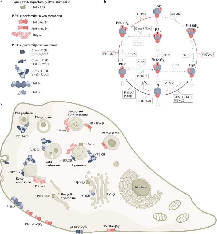
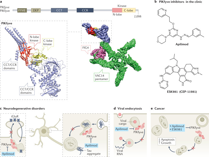
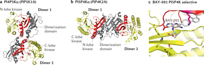
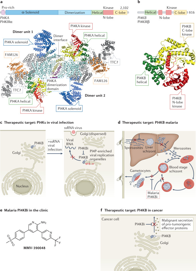
Lipid phosphoinositides are master regulators of almost all aspects of a cell's life and death and are generated by the tightly regulated activity of phosphoinositide kinases. Although extensive efforts have focused on drugging class I phosphoinositide 3-kinases (PI3Ks), recent years have revealed opportunities for targeting almost all phosphoinositide kinases in human diseases, including cancer, immunodeficiencies, viral infection and neurodegenerative disease. This has led to widespread efforts in the clinical development of potent and selective inhibitors of phosphoinositide kinases. This Review summarizes our current understanding of the molecular basis for the involvement of phosphoinositide kinases in disease and assesses the preclinical and clinical development of phosphoinositide kinase inhibitors.


Conflict of interest statement

J.E.B. reports personal fees from Scorpion Therapeutics and Olema Oncology; and research grants from Novartis. J.T., B.M.E. and G.R.V.H. declare no competing interests.

Figures

Fig. 1
Fig. 1. Phosphoinositides and the phosphoinositide kinases that generate them.
a, All phosphoinositide kinases (PIKs) encoded by the human genome, grouped by evolutionary relatedness. Families were initially grouped around activity, before specific enzymes had been cloned or complete identification of the specific regio-isomer substrates and products. Hence, the phosphoinositide 3-kinase (PI3K) superfamily incorporates the type I PIKs (now known to be PI3Ks) and type III PIKs (now known to be PI4KA and PI4KB, and still referred to as PI4KIIIα and PI4KIIIβ at the protein level). Class III PI3K VPS34 exists as two distinct heterotetramers, differing in a single subunit between complexes I and II (referred to as VPS34 CI/CII). Cloning of the type II PIKs (PI4K2A, PI4K2B) revealed them to be an evolutionarily distinct family of enzymes. The phosphatidylinositol phosphate kinases (PIPKs), are now known to be three subfamilies, each catalysing a specific hydroxyl phosphorylation on different substrates. b, Substrate and catalytic activity of PIKs. The production and turnover of phosphoinositides are mediated by the coordinated action of lipid kinases, phosphatases and lipases. All phosphoinositide species are generated from phosphatidylinositol (PI). Three different hydroxyls on the inositol ring of PI can be phosphorylated, at the D3, D4 and D5 positions. This leads to the generation of seven phosphoinositides: three mono-phosphorylated PIPs, phosphatidylinositol 3-phosphate (PI3P), PI4P and PI5P; three bis-phosphorylated PIP2s, phosphatidylinositol 4,5-bisphosphate (PI(4,5)P2), PI(3,5)P2, PI(3,4)P2 and the single tris-phosphorylated PIP3 phosphatidylinositol 3,4,5-trisphosphate (PI(3,4,5)P3 or PIP3). Grey lines indicate families of phosphatases that remove indicated phosphates, usually opposing a kinase reaction. Note that, like the kinases, the phosphatases were named on the basis of activity, phenotype or homology. These often predated definitive identification of specific catalytic activity, so the names are now somewhat arbitrary. For many enzymes, alternative substrates or catalytic activities have been reported in the test tube or in cells. However, we focus on the major pathways and activities that support the biology and pathology discussed in the text. c, Subcellular distribution of PIKs. We focus on membranes where most activity is reported, which does not necessarily reflect steady-state distribution of the enzymes themselves. For example, the PI5P4K enzymes are mostly localized in the cytosol and/or nucleoplasm. FIG4, FIG4 phosphoinositide 5-OH phosphatase; INPP4, inositol polyphosphatase 4-OH phosphatase; INPP5, inositol polyphosphatase 5-OH phosphatase; MTMR, mytotubularin-related; PTEN, phosphatase and tensin homologue; SAC, SAC phosphoinositide phosphatase.
Fig. 2
Fig. 2. Structure–function, inhibition and therapeutic targeting of PIKfyve.
a, Domain architecture of PIKfyve. The predicted structure of a fragment of the CCR/CCT and kinase domains (alphafold model of Q9Y2I7, all regions with pLDDT <50 removed) from the cryo-electron microscopy (cryo-EM) density is shown with the domains coloured according to the domain schematic. The cryo-EM density of the complex of PIKfyve with VAC14 and FIG4 is also shown, with the VAC14 pentamer coloured green, FIG4 coloured pink and the PIKfyve coloured as in the schematic. VAC14 in isolation forms a symmetrical pentamer, with oligomerization mediated by the C terminus. FIG4 can form a complex with VAC14 in the absence of PIKfyve, and it binds at the end of two VAC14 arms, leading to distortion of the symmetry of the VAC14 pentamer. PIKfyve’s association with VAC14 is strongly dependent on the presence of FIG4 (ref.). A single copy of PIKfyve binds to the opposite sides of the VAC14 arm bound to FIG4. Multiple cryo-EM maps were compiled to generate this figure (EMD: 22631, EMD: 22647, EMD: 22634). b, PIKfyve selective inhibitors (apilimod and ESK981) currently in clinical trials for cancer and viral infection. c, PIKfyve as a target for neurodegenerative disorders. Inhibition of PIKfyve prevents endocytic recycling of ionotropic glutamate receptors to the synapse, reducing excitotoxic death of glutamatergic neurons. It also prevents endocytic trafficking of tau or α-synuclein aggregates to the lysosome. d, PIKfyve as a target for viral infection. Again, disruption of PIKfyve activity prevents endocytic trafficking of endocytosed virus, preventing its escape into the cytoplasm from endolysosomes. e, PIKfyve as a target for cancer. PIKfyve inhibition prevents maturation and fusion of late autophagosomes with lysosomes, preventing the anti-apoptotic and pro-growth effects of autophagy in cancer cells.
Fig. 3
Fig. 3. Structure–function, and inhibition of the PI4P5Ks and PI5P4Ks.
a, Structure of the zebrafish homologue of phosphoinositide 4-phosphate 5-kinase-α (PI4P5Kα), with domains annotated on the figure. PI4P5Kα (PDB:4TZ7) shows a putative dimeric interface composed of the dimerization domain as well as the N-lobe of the kinase domain, which is unique compared with the phosphoinositide 5-phosphate 4-kinases (PI5P4Ks; see panel b). In contrast to the stable PI5P4K dimers, PI4P5Ks exist in a monomer–dimer equilibrium in solution, with dimerization promoted by binding to PI(4,5)P2-containing membrane surfaces, leading to enhanced catalytic efficiency. Like all lipid kinases, the kinase domain contains an activation loop that determines substrate specificity and also has a role in membrane recruitment. Swapping the activation loops between the type I PI4P5Ks and the type II PI5P4Ks led to not only swapped substrate specificity between PI4P and PI5P but also modified subcellular localization,. b, Structure of PI5P4Kα dimer, in which, in contrast to the PI4P5K, dimerization is putatively mediated solely by the dimerization domain (PDB: 6YM5). For type II PI5P4Ks, catalytic activity and PI5P substrate binding is carried out by the kinase domain, while homo- and heterodimerization with other type II PI5P4Ks is driven solely by the dimerization domain. This differs from the type I PI4P5Ks, which have a unique dimerization interface composed of both the kinase and dimerization domains, with dimerization required for PI4P5K lipid kinase activity (panel a). The difference in the dimerization interface between PI4P4Ks and PI5P4Ks allows for the potential formation of complexes between type I and type II phosphatidylinositol phosphate (PIP) kinases, with their roles being unknown, although preliminary evidence suggests a potential regulatory role,. c, Structure of PI5P4Kα bound to the selective inhibitor BAY-091, with the domains coloured according to panel b (PDB: 6YM5) and residues that make crucial interactions in determining selectivity shown as sticks.
Fig. 4
Fig. 4. Structure–function, inhibition and therapeutic targeting of PI4KA and PI4KB.
a, Domain architecture of phosphatidylinositol 4-kinase A (PI4KA). The architecture of the dimer of heterotrimers of PI4KA–TTC7–FAM126B (PDB: 6BQ1, with the solenoid region generated from the predicted α-fold model), with the domains coloured according to the domain schematic. The dimer interface between the two heterotrimers of PI4KA–TTC7–FAM126 is highlighted by a box, with regions that directly contact the other dimer unit coloured differently to highlight the dimer interface. b, Domain architecture of PI4KB. The structure of PI4KB (PDB: 4D0L), with the domains coloured according to panel domain schematic. c, Inhibition of PI4KB as an antiviral for positive-strand single-stranded RNA viruses (+ssRNA). Multiple picornaviruses require PI4KB as a host factor to generate PI4P-enriched viral replication organelles after viral infection. PI4P in these organelles recruits additional cellular machinery and restructures the lipid environment to generate a platform optimal for viral replication. Disruption of PI4KB either genetically or pharmacologically can prevent viral replication. d, Inhibition of the malarial homologue of PI4KB as an antimalarial therapeutic. The life cycle of malaria in both the vector (mosquito) and host (human) is indicated. The various life cycle stages of the Plasmodium species that cause malaria are annotated, and where malarial PI4KB inhibitors (PI4KBi; KDU691, KAI407, MMV390048) have shown efficacy are shown. PI4KBi have shown particular promise in the prevention of the multi-nucleated schizont stages in blood and liver by preventing membrane trafficking from the Golgi. e, Malarial PI4KBi currently in clinical trials. f, Inhibition of PI4KB as an anticancer therapeutic. PI4P generated by PI4KB plays a crucial part in malignant secretion of pro-tumorigenic effector proteins from cancer cells that contain a chromosome 1q region that is frequently amplified in diverse cancers. PI4P enhances secretion through activating Golgi phosphoprotein 3 (GOLPH3)-dependent vesicular release from the Golgi, with inhibition of PI4KB using highly selective PI4KIIIβ-IN-10 derivatives (PI4KBi) preventing this secretion. Panel a adapted with permission from ref., Elsevier.
Fig. 5
Fig. 5. Structure–function, inhibition and therapeutic targeting of class II PI3Ks.
a, Domain architecture of class II PI3Ks. b, The combined model of PI3KC2α with the domains coloured according to the domain schematic. The model is derived from both cryogenic electron microscopy (cryo-EM) and X-ray data (compiled from PDB: 7BI2, 6BTY). The interface of the C-terminal C2 domain (C-C2) with the Ras-binding domain (RBD) was generated by docking the structure of the C-C2 (PDB: 6BTY) into the cryo-EM density of EMD-12191. The putative PX interface on the kinase domain is based on HDX-MS data. c, The model of PIK3C2α activation, in which the PX and C-C2 domains inhibit class II PI3K activity in the closed configuration, and upon lipid binding of both these domains, class II PI3K adopts an open active configuration, allowing it to bind to lipid substrate, leading to opening of both the kinase and RBD domains. There are additional protein-binding partners that have important roles in activating and switching off class II PI3K signalling, but which are omitted here for clarity of the inactive and active states. d, Structure of PI3KC2α bound to the selective inhibitor PITCOIN3 (PDB: 7Z75), with the domains coloured according to the domain schematic in panel a. e, Inhibition of PI3KC2α as an anti-thrombolytic: genetic or pharmacological inhibition of PI3KC2α (PI3KC2αi) is reported to disrupt attachment of the membrane skeleton in platelets, disrupting the extensive infolding of the plasma membrane known as the open canicular system. This impedes the formation of protrusions and filopodia in activated platelets, thus reducing the formation of thrombi under arterial shear stress. This has been observed in mice, as well as in humans ex vivo. f, PI3KC2α as a target for tumour angiogenesis. Genetic disruption of PI3KC2α has been shown to potently inhibit angiogenesis; in adult mice, this can severely disrupt vascularization of tumours. HBD, helical bundle domain; N-C2, N-terminal C2 domain.
Fig. 6
Fig. 6. Structure–function, inhibition and therapeutic targeting of class III PI3Ks.
a, Domain architecture of VPS34. The architecture of the tetramer of complex II (CII) of VPS34 is shown (PDB: 7BL1), with VPS34 domains coloured according to the domain schematic. The additional UVRAG, BECLIN-1 and VPS15 subunits are indicated, with the binding site of the VPS34 CII activator RAB5 shown (RAB5 shown as a transparent surface). b, Structure of VPS34 bound to the class III PI3K selective inhibitors PIK-III and SAR405 (PDB: 4PH4, 4OYS). The hydrophobic pocket surrounding the -CF3 (SAR405) or cyclo-propyl (PIK-III) groups that is crucial in VPS34 selectivity is annotated. c, Complex-selective activation of the VPS34 CI and VPS34 CII by either RAB1 or RAB5 GTPase, respectively. RAB1, present at the autophagosome, recruits and activates VPS34 CI, whereas RAB5, present on endosomal membranes, recruits and activates VPS34 CII. d, VPS34 inhibitors (VPS34i) target autophagy in cancer. Pharmacological block of VPS34 disrupts VPS34 CI, disrupting autophagosome biogenesis. This precludes a necessary adaptation and mechanism of resistance for the cancer cell in the stressful environment of the tumour bed.

References

    1. Balla T. Phosphoinositides: tiny lipids with giant impact on cell regulation. Physiol. Rev. 2013;93:1019–1137. doi: 10.1152/physrev.00028.2012. - DOI - PMC - PubMed
    1. Dickson EJ, Hille B. Understanding phosphoinositides: rare, dynamic, and essential membrane phospholipids. Biochem. J. 2019;476:1–23. doi: 10.1042/BCJ20180022. - DOI - PMC - PubMed
    1. Schink KO, Tan K-W, Stenmark H. Phosphoinositides in control of membrane dynamics. Annu. Rev. Cell Dev. Biol. 2016;32:143–171. doi: 10.1146/annurev-cellbio-111315-125349. - DOI - PubMed
    1. Hasegawa J, Strunk BS, Weisman LS. PI5P and PI(3,5)P2: minor, but essential phosphoinositides. Cell Struct. Funct. 2017;42:49–60. doi: 10.1247/csf.17003. - DOI - PMC - PubMed
    1. Gozzelino L, De Santis MC, Gulluni F, Hirsch E, Martini M. PI(3,4)P2 signaling in cancer and metabolism. Front. Oncol. 2020;10:360. doi: 10.3389/fonc.2020.00360. - DOI - PMC - PubMed

Publication types

Substances