. 2022 Nov 15;6(1):57.
doi: 10.1186/s41747-022-00307-8.

Cerebellar irradiation does not cause hyperactivity, fear, and anxiety-related disorders in the juvenile rat brain

Affiliations

Cerebellar irradiation does not cause hyperactivity, fear, and anxiety-related disorders in the juvenile rat brain

Yafeng Wang et al. Eur Radiol Exp. .

Abstract

Background: The cerebellum is involved in hyperactivity, fear, and anxiety disorders that could be induced by whole-brain irradiation (WBI). However, whether cerebellar irradiation alone (CIA) could induce these disorders is unknown. We investigated the effect of CIA in an animal model.

Methods: Eleven-day-old rat pups underwent a single 3-Gy dose of either WBI (n = 28) or CIA (n = 20), while 34 rat pups were sham-irradiated (controls). Cell death was evaluated in the subgranular zone of the hippocampus by counting pyknotic cells after haematoxylin/eosin staining at 6 h after irradiation for 10, 8, and 9 pups, respectively. Behavioural changes were evaluated via open-field test at 6 weeks for 18, 12, and 25 pups, respectively. Unpaired two-tailed t-test and one-way and two-way repeated ANOVA were used.

Results: Massive cell death in cerebellar external granular layer was detected at 6 h after CIA (1,419 ± 211 mm, mean ± S.E.M. versus controls (68 ± 12 mm) (p < 0.001)), while no significant difference between CIA (1,419 ± 211 mm) versus WBI (1,433 ± 107 mm) (p = 0.955) was found. At open-field behavioural test, running distance, activity, wall distance, middle zone visit times, and duration were higher for WBI versus controls (p < 0.010), but no difference between CIA and controls was found (p > 0.05).

Conclusions: Although the cerebellum is involved in hyperactivity, fear, and anxiety disorders, CIA did not induce these disorders, indicating that WBI-induced cerebellar injury does not directly cause these behavioural abnormalities after WBI. Thus, targeting the cerebellum alone may not be enough to rescue or reduce these behavioural abnormalities after WBI.

Keywords: Anxiety; Cerebellum; Hyperactivity; Models (animal); Radiotherapy.


Conflict of interest statement

The authors declare that they have no competing interests.

Figures

Fig. 1
Fig. 1
Schematic view of the study process. The pups were subjected to irradiation at postnatal day 11. A total of 27 pups (9 for control group, 10 for whole-brain irradiation (WBI) group, and 8 for cerebellar irradiation alone were killed at 6 hours for cell death evaluation. A total of 55 pups were used for behavioural evaluation at 6 weeks after irradiation. IR, Irradiation.
Fig. 2
Fig. 2
Cell death in the hippocampal subgranular zone (SGZ) after whole-brain irradiation (WBI) and cerebellar irradiation alone (CIA). Representative black-and-white images of haematoxylin and eosin staining showing the nuclear morphology in the hippocampal SGZ of a control rat (a), a rat subjected to WBI (b), and a rat subjected to CIA (c). d Graph showing the dead cell density in the control, WBI, and CIA groups; each dot represents one mouse. The data are presented as the mean ± standard error of the mean (n = 8−10). ***p < 0.001 by one-way Analysis of variance (ANOVA). IR, Irradiation.
Fig. 3
Fig. 3
Cell death in the cerebellar external granular layer. Representative black-and-white images of haematoxylin and eosin staining showing the nuclear morphology in the cerebellum of a control rat (a), a rat subjected to whole-brain irradiation (WBI) (b), and a rat subjected to cerebellar irradiation alone (CIA) (c). d Graph showing the dead cell density in the control, WBI, and CIA groups; each dot represents one mouse. The data are presented as the mean ± standard error of the mean (n = 8−10). ***p < 0.001 by one-way ANOVA. IR, Irradiation.
Fig. 4
Fig. 4
Assessment of activity in the open-field test. a The upper graphs show the running distance every 5 min for a total of 20 min. ###p < 0.001 by two-way repeated ANOVA comparing the control and whole-brain irradiation (WBI) groups (n = 16−18). N.S. stands for no significant difference by two-way ANOVA comparing the control and cerebellar irradiation groups (n = 9−12). The lower graphs show the distance run over 20 min. ** p < 0.01 by unpaired two-tailed Student t-test comparing the control and WBI groups (n = 16−18). N.S. stands for no significant difference by unpaired two-tailed Student t-test comparing the control and cerebellar irradiation alone (CIA) groups (n = 10−12). b The upper graph shows the activity every 5 min for a total of 20 min. ###p < 0.001 by two-way ANOVA comparing the control and WBI groups (n = 16–18). N.S. stands for no significant difference by two-way ANOVA comparing the control and CIA groups (n = 9−12). The lower graphs show the distance run over 20 min. *p < 0.05 by unpaired two-tailed Student t-test comparing the control and WBI groups (n = 16−18). N.S. stands for no significant difference by unpaired two-tailed Student t-test comparing the control and CIA groups (n = 10−12). For all graphs, the data are presented as the mean ± standard error of the mean. IR, Irradiation.
Fig. 5
Fig. 5
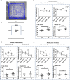
Assessment of fear and anxiety from the open-field test. a A picture showing the open-field arena. The blue line is the running trace of the rats, and the white rectangle is the middle zone. b A schematic showing the dimensions of the open-field arena. c The upper graphs show the distance from the wall every 5 min for a total of 20 min. ##p < 0.01 by two-way ANOVA comparing the control and whole-brain irradiation (WBI) groups (n = 16−18). N.S. stands for no significant difference by two-way ANOVA comparing the control and cerebellar irradiation alone (CIA) groups (n = 9−12). The lower graphs show the wall distance traveled over 20 min. ** p< 0.01 by unpaired two-tailed Student t-test comparing the control and WBI groups (n = 16−18). N.S. stands for no significant difference by unpaired two-tailed Student t-test between the control and CIA groups (n = 10−12). d The upper graphs show the time spent in the middle zone every 5 min for a total of 20 min. ##p < 0.01 by two-way repeated ANOVA comparing the control and WBI groups (n = 16−18). N.S. stands for no significant difference by two-way ANOVA comparing the control and CIA groups (n = 9−12). The lower graphs show the time spent in the middle zone over 20 min. **p < 0.01 by unpaired two-tailed Student’s t-test comparing the control and WBI groups (n = 16−18). N.S. stands for no significant difference by unpaired two-tailed Student t-test (n = 10−12). e The upper graphs show the number of times the middle zone was visited for every 5-min interval for a total of 20 min. ##p < 0.01 by two-way ANOVA comparing the control and WBI groups (n =16−18). N.S. stands for no significant difference by two-way ANOVA comparing the control and CIA groups (n = 9−12). The lower graphs show the number of visits to the middle zone over 20 min. **p < 0.01 by unpaired two-tailed Student t-test comparing the control and WBI groups (n = 16−18). N.S. stands for no significant difference by unpaired two-tailed Student t-test (n = 10−12). For all graphs, the data are presented as the mean ± standard error of the mean. IR, Irradiation.

Similar articles

Cited by

References

    1. Lassaletta A, Bouffet E, Mabbott D, Kulkarni AV. Functional and neuropsychological late outcomes in posterior fossa tumors in children. Childs Nerv Syst. 2015;31:1877–1890. doi: 10.1007/s00381-015-2829-9. - DOI - PubMed
    1. Marazziti D, Baroni S, Catena-Dell’Osso M, et al. Cognitive, psychological and psychiatric effects of ionizing radiation exposure. Curr Med Chem. 2012;19:1864–1869. doi: 10.2174/092986712800099776. - DOI - PubMed
    1. Khatua S, Sadighi ZS, Pearlman ML, Bochare S, Vats TS. Brain tumours in children--current therapies and newer directions. Indian J Pediatr. 2012;79:922–927. doi: 10.1007/s12098-012-0689-9. - DOI - PubMed
    1. DeNunzio NJ, Yock TI. Modern radiotherapy for paediatric brain tumours. Cancers. 2020;12:1533. doi: 10.3390/cancers12061533. - DOI - PMC - PubMed
    1. Scaringi C, Agolli L, Minniti G. Technical advances in radiation therapy for brain tumours. Anticancer Res. 2018;38:6041–6045. doi: 10.21873/anticanres.12954. - DOI - PubMed

Publication types

LinkOut - more resources