

Epigenome-wide analysis of T-cell large granular lymphocytic leukemia identifies BCL11B as a potential biomarker
- PMID: 36376973
- PMCID: PMC9664638
- DOI: 10.1186/s13148-022-01362-z
Epigenome-wide analysis of T-cell large granular lymphocytic leukemia identifies BCL11B as a potential biomarker
Erratum in
-
Publisher Correction: Epigenome-wide analysis of T-cell large granular lymphocytic leukemia identifies BCL11B as a potential biomarker.Clin Epigenetics. 2023 Jan 6;15(1):3. doi: 10.1186/s13148-022-01405-5. Clin Epigenetics. 2023. PMID: 36609404 Free PMC article. No abstract available.
Abstract
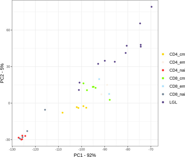
Background: The molecular pathogenesis of T-cell large granular lymphocytic leukemia (T-LGLL), a mature T-cell leukemia arising commonly from T-cell receptor αβ-positive CD8+ memory cytotoxic T cells, is only partly understood. The role of deregulated methylation in T-LGLL is not well known. We analyzed the epigenetic profile of T-LGLL cells of 11 patients compared to their normal counterparts by array-based DNA methylation profiling. For identification of molecular events driving the pathogenesis of T-LGLL, we compared the differentially methylated loci between the T-LGLL cases and normal T cells with chromatin segmentation data of benign T cells from the BLUEPRINT project. Moreover, we analyzed gene expression data of T-LGLL and benign T cells and validated the results by pyrosequencing in an extended cohort of 17 patients, including five patients with sequential samples.
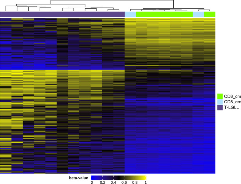
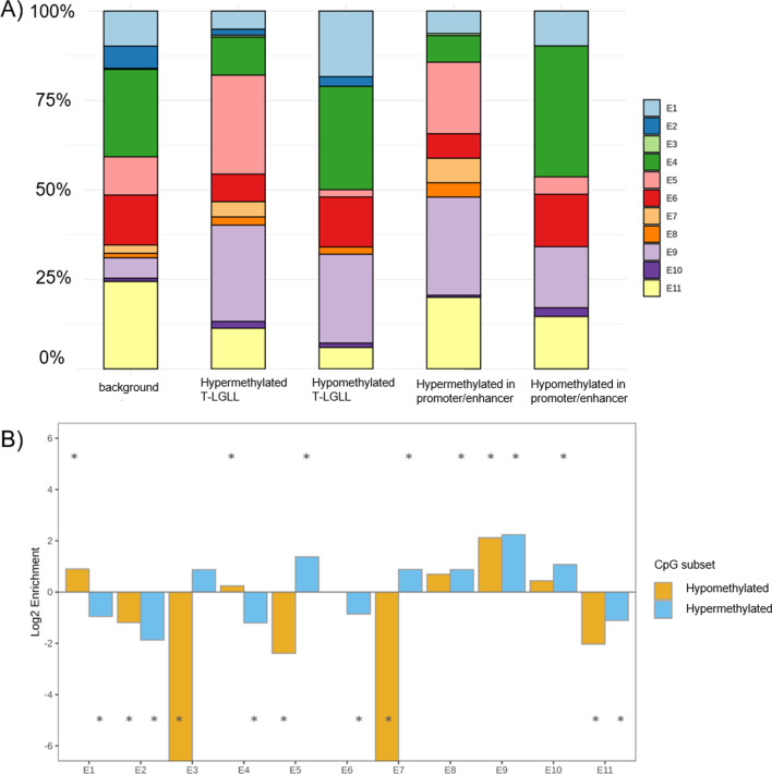
Results: We identified dysregulation of DNA methylation associated with altered gene expression in T-LGLL. Since T-LGLL is a rare disease, the samples size is low. But as confirmed for each sample, hypermethylation of T-LGLL cells at various CpG sites located at enhancer regions is a hallmark of this disease. The interaction of BLC11B and C14orf64 as suggested by in silico data analysis could provide a novel pathogenetic mechanism that needs further experimental investigation.
Conclusions: DNA methylation is altered in T-LGLL cells compared to benign T cells. In particular, BCL11B is highly significant differentially methylated in T-LGLL cells. Although our results have to be validated in a larger patient cohort, BCL11B could be considered as a potential biomarker for this leukemia. In addition, altered gene expression and hypermethylation of enhancer regions could serve as potential mechanisms for treatment of this disease. Gene interactions of dysregulated genes, like BLC11B and C14orf64, may play an important role in pathogenic mechanisms and should be further analyzed.
Keywords: BCL11B; DNA methylation; Large granular lymphocytic leukemia; Pyrosequencing; T-LGLL.
© 2022. The Author(s).
Conflict of interest statement
The authors declare no competing financial interest.
Figures


References
-
- Bigouret V, Hoffmann T, Arlettaz L, Villard J, Colonna M, Ticheli A, et al. Monoclonal T-cell expansions in asymptomatic individuals and in patients with large granular leukemia consist of cytotoxic effector T cells expressing the activating CD94:NKG2C/E and NKD2D killer cell receptors. Blood. 2003;101:3198–3204. - PubMed
-
- Lamy L. Large granular lymphocyte leukemia. Cancer Control. 1998;5:25–33. - PubMed
-
- Viny AD, Lichtin A, Pohlman B, Loughran T, Maciejewski J. Chronic B-cell dyscrasias are an important clinical feature of T-LGL leukemia. Leuk Lymphoma. 2008;49:932–938. - PubMed
-
- Sun H, Wei S, Yang L. Dysfunction of immune system in the development of large granular lymphocyte leukemia. Hematology. 2019;24:139–147. - PubMed
Publication types
MeSH terms
Substances
LinkOut - more resources
Full Text Sources
Research Materials
