. 2022 Nov 14;17(1):31.
doi: 10.1186/s13062-022-00345-7.

Dual-labeled nanoparticles based on small extracellular vesicles for tumor detection

Affiliations

Dual-labeled nanoparticles based on small extracellular vesicles for tumor detection

Ana Santos-Coquillat et al. Biol Direct. .

Abstract

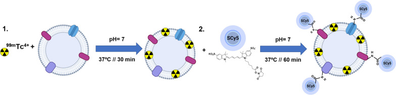
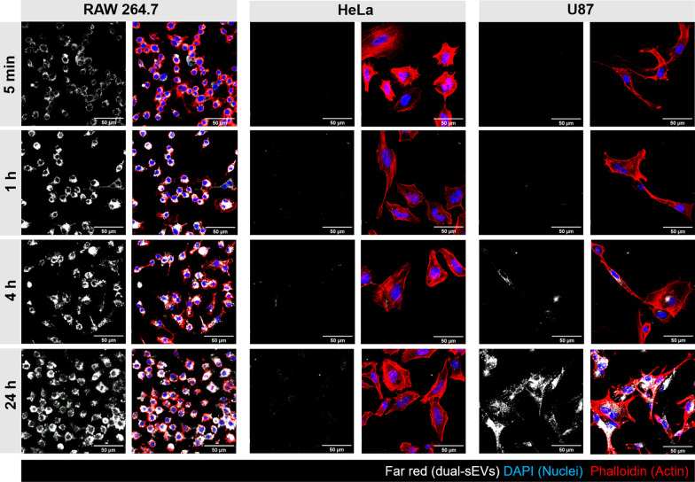
Background: Small extracellular vesicles (sEVs) are emerging natural nanoplatforms in cancer diagnosis and therapy, through the incorporation of signal components or drugs in their structure. However, for their translation into the clinical field, there is still a lack of tools that enable a deeper understanding of their in vivo pharmacokinetics or their interactions with the cells of the tumor microenvironment. In this study, we have designed a dual-sEV probe based on radioactive and fluorescent labeling of goat milk sEVs.

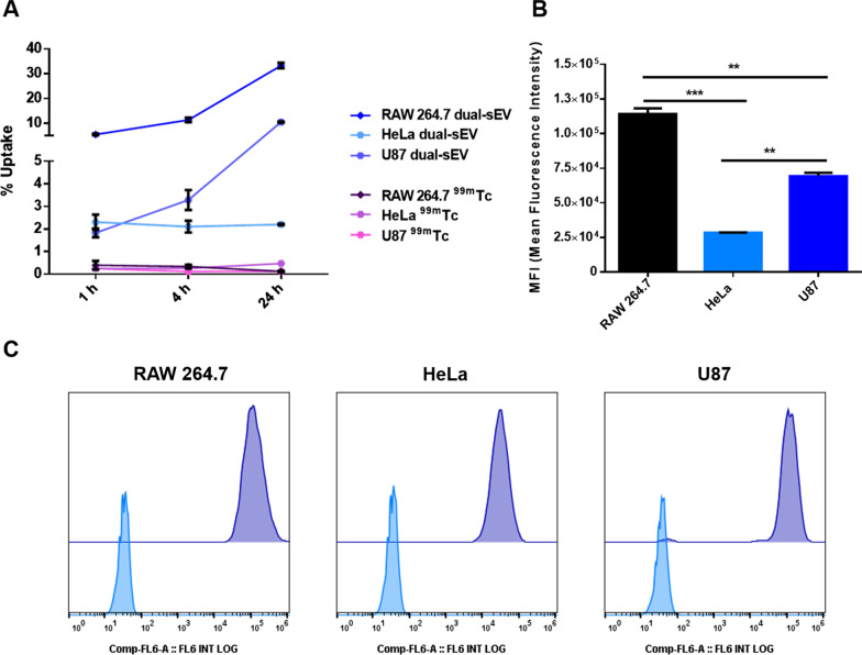
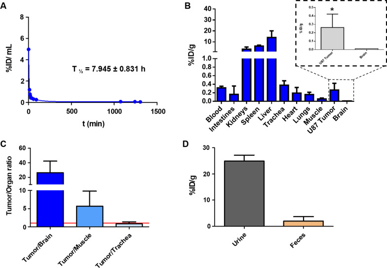
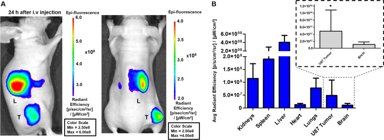
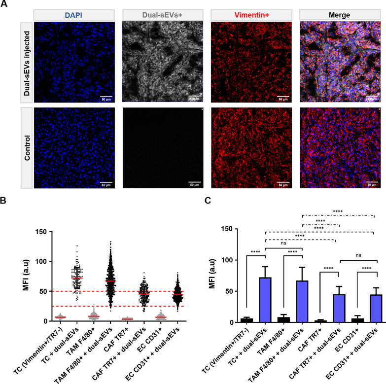
Results: The imaging nanoprobe was tested in vitro and in vivo in a model of glioblastoma. In vitro assessment of the uptake of the dual probe in different cell populations (RAW 264.7, U87, and HeLa) by optical and nuclear techniques (gamma counter, confocal imaging, and flow cytometry) revealed the highest uptake in inflammatory cells (RAW 264.7), followed by glioblastoma U87 cells. In vivo evaluation of the pharmacokinetic properties of nanoparticles confirmed a blood circulation time of ~ 8 h and primarily hepatobiliary elimination. The diagnostic capability of the dual nanoprobe was confirmed in vivo in a glioblastoma xenograft model, which showed intense in vivo uptake of the SEV-based probe in tumor tissue. Histological assessment by confocal imaging enabled quantification of tumor populations and confirmed uptake in tumor cells and tumor-associated macrophages, followed by cancer-associated fibroblasts and endothelial cells.

Conclusions: We have developed a chemical approach for dual radioactive and fluorescent labeling of sEVs. This methodology enables in vivo and in vitro study of these vesicles after exogenous administration. The dual nanoprobe would be a promising technology for cancer diagnosis and a powerful tool for studying the biological behavior of these nanosystems for use in drug delivery.

Keywords: Diagnosis; Extracellular vesicles; Molecular imaging; Oncology; Optical imaging; SPECT.


Conflict of interest statement

The authors declare no competing interests.

Figures

Fig. 1
Fig. 1
Synthesis of dual-sEVs. 1 Radioactive labeling of sEVs with 99mTc (IV). 2 Fluorescent labeling of the resulting product with SCy5 fluorophore. Illustration made with Biorender
Fig. 2
Fig. 2
Physicochemical characterization of dual-sEVs. A Radioactive (red) and UV/VIS (blue) HPLC chromatograms of dual-sEVs (normalized intensity), and UV/VIS HPLC chromatogram of non-labeled sEVs (green). B Radio TLC chromatogram (counts/mm × 1000) of dual-sEVs. C Fluorimetric analysis (relative fluorescent units; RFU) of the maximum emission peak of dual-sEVs
Fig. 3
Fig. 3
Physicochemical characterization of dual-sEVs. A Nanoparticle tracking analysis with concentration (particles/mL) and size of labeled sEVs (nm). B Transmission electron microscopy images showing the morphology and size of the sEVs
Fig. 4
Fig. 4
In vitro assessment of radioactive and optical uptake of dual-sEVs by gamma counter and flow cytometry. A Radioactive uptake for RAW 264.7, HeLa, and U87 cells after 1 h, 4 h, and 24 h of dose addition. 99mTc was used as a control. Data are represented as mean ± standard deviation (SD). B Median fluorescence intensity (MFI) inside the cells was evaluated at 24 h. * p < 0.05, ** p < 0.01, *** p < 0.001. Data are represented as mean ± SD. C Flow cytometry diagrams of control cells (RAW 264.7, HeLa, and U87 cells, in blue) and treated cells (RAW 264.7, HeLa, and U87 cells, in purple)
Fig. 5
Fig. 5
Optical uptake of dual-sEVs by confocal microscopy in RAW 264.7, HeLa, and U87 cells at 5 min, 1 h, 4 h, and 24 h after the administration of 5 μg/mL of dual-sEVs. Blue, DAPI; red, phalloidin; and white, dual-sEVs
Fig. 6
Fig. 6
In vivo and ex vivo assessment of dual-sEVs by nuclear techniques. A Blood half-life. B Ex vivo biodistribution of dual-sEVs in a U87 xenograft mouse model 24 h after tracer injection. Detailed radioactivity in the brain (control organ) compared to U87 tumor tissue. * p < 0.05. C Ratio of dual-SEV uptake in U87 tumor tissue to non-target tissues at 24 h. D Excretion profile of dual-sEVs in urine and feces collected from the animal 24 h post-injection. Radioactivity in tissues is expressed as % ID/g. Data are represented as mean ± SD
Fig. 7
Fig. 7
In vivo optical imaging of dual-sEVs. A In vivo optical imaging of tumor-bearing mice 24 h after i.v. injection in the lateral (left) and prone positions (right). B Ex vivo biodistribution of the excised organs (brain, spleen, kidneys, liver, tumor, heart, lungs; n = 11). Detailed quantification of the brain (control organ) compared to U87 tumor tissue. * p < 0.05. Data is represented as mean ± SD
Fig. 8
Fig. 8
Histological analysis of the tumor microenvironment. A Confocal microscopy of tumor tissue with injection of dual-sEVs (white). Blue, DAPI; red, vimentin + . B Quantification of the uptake by control and injected (dual-sEVs +) populations: TCs, vimentin + /TR7-; TAMs, F4/80 + ; CAFs, TR7 + ; and ECs, CD31 + . C Mean uptake values in the populations. Data are represented as mean ± SD. **** p < 0.0001

References

    1. Weissleder R, Nahrendorf M. Advancing biomedical imaging. Proc Natl Acad Sci U S A. 2015;112:14424–14428. - PMC - PubMed
    1. O'Connor JPB, Aboagye EO, Adams JE, Aerts HJWL, Barrington SF, Beer AJ, Boellaard R, Bohndiek SE, Brady M, Brown G, et al. Imaging biomarker roadmap for cancer studies. Nat Rev Clin Oncol. 2017;14:169–186. - PMC - PubMed
    1. Louie A. Multimodality imaging probes: design and challenges. Chem Rev. 2010;110:3146–3195. - PMC - PubMed
    1. Liu M, Anderson R-C, Lan X, Conti PS, Chen K. Recent advances in the development of nanoparticles for multimodality imaging and therapy of cancer. Med Res Rev. 2020;40:909–930. - PubMed
    1. Hapuarachchige S, Artemov D. Theranostic pretargeting drug delivery and imaging platforms in cancer precision medicine. Front Oncol. 2020;10:1131. - PMC - PubMed

Publication types

LinkOut - more resources