

Primary non-Hodgkin lymphoma of the chiasm and optic tract in a nonimmunocompromised patient: illustrative case
- PMID: 36377128
- PMCID: PMC9664242
- DOI: 10.3171/CASE22340
Primary non-Hodgkin lymphoma of the chiasm and optic tract in a nonimmunocompromised patient: illustrative case
Abstract
Background: Chiasmatic and optic track lymphoma as the primary lesion of the central nervous system (CNS) is extremely rare.
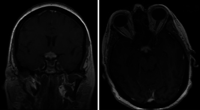
Observations: The authors report a case of a previously healthy 62-year-old woman who presented with quick and progressive visual impairment leading to bilateral blindness. Brain imaging studies suggested glioma or lymphoma of the chiasm and the posterior visual pathway. Postoperative examination revealed low-grade malignant B-cell lymphoma. No evidence of extracranial lymphoma was found, so a final diagnosis of primary CNS lymphoma (PCNSL) was made.
Lessons: To the authors' knowledge, PCNSL confined to the optic chiasm has rarely been reported in nonimmunocompromised patients. The present case of lymphoma affecting the optic chiasm and optic tract is extremely rare.
Keywords: chiasmatic involvement; non-Hodgkin lymphoma; optic nerve involvement; primary central nervous system lymphoma.
Conflict of interest statement
Figures


References
-
- Armitage JO, Gascoyne RD, Lunning MA, Cavalli F. Non-Hodgkin lymphoma. Lancet. 2017;390(10091):298–310. - PubMed
-
- Corn BW, Marcus SM, Topham A, Hauck W, Curran WJ., Jr Will primary central nervous system lymphoma be the most frequent brain tumor diagnosed in the year 2000? Cancer. 1997;79(12):2409–2413. - PubMed
-
- Eby NL, Grufferman S, Flannelly CM, Schold SC, Jr, Vogel FS, Burger PC. Increasing incidence of primary brain lymphoma in the US. Cancer. 1988;62(11):2461–2465. - PubMed
-
- Kim UR, Shah AD, Arora V, Solanki U. Isolated optic nerve infiltration in systemic lymphoma—a case report and review of literature. Ophthal Plast Reconstr Surg. 2010;26(4):291–293. - PubMed
LinkOut - more resources
Full Text Sources
