. 2022 Dec 20;13(6):e0285422.
doi: 10.1128/mbio.02854-22. Epub 2022 Nov 15.

Postinfluenza Environment Reduces Aspergillus fumigatus Conidium Clearance and Facilitates Invasive Aspergillosis In Vivo

Affiliations

Postinfluenza Environment Reduces Aspergillus fumigatus Conidium Clearance and Facilitates Invasive Aspergillosis In Vivo

Ko-Wei Liu et al. mBio. .

Abstract

Aspergillus fumigatus is a human fungal pathogen that is most often avirulent in immunecompetent individuals because the innate immune system is efficient at eliminating fungal conidia. However, recent clinical observations have shown that severe influenza A virus (IAV) infection can lead to secondary A. fumigatus infections with high mortality. Little is currently known about how IAV infection alters the innate antifungal immune response. Here, we established a murine model of IAV-induced A. fumigatus (IAV-Af) superinfection by inoculating mice with IAV followed 6 days later by A. fumigatus conidia challenge. We observed increased mortality in the IAV-Af-superinfected mice compared to mice challenged with either IAV or A. fumigatus alone. A. fumigatus conidia were able to germinate and establish a biofilm in the lungs of the IAV-Af superinfection group, which was not seen following fungal challenge alone. While we did not observe any differences in inflammatory cell recruitment in the IAV-Af superinfection group compared to single-infection controls, we observed defects in Aspergillus conidial uptake and killing by both neutrophils and monocytes after IAV infection. pHrodo Green zymosan bioparticle (pHrodo-zymosan) and CM-H2DCFDA [5-(and-6)-chloromethyl-2',7'-dichlorodihydrofluorescein diacetate] staining, indicators of phagolysosome maturation and reactive oxygen species (ROS) production, respectively, revealed that the fungal killing defect was due in part to reduced phagolysosome maturation. Collectively, our data demonstrate that the ability of neutrophils and monocytes to kill and clear Aspergillus conidia is strongly reduced in the pulmonary environment of an IAV-infected lung, which leads to invasive pulmonary aspergillosis and increased overall mortality in our mouse model, recapitulating what is observed clinically in humans. IMPORTANCE Influenza A virus (IAV) is a common respiratory virus that causes seasonal illness in humans, but can cause pandemics and severe infection in certain patients. Since the emergence of the 2009 H1N1 pandemic strains, there has been an increase in clinical reports of IAV-infected patients in the intensive care unit (ICU) developing secondary pulmonary aspergillosis. These cases of flu-Aspergillus superinfections are associated with worse clinical outcomes than secondary bacterial infections in the setting of IAV. To date, we have a limited understanding of the cause(s) of secondary fungal infections in immunocompetent hosts. IAV-induced modulation of cytokine production and innate immune cellular function generates a unique immune environment in the lung, which could make the host vulnerable to a secondary fungal infection. Our work shows that defects in phagolysosome maturation in neutrophils and monocytes after IAV infection impair the ability of these cells to kill A. fumigatus, thus leading to increased fungal germination and growth and subsequent invasive aspergillosis. Our work lays a foundation for future mechanistic studies examining the exact immune modulatory events occurring in the respiratory tract after viral infection leading to secondary fungal infections.

Keywords: Aspergillus fumigatus; antifungal immunity; host-pathogen interactions; influenza A virus; innate immunity; invasive pulmonary aspergillosis; monocytes; neutrophils; pathogenesis; phagocytosis; phagolysosome; phagolysosome maturation; superinfection.


Conflict of interest statement

The authors declare no conflict of interest.

Figures
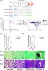
FIG 1
FIG 1
Influenza A virus infection aggravates invasive aspergillosis disease progression. (A) Schematic design of IAV-Af infection mouse model. C57BL/6J mice were inoculated with 100 EID50 of A/PR/8/34 (IAV) or PBS at day 0 followed by 3.4 × 10⁷ CEA10 conidia or PBS at day 6. Mice were euthanized at either 8, 24, 36, or 48 h post-CEA10 inoculation. (B) Survival curve (left) of mice with PBS inoculation, IAV single infection, CEA10 single infection, and IAV-CEA10 superinfection (n = 10) and the weights (right) of mice in each group. Two independent experiments were performed, and data are shown as the representative results. (C) For quantification of pathogen load, RNA was isolated from the lungs of mice exposed to 6 days of either IAV or PBS followed by 36 h of CEA10 or PBS inoculation. Fungal burden was examined by qRT-PCR on A. fumigatus 18S rRNA (n = 7). (D) Viral load was examined by qRT-PCR on viral matrix protein (n = 7). Panels C and D are representative of three independent experiments. (E) For lung histology, mice were euthanized after 6 days of IAV or PBS incubation followed by 48 h postinoculation with CEA10 or PBS. Representative histology images of mice lungs were observed with (E) GMS staining and (F) H&E staining. Two independent experiments were performed with n = 5 per experiment. The log rank test and Gehan-Breslow-Wilcoxon test were performed for statistical analysis of the survival curve, and nonparametric analyses were performed (Mann-Whitney for single comparisons) for the pathogen load. All error bars represent standard deviations. NS, not significant at P > 0.05; *, P ≤ 0.05; **, P ≤ 0.01; ***, P ≤ 0.001; ****, P ≤ 0.0001.
FIG 2
FIG 2
Influenza A virus infection does not affect lung cellularity during A. fumigatus infection. C57BL/6J mice were inoculated with 100 EID50 of A/PR/8/34 (IAV) or PBS at day 0 followed by 3.4 × 10⁷ CEA10 conidia or PBS at day 6. Mice were euthanized at 36 h postinoculation with CEA10 or PBS for lung cellularity experiments. Three independent experiments were performed, and data are shown as the combined results (PBS/PBS group, n = 7; IAV/PBS group, n = 8; PBS/CEA10 group, n = 22; IAV/CEA10 group, n = 22). All lung cell numbers were acquired by flow cytometry as indicated: (A) total lung cells, (B) neutrophils (CD45+ Ly6G+ CD11b+), (C) alveolar macrophages (aMac) (Ly6G CD103 SiglecF+ CD11b+), (D) interstitial macrophages (iMac) (Ly6G CD103 SiglecF CD11bhi CD64+), (E) monocytes (Ly6G CD103 SiglecF CD11bhi CD64 MHC-II), (F) CD103+ cDC1s (MHC-II+ CD11c+ CD11b CD103+), (G) CD11b+ cDC2s (MHC-IIhi CD11chi CD103 CD11b+), (H) pDCs (MHC-II+ CD11c+ CD11b CD103 CD317+). The Kruskal-Wallis test with Dunn’s multiple comparisons was performed for statistical analyses. All error bars represent standard deviations. NS, not significant at P > 0.05; *, P ≤ 0.05; **, P ≤ 0.01; ***, P ≤ 0.001; ****, P ≤ 0.0001.
FIG 3
FIG 3
Defects in leukocyte-mediated fungal killing post-influenza A virus infection. C57BL/6J mice were inoculated with 100 EID50 of A/PR/8/34 (IAV) or PBS at day 0 followed by 3.4 × 10⁷ FLARE (mRFP+/AF633+) conidia or PBS at day 6. Mice were euthanized at 36 h postinoculation with FLARE conidia or PBS. The percentage of cells positive for conidial tracer (AF633+) and conidial viability within the immune cells (mRFP+/AF633+) were analyzed. Phagocytosis and conidial viability were examined in (A) neutrophils (CD45+ Ly6G+ CD11b+), (B) monocytes (Ly6G CD103 SiglecF CD11bhi CD64 MHC-II), (C) alveolar macrophages (aMac) (Ly6G CD103 SiglecF+ CD11b+), (D) interstitial macrophages (iMac) (Ly6G CD103 SiglecF CD11bhi CD64+), and (E) CD11b+ cDC2s (MHC-IIhi CD11chi CD103 CD11b+). (F) The viability of FLARE conidia within immune cells in the lung suspension was assessed by CFU. (G) The viability of free FLARE conidia in the lung suspension is shown as the percentage of mRFP+ cells in the free conidial population (AF633+ FSClow SSClow). Three independent experiments were performed, and data are shown as the combined results (PBS/CEA10 group, n = 22; IAV/CEA10 group, n = 21). The Mann-Whitney test with single comparisons was performed. All error bars represent standard deviations. NS, not significant at P > 0.05; *, P ≤ 0.05; **, P ≤ 0.01; ***, P ≤ 0.001; ****, P ≤ 0.0001.
FIG 4
FIG 4
Postinfluenza immunity does not hinder neutrophil or monocyte ROS production. C57BL/6J mice were inoculated with 100 EID50 of A/PR/8/34 (IAV) or PBS at day 0 followed by 3.4 × 10⁷ CEA10 conidia or PBS at day 6. Mice were euthanized at 8 h postinoculation with CEA10 or PBS for ROS measurement. Lung cell suspensions were stained with CM-H2DCFDA for 30 min and then stained for neutrophils and monocytes. ROS production was measured by the signal from CM-H2DCFDA staining in (A) neutrophils (Ly6G+) and (B) monocytes (Ly6G SiglecF CD11bhi CD64 MHC-II). Two independent experiments were performed, and data are shown as the combined results (PBS/PBS group, n = 8; IAV/PBS group, n = 8; PBS/CEA10 group, n = 12; IAV/CEA10 group, n = 11). The Kruskal-Wallis test with Dunn’s multiple comparisons was performed for statistical analyses. All error bars represent standard deviations. NS, not significant at P > 0.05; *, P ≤ 0.05; **, P ≤ 0.01; ***, P ≤ 0.001; ****, P ≤ 0.0001.
FIG 5
FIG 5
Reduced phagolysosome maturation can be detected in neutrophils and monocytes during early viral infection. C57BL/6J mice were inoculated with 100 EID50 of A/PR/8/34 (IAV) or PBS at day 0 followed by 3.4 × 10⁷ CEA10 conidia or PBS at day 6. Mice were euthanized at 8 h postinoculation with CEA10 or PBS for phagolysosome maturation analysis. Lung cell suspensions were incubated with pHrodo-zymosan for 2 h and then stained for neutrophils and monocytes. The phagolysosome maturation level was measured by the signal from the color change of pHrodo-zymosan in (A) neutrophils (Ly6G+) and (B) monocytes (Ly6G SiglecF CD11bhi CD64 MHC-II). Two independent experiments were performed, and data are shown as the combined results (PBS/PBS group, n = 8; IAV/PBS group, n = 8; PBS/CEA10 group, n = 12; IAV/CEA10 group, n = 11). The Kruskal-Wallis test with Dunn’s multiple comparisons was performed for statistical analyses. All error bars represent standard deviations. NS, not significant at P > 0.05; *, P ≤ 0.05; **, P ≤ 0.01; ***, P ≤ 0.001; ****, P ≤ 0.0001.
FIG 6
FIG 6
Functional neutrophils from the post-IAV environment show decreasing phagolysosome maturation and conidial killing. C57BL/6J mice were inoculated with 100 EID50 of A/PR/8/34 (IAV) or PBS at day 0 followed by 3.4 × 10⁷ FLARE (mRFP+/AF633+) conidia or PBS at day 6. Mice were euthanized at 36 h postinoculation with FLARE conidia or PBS. Lung cell suspensions were incubated with pHrodo-zymosan for 2 h and then stained for neutrophils. (A) Representative confocal images of neutrophil labeling (Ly6G-Pb [white]), mature phagolysosome (pHrodo-zymosan [green]), labeled conidia (AF633 [yellow]), and conidial viable marker (mRFP [pink]). The right images feature labeled conidia, neutrophils, and pHrodo-zymosan signal. The left images feature labeled conidia and RFP to indicate fungal viability. Fungal conidia with no RFP signal are considered “dead.” (B) Currently functional neutrophils are indicated as pHrodo-zymosan+ cells. (C) The number of mature phagolysosomes is shown by the geometric mean fluorescent intensity (gMFI) of pHrodo-zymosan in Ly6G+ pHrodo-zymosan+ cells. The percentage of cells positive for conidia (AF633+) and conidial viability within the immune cells (mRFP+/AF633+) were analyzed as (D) the percentage of conidial uptake in Ly6G+ pHrodo-Zymosan+ cells and (E) the percentage of viable conidia in Ly6G+ pHrodo-zymosan+ cells. This repeated experiment was done as the combination of FLARE experiment (Fig. 3) and phagolysosome maturation measurement (Fig. 5) (PBS/CEA10 group, n = 6; IAV/CEA10 group, n = 5). The Mann-Whitney test with single comparisons was performed. All error bars represent standard deviations. NS, not significant at P > 0.05; *, P ≤ 0.05; **, P ≤ 0.01; ***, P ≤ 0.001; ****, P ≤ 0.0001.
FIG 7
FIG 7
Influenza A virus infection increases transcripts of genes associated with an inflammatory response but reduces transcript levels of known fungal pattern recognition receptors. C57BL/6J mice were inoculated with 100 EID50 of A/PR/8/34 (IAV) or PBS at day 0 followed by 3.4 × 10⁷ CEA10 conidia or PBS at day 6. Mice were euthanized at 8 h postinoculation with CEA10 or PBS for antifungal gene transcript analysis. Antifungal gene transcript levels were measured by qRT-PCR with RNA from lung cell suspensions. (A) Increased (red) or decreased (green) transcript levels of genes associated with antifungal responses in the IAV/CEA10 group compared to the PBS/CEA10 group are represented by the heat map (PBS/CEA10 group, n = 3; IAV/CEA10 group, n = 3). (B) A volcano plot shows the distribution of fold changes of antifungal gene transcript levels in the IAV/CEA10 group compared to the PBS/CEA10 group. Genes with an increase in fold changes of >2 are shown in red, and genes with a decrease in fold changes of >2 are shown in blue. The P value threshold of 0.05 (Student’s t test) is indicated by the line in the plot.

References

    1. Margalit A, Kavanagh K. 2015. The innate immune response to Aspergillus fumigatus at the alveolar surface. FEMS Microbiol Rev 39:670–687. doi:10.1093/femsre/fuv018. - DOI - PubMed
    1. Kauffman HF. 2003. Immunopathogenesis of allergic bronchopulmonary aspergillosis and airway remodeling. Front Biosci 8:190–196. doi:10.2741/990. - DOI - PubMed
    1. Pashley CH. 2014. Fungal culture and sensitisation in asthma, cystic fibrosis and chronic obstructive pulmonary disorder: what does it tell us? Mycopathologia 178:457–463. doi:10.1007/s11046-014-9804-y. - DOI - PubMed
    1. Dagenais TRT, Keller NP. 2009. Pathogenesis of Aspergillus fumigatus in invasive aspergillosis. Clin Microbiol Rev 22:447–465. doi:10.1128/CMR.00055-08. - DOI - PMC - PubMed
    1. El-Baba F, Gao Y, Soubani AO. 2020. Pulmonary aspergillosis: what the generalist needs to know. Am J Med 133:668–674. doi:10.1016/j.amjmed.2020.02.025. - DOI - PubMed