The right coronary artery in the heart of chinchilla (Chinchilla laniger Molina)
- PMID: 36380084
- PMCID: PMC10209256
- DOI: 10.1007/s11259-022-10035-4
The right coronary artery in the heart of chinchilla (Chinchilla laniger Molina)
Abstract
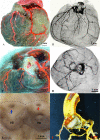
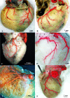
The pattern of normal coronary vascularization in a mammalian heart includes the presence of both right and left coronary arteries. According to the literature data, the presence of single major coronary arteries is mainly related to cardiac abnormalities. Previously it has been reported that the right coronary artery is absent in the coronary vascularization of the heart in the chinchilla. Our research was carried out on thirty chinchillas (Chinchilla laniger Molina). The coronary vessels were filled with colored latex to render them visible. The examinations were supplemented additionally with the use of microcomputed tomography with arterial contrast. Our study demonstrates its undoubtedly presence of the right coronary artery. In all subjects the right coronary artery was present, as was the left coronary artery. Two types of right coronary artery were found. Our results indicate that the normal pattern of coronary vascularization of heart in chinchilla includes both the right and left coronary arteries. An open question remains the presence of single coronary artery is a normal pattern of cardiac arterial vascularization in chinchilla.
Keywords: Anatomy; Chinchilla; Coronary arterial system.
© 2022. The Author(s).
Conflict of interest statement
The authors declare no competing interests.
The authors have no conflicts of interest to declare that are relevant to the content of this article. The research conformed with the requirements of Act for the Protection of Animals Used for Scientific or Educational Purposes (15 January 2015). According with Polish Law: “Studies of tissues obtained post-mortem do not require the approval of an ethics committee”.
Figures


References
-
- Aksoy G, Karadag H. An anatomic investigation on the heart and coronary arteries in the domestic cat and White New Zealand Rabbits. Vet Bil Derg. 2002;18:33–40.
-
- Barszcz K, Kupczyńska M, Wąsowicz M, Czubaj N, Sokołowski W. Patterns of the arterial vascularization of the dog’s heart. Med Weter. 2013;69:531–534.
-
- Barszcz K, Kupczyńska M, Klećkowska.Nawrot J, Janczak P, Krasucki K, Wąsowicz M (2014) Arterial coronary circulation in cats. Med Weter 70(6):373-377
-
- Barszcz K, Kupczyńska M, Polguj M, Klećkowska-Nawrot J, Janeczek M, Goździewska-Harłajczuk K, Dzierzęcka M, Janczyk P. Morphometry of the coronary ostia and the structure of coronary arteries in the shorthair domestic cat. PLoS ONE. 2017;12(10):e0186177. doi: 10.1371/journal.pone.0186177. - DOI - PMC - PubMed
MeSH terms
LinkOut - more resources
Full Text Sources

