. 2023 Apr;280(4):1841-1854.
doi: 10.1007/s00405-022-07729-5. Epub 2022 Nov 16.

Pepsin enhances glycolysis to promote malignant transformation of vocal fold leukoplakia epithelial cells with dysplasia

Affiliations

Pepsin enhances glycolysis to promote malignant transformation of vocal fold leukoplakia epithelial cells with dysplasia

Haitong Li et al. Eur Arch Otorhinolaryngol. 2023 Apr.

Abstract

Purpose: The mechanism underlying malignant transformation of vocal fold leukoplakia (VFL) and the precise role of the expression of pepsin in VFL remain unclear. This study aimed to investigate the effects of acidified pepsin on VFL epithelial cell growth and migration, and also identify pertinent molecular mechanisms.

Methods: Immunochemistry and Western blotting were performed to measure glucose transporter type 1 (GLUT1), monocarboxylate transporters 4 (MCT4), and Hexokinase-II (HK-II) expressions. Cell viability, cell cycle, apoptosis, and migration were investigated by CCK-8 assay, flow cytometry and Transwell chamber assay, respectively. Glycolysis-related contents were determined using the corresponding kits. Mitochondrial HK-II was photographed under a confocal microscope using Mito-Tracker Red.

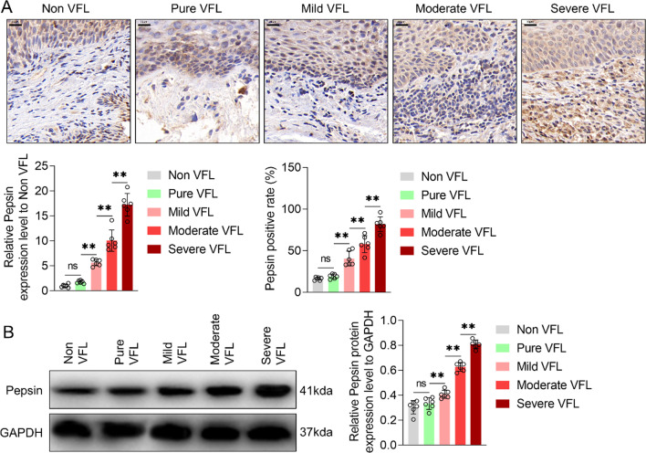
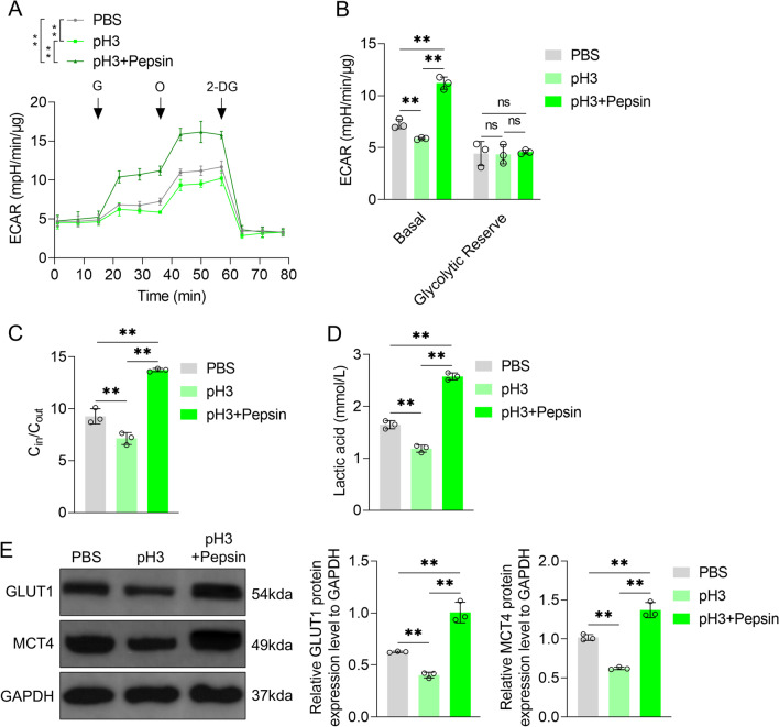
Results: It was found: the expression of pepsin and proportion of pepsin+ cells in VFL increased with the increased dysplasia grade; acidified pepsin enhanced cell growth and migration capabilities of VFL epithelial cells, reduced mitochondrial respiratory chain complex I activity and oxidative phosphorylation, and enhanced aerobic glycolysis and GLUT1 expression in VFL epithelial cells; along with the transfection of GLUT1 overexpression plasmid, 18FFDG uptake, lactate secretion and growth and migration capabilities of VFL epithelial cell were increased; this effect was partially blocked by the glycolysis inhibitor 2-deoxy-glucose; acidified pepsin increased the expression of HK-II and enhanced its distribution in mitochondria of VFL epithelial cells.

Conclusion: It was concluded that acidified pepsin enhances VFL epithelial cell growth and migration abilities by reducing mitochondrial respiratory complex I activity and promoting metabolic reprogramming from oxidative phosphorylation to aerobic glycolysis.

Keywords: GLUT1; Glycolysis; HK-II; Laryngeal cancer; Malignant transformation; Pepsin; Vocal fold leukoplakia.


Figures

Fig. 1
Fig. 1
Pepsin protein content in vocal fold leukoplakia (VFL) with different grades of dysplasia. A Immunochemistry and B western blotting were performed to measure pepsin protein content. Proportion of pepsin+ cells was also detected. GAPDH was served as the internal reference. Bar graphs show strip gray values. non-VFL normal adjacent tissue of VFL, pure VFL VFL without dysplasia, mild VFL VFL with mild dysplasia, moderate VFL VFL with moderate dysplasia, severe VFL VFL with severe dysplasia. Scale, 20 μm. One-way ANOVA was used for statistical analysis. ns, P > 0.05; **P < 0.01. n = 6 cases/group
Fig. 2
Fig. 2
Effects of acidified pepsin on the growth and migration of VFL epithelial cells. A Effects of acidified medium (pH 3 or 5) and different acidified pepsin concentrations (pH 3 or 5; 0.01, 0.1, 1 mg/mL) on the activity of VFL epithelial cells were detected by CCK-8 assay. B PI staining combined with flow cytometry was used to detect the effects of acidified medium (pH 3) and acidified pepsin (pH 3, 0.1 mg/mL) on cell cycle distribution. C Effects of acidified medium (pH 3) or acidified pepsin (pH 3, 0.1 mg/mL) on mitochondrial membrane potential were detected by JC-1 probe and flow cytometry. D Effects of acidified medium (pH 3) and acidified pepsin (pH 3, 0.1 mg/mL) on caspase-3 shear and Bax and Bcl-2 expression were detected by Western blotting. GAPDH was served as the internal reference. On the right, strip gray values are shown. E Transwell chamber assay to detect the effects of acidified medium (pH 3) and acidified pepsin (pH 3, 0.1 mg/mL) on the migration ability of VFL epithelial cells. Scale, 100 μm. One-way ANOVA was used for statistical analysis. ns, P > 0.05; **P < 0.01
Fig. 3
Fig. 3
Effects of acidified pepsin on respiratory chain complex I activity and oxidative phosphorylation of VFL epithelial cells. A Effects of acidified medium (pH 3) and acidified pepsin (pH 3, 0.1 mg/mL) on mitochondrial respiratory chain complex I activity and B oxygen consumption rate (OCR). OCR of cells treated with ATP synthase inhibitor Oligomycin (O, 1 μM) or uncoupling agent carbonyl cyanide p-trifluoromethoxyphenylhydrazone (FCCP, F, 300 nM) or electron transport chain inhibitor rotenone/antimycin A (A, 1 μM) was detected. C Basic, ATP-dependent [difference between basic OCR and oligomycin-inhibited OCR], and Respiratory Reserve OCR [difference between FCCP-induced OCR and basal OCR] in B. One-way ANOVA was used for statistical analysis. *P < 0.05; **P < 0.01
Fig. 4
Fig. 4
Effects of acidified pepsin on aerobic glycolysis of VFL epithelial cells. A Effects of acidified medium (pH 3) and acidified pepsin (pH 3, 0.1 mg/mL) on extracellular acidification rate (ECAR) of VFL epithelial cells treated with Glucose (G, 10 mM) or Oligomycin (O, 1 μM) or 2-DG (80 mM). B Basic ECAR and glycolytic reserve ECAR (difference between oligomycin-induced ECAR and basic ECAR) in A. C 18F-FDG was used to detect the effects of acidified medium (pH 3) and acidified pepsin (pH 3, 0.1 mg/mL) on glucose uptake. D Effects of acidified medium (pH 3) and acidified pepsin (pH 3, 0.1 mg/mL) on lactic acid and E glucose transporter type 1 (GLUT1), monocarboxylate transporters 4 (MCT4) protein contents, as detected by Western blotting. GAPDH was served as the internal reference. On the right, strip gray values are shown. One-way ANOVA was used for statistical analysis. **P < 0.01
Fig. 5
Fig. 5
Effects of GLUT1 overexpression plasmid on glycolysis of VFL epithelial cells. A Effects of transfection with GLUT1 overexpression plasmid on GLUT1 protein content in VFL epithelial cells, as detected by Western blotting. GAPDH was served as the internal reference. On the right, strip gray values are shown. B Effects of transfection with GLUT1 overexpression plasmid on glucose uptake and C lactic acid content. t test was used for statistical analysis. **P < 0.01
Fig. 6
Fig. 6
Effects of GLUT1 overexpression plasmid and 2-deoxy-glucose (2-DG) on glycolysis and growth and migration of VFL epithelial cells. A Effects of GLUT1 overexpression plasmid and 2-DG (5 mM) on the content of lactic acid and B activity of VFL epithelial cells. C PI dye combined with flow cytometry was used to detect the effects of GLUT1 overexpression plasmid and 2-DG (5 mM) on cell cycle distribution. D Effects of GLUT1 overexpression plasmid and 2-DG (5 mM) on caspase-3 shear and Bax and Bcl-2 expression were detected by Western blotting. GAPDH was served as the internal reference. On the right, strip gray values are shown. E Transwell chamber assay was performed to detect the effects of GLUT1 overexpression plasmid and 2-DG (5 mM) on the migration ability of VFL epithelial cells. Scale, 100 μm. One-way ANOVA was used for statistical analysis. **P < 0.01
Fig. 7
Fig. 7
Effects of acidified pepsin on Hexokinase-II (HK-II) expression and mitochondrial translocation in VFL epithelial cells. A Effects of acidified medium (pH 3) and acidified pepsin (pH 3, 0.1 mg/mL) on the amount of HK-II protein, as detected by Western blotting. GAPDH was served as the internal reference. On the right, strip gray values have been plotted. B Effects of acidified medium (pH 3) and acidified pepsin (pH 3, 0.1 mg/mL) on fluorescence expression intensity of HK-II in the mitochondria of VFL epithelial cells, as observed under a laser confocal microscope. Scale, 20 μm. One-way ANOVA was used for statistical analysis. **P < 0.01

References

    1. Parker NP. Vocal fold leukoplakia: incidence, management, and prevention. Curr Opin Otolaryngol Head Neck Surg. 2017;25:464–468. doi: 10.1097/MOO.0000000000000406. - DOI - PubMed
    1. van Hulst AM, Kroon W, van der Linden ES, et al. Grade of dysplasia and malignant transformation in adults with premalignant laryngeal lesions. Head Neck. 2016;38(Suppl 1):E2284–2290. doi: 10.1002/hed.24185. - DOI - PubMed
    1. Scheifele C, Reichart PA. Oral leukoplakia in manifest squamous epithelial carcinoma. A clinical prospective study of 101 patients. Mund Kiefer Gesichtschir. 1998;2:326–330. doi: 10.1007/s100060050081. - DOI - PubMed
    1. Reibel J. Prognosis of oral pre-malignant lesions: significance of clinical, histopathological, and molecular biological characteristics. Crit Rev Oral Biol Med. 2003;14:47–62. doi: 10.1177/154411130301400105. - DOI - PubMed
    1. Kostev K, Jacob LEC, Kalder M, Sesterhenn AM, Seidel DU. Association of laryngeal cancer with vocal cord leukoplakia and associated risk factors in 1,184 patients diagnosed in otorhinolaryngology practices in Germany. Mol Clin Oncol. 2018;8:689–693. doi: 10.3892/mco.2018.1592. - DOI - PMC - PubMed

LinkOut - more resources