

Intestinal epithelium in early life
- PMID: 36380094
- PMCID: PMC10329854
- DOI: 10.1038/s41385-022-00579-8
Intestinal epithelium in early life
Abstract
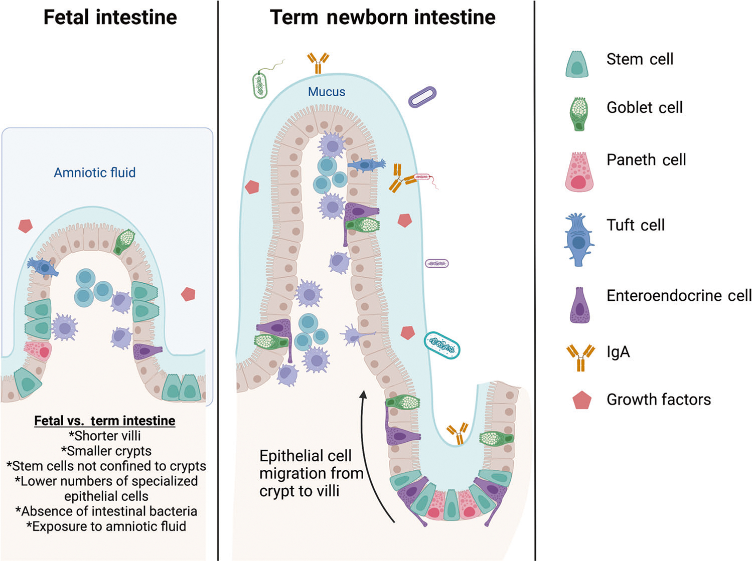
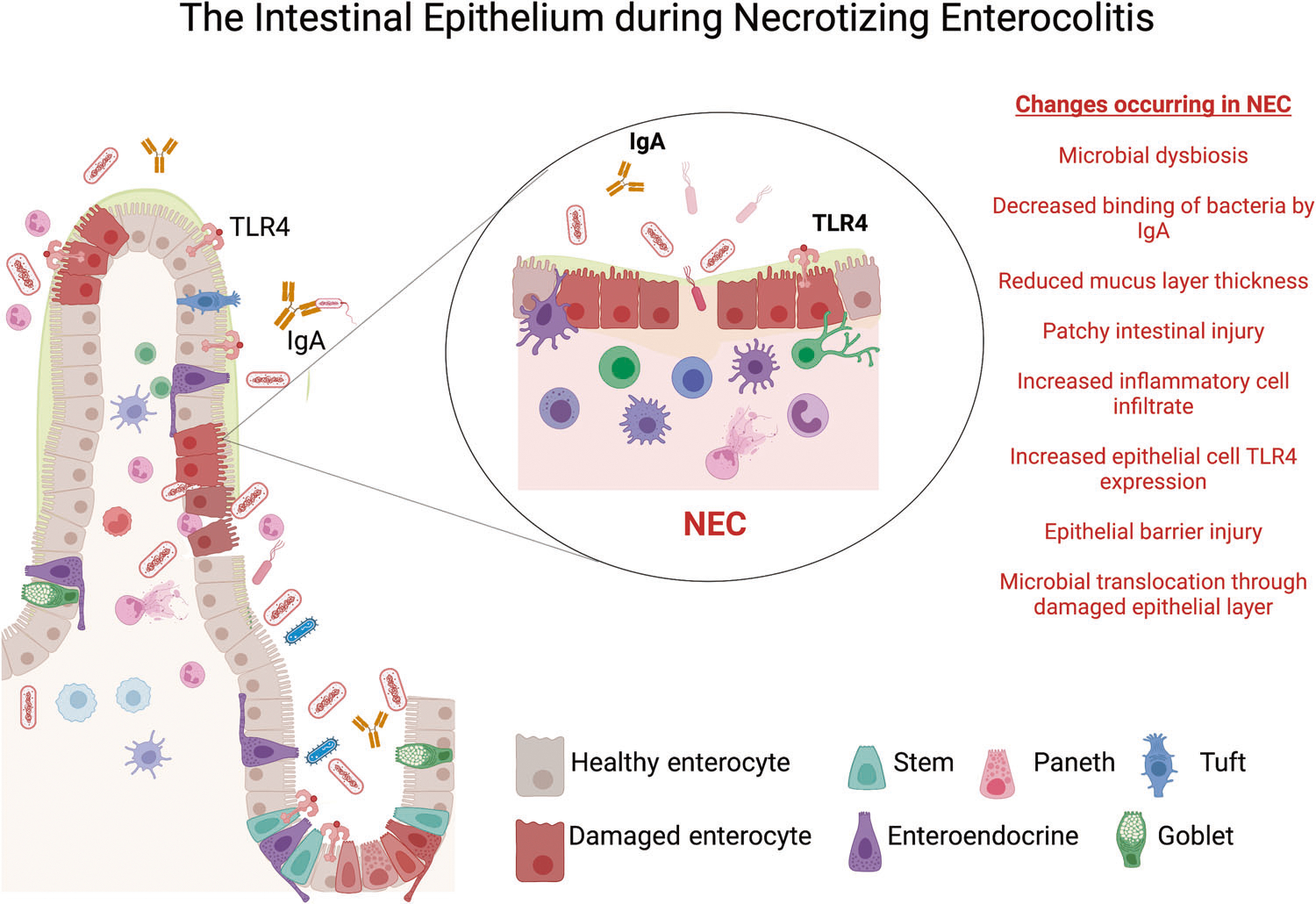
Rapid development of the fetal and neonatal intestine is required to meet the growth requirements of early life and form a protective barrier against external insults encountered by the intestinal mucosa. The fetus receives nutrition via the placenta and is protected from harmful pathogens in utero, which leads to intestinal development in a relatively quiescent environment. Upon delivery, the intestinal mucosa is suddenly tasked with providing host defense and meeting nutritional demands. To serve these functions, an array of specialized epithelial cells develop from intestinal stem cells starting in utero and continuing postnatally. Intestinal disease results when these homeostatic processes are interrupted. For preterm neonates, the most common pathology resulting from epithelial barrier dysfunction is necrotizing enterocolitis (NEC). In this review, we discuss the normal development and function of the intestinal epithelium in early life as well as how disruption of these processes can lead to NEC.
© 2022. The Author(s), under exclusive licence to Society for Mucosal Immunology.
Conflict of interest statement
COMPETING INTERESTS
The authors declare no competing interests.
Figures
Similar articles
-
Erythropoietin protects epithelial cells from excessive autophagy and apoptosis in experimental neonatal necrotizing enterocolitis.PLoS One. 2013 Jul 25;8(7):e69620. doi: 10.1371/journal.pone.0069620. Print 2013. PLoS One. 2013. PMID: 23936061 Free PMC article.
-
The decrease of fucosylation in intestinal epithelium is related to the development of necrotizing enterocolitis.Mol Immunol. 2021 Mar;131:23-32. doi: 10.1016/j.molimm.2020.12.036. Epub 2021 Jan 16. Mol Immunol. 2021. PMID: 33465592
-
Tumor necrosis factor-alpha and apoptosis signal-regulating kinase 1 control reactive oxygen species release, mitochondrial autophagy, and c-Jun N-terminal kinase/p38 phosphorylation during necrotizing enterocolitis.Oxid Med Cell Longev. 2009 Nov-Dec;2(5):297-306. doi: 10.4161/oxim.2.5.9541. Oxid Med Cell Longev. 2009. PMID: 20716917 Free PMC article.
-
Pathogenesis of neonatal necrotizing enterocolitis.Pediatr Surg Int. 2015 Jun;31(6):509-18. doi: 10.1007/s00383-015-3697-9. Epub 2015 Apr 9. Pediatr Surg Int. 2015. PMID: 25854935 Review.
-
The role of the intestinal barrier in the pathogenesis of necrotizing enterocolitis.Shock. 2007 Feb;27(2):124-33. doi: 10.1097/01.shk.0000239774.02904.65. Shock. 2007. PMID: 17224785 Review.
Cited by
-
REGγ-mediated Barrier Disruption and NF-κB Activation Aggravates Intestinal Inflammation in Necrotizing Enterocolitis.Inflammation. 2025 Aug;48(4):2476-2493. doi: 10.1007/s10753-024-02203-2. Epub 2024 Dec 10. Inflammation. 2025. PMID: 39653983
-
Alginate Oligosaccharides Enhance Antioxidant Status and Intestinal Health by Modulating the Gut Microbiota in Weaned Piglets.Int J Mol Sci. 2024 Jul 23;25(15):8029. doi: 10.3390/ijms25158029. Int J Mol Sci. 2024. PMID: 39125598 Free PMC article.
-
Modulation of intestinal TLR4 expression in infants with neonatal opioid withdrawal syndrome.J Perinatol. 2024 Aug;44(8):1125-1131. doi: 10.1038/s41372-023-01859-2. Epub 2023 Dec 27. J Perinatol. 2024. PMID: 38151596 Free PMC article.
-
HAMLET in human milk is resistant to digestion and carries essential free long chain polyunsaturated fatty acids and oleic acid.Food Chem. 2023 Nov 30;427:136752. doi: 10.1016/j.foodchem.2023.136752. Epub 2023 Jun 28. Food Chem. 2023. PMID: 37392621 Free PMC article.
-
Absence of gut microbiota reduces neonatal survival and exacerbates liver disease in Cyp2c70-deficient mice with a human-like bile acid composition.Clin Sci (Lond). 2023 Jul 14;137(13):995-1011. doi: 10.1042/CS20230413. Clin Sci (Lond). 2023. PMID: 37384590 Free PMC article.
References
-
- Condino AA et al. Abnormal intestinal histology in neonates with congenital anomalies of the gastrointestinal tract. Biol. Neonate 85, 145–150 (2004). - PubMed
-
- Trahair JF, Harding R, Bocking AD, Silver M & Robinson PM The role of ingestion in the development of the small intestine in fetal sheep. Q. J. Exp. Physiol. 71, 99–104 (1986). - PubMed
Publication types
MeSH terms
Grants and funding
LinkOut - more resources
Full Text Sources
