. 2023;15(1):297-316.
doi: 10.1159/000527008. Epub 2022 Nov 15.

IRF1 Is Required for MDA5 (IFIH1) Induction by IFN-α, LPS, and poly(I:C) in Murine Macrophages

Affiliations

IRF1 Is Required for MDA5 (IFIH1) Induction by IFN-α, LPS, and poly(I:C) in Murine Macrophages

Iris Aparici-Herraiz et al. J Innate Immun. 2023.

Erratum in

  • Erratum.
    [No authors listed] [No authors listed] J Innate Immun. 2025;17(1):287. doi: 10.1159/000545776. Epub 2025 Jun 11. J Innate Immun. 2025. PMID: 40499528 Free PMC article. No abstract available.

Abstract

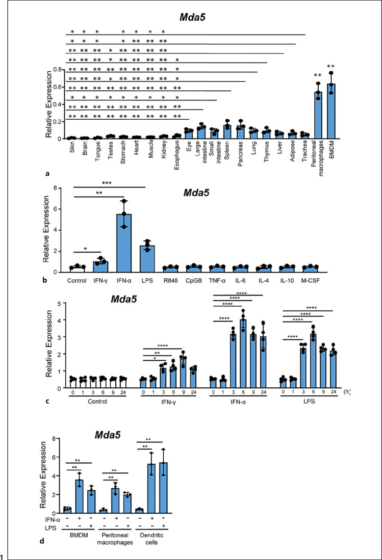
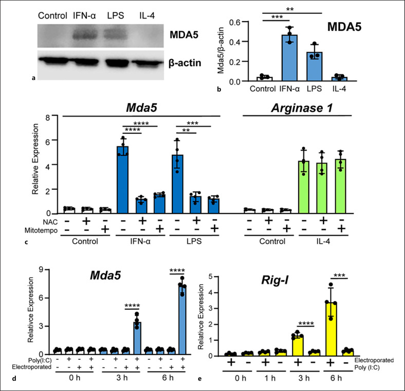
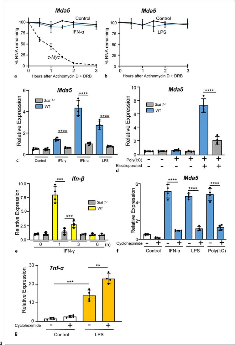
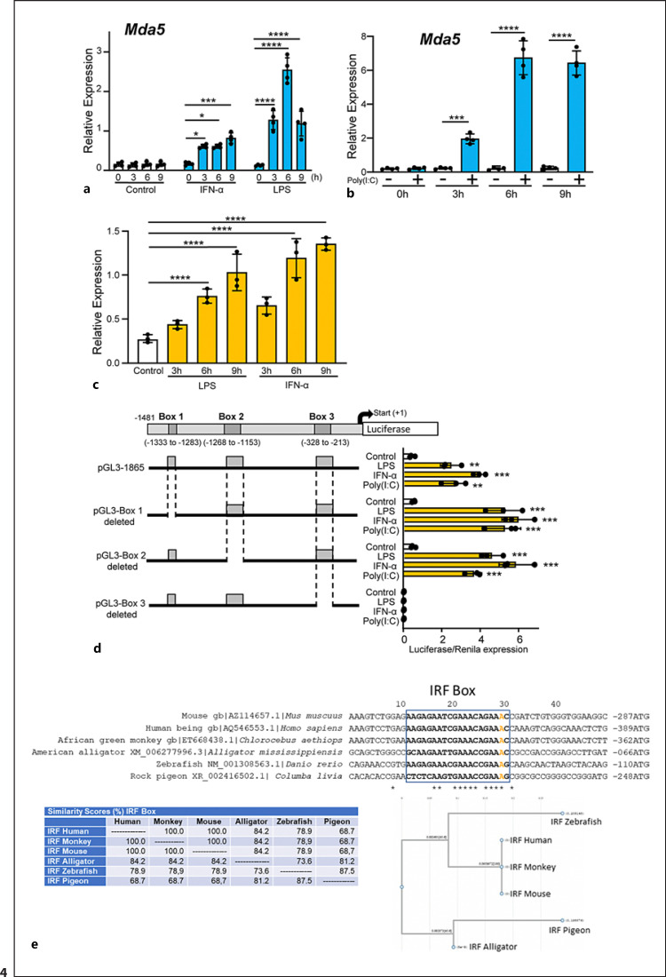
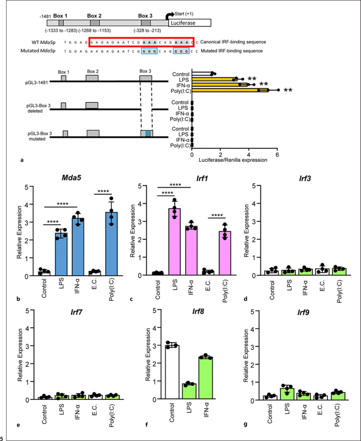
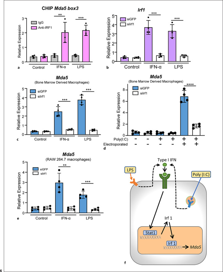
Melanoma differentiation-associated protein 5 (MDA5) induces type I interferons (IFNs) after the recognition of viral RNA. In addition, gain-of-function mutations in the interferon induced with helicase C domain 1 (IFIH1) gene, which encodes MDA5, lead to type I interferonopathies. Here, we show that Mda5 is highly expressed in murine macrophages and is regulated by pro-inflammatory stimuli such as the cytokines IFN-α and IFN-γ, the TLR ligand LPS, and a mimic of dsRNA, poly(I:C). Mda5 induction is mediated through the production of reactive oxygen species. The induction by IFN-α or LPS occurs at the transcriptional level since the Mda5 mRNA half-life before and after induction is very stable. Interestingly, STAT1 is required for Mda5 induction by IFN-α, LPS, or poly(I:C). The time course of induction of at least 3 h and the need for protein synthesis indicate that Mda5 requires an intermediate protein for transcription. In transient transfection experiments, we found that a 105-bp fragment of this gene, between -1153 and -1258 bp relative to the transcription start site, is required for transcription. In this specific region, we observed a sequence containing an IRF-binding motif, which, when mutated, abolishes the induction of Mda5. This sequence is strongly conserved in the IFIH1 promoters of eutherian mammals and in other distant species. Kinetic experiments, chromatin immunoprecipitation assays, and gene-silencing experiments revealed that IRF1 is required for induction of Mda5 expression.

Keywords: Gene regulation; Inflammation; Interferons; Macrophages; Reactive oxygen species.


Conflict of interest statement

The authors declare that the research was conducted in the absence of any commercial or financial relationships that could be construed as a potential conflict of interest.

Figures

Fig. 1
Fig. 1
Selective tissue expression of Mda5. a Tissues from three mice were used to obtain RNA, and Mda5 expression was determined by RT-PCR (independent experiments, n = 3). For comparison, we used BMDMs and peritoneal macrophages that were significantly increased in relation to all the tissues (p < 0.01). b Seven-day-old BMDMs were starved of growth factors for 16–18 h and then incubated for 24 h with different stimuli at a concentration of 10 ng/mL with the exception of R848 and CpG which was 2.5 μg/mL. Mda5 expression was then determined by RT-PCR (n = 3). Controls were untreated cells. c Time course of Mda5 expression. BMDMs were incubated with the indicated stimuli, and Mda5 induction was measured by RT-PCR (n = 4). d Mda5 expression is induced in BMDM, peritoneal macrophages and DCs by IFN-α or LPS. Cells were incubated for 24 h with the indicated stimuli and Mda5 expression was then determined by RT-PCR (n = 3). Each experiment was performed in triplicate, and the results are shown as the mean ± SD. *p < 0.05, **p < 0.01, ***p < 0.001, and ****p < 0.0001 in relation to each stimulated sample compared to controls in each experiment. Data were analyzed using the unpaired Student's t test with the exception of c that was calculated using a two-way ANOVA test followed by a Bonferroni correction.
Fig. 2
Fig. 2
Protein and mRNA expression of MDA5. a Representative blot of MDA5 protein expression in BMDMs. Total protein extracts from BMDMs treated for 24 h with the indicated stimuli were subjected to western blotting to determine MDA5 expression. β-actin was used as the loading control. b Quantitation of MDA5 expression in three independent experiments in relation to β-actin expression (n = 3). c BMDMs were incubated for 1 h with media, NAC (20 mM) or Mito-TEMPO (50 μM). The BMDMs were then treated with IFN-α or LPS for 3h and Mda5 expression was determined (n = 4). As control, BMDMs were treated with IL-4 and Arginase 1 was determined (n = 4). d BMDMs were electroporated with poly(I:C) (2 μg in 100 μL), while controls were non-electroporated BMDMs. The mock transfection controls were the electroporated cells with media. Mda5 expression was determined at the indicated times (n = 4). e Similar experiment to that in (d), but Rig-I expression was determined instead (n = 4). Each experiment was performed in triplicate, and the results are shown as the mean ± SD. **p < 0.01, ***p < 0.001, and *p < 0.0001 in relation to each stimulated sample compared to controls in each experiment. Data were analyzed using the unpaired Student's t test.
Fig. 3
Fig. 3
Mda5 expression is stable and the expression is dependent on STAT1. a BMDMs were treated with or without IFN-α for 6 h, then DRB (20 g/mL) and actinomycin D (5 g/mL) were added. Mda5 expression was then measured by RT-PCR at the indicated time points. Cell viability was 95% for all culture conditions. As a control, we also determined the half-life of c-myc (n = 3). b Similar experiment to that in (a), but LPS was used instead as the Mda5 activator (n = 3). c BMDMs from Stat1 knockout mice and the corresponding wild-type counterparts were isolated and stimulated with IFN-γ, IFN-α, or LPS for 6 h. Mda5 expression was analyzed by RT-PCR (n = 4). d Similar experiment to that in (c), but using electroporated poly(I:C) as the activator (n = 4). e Similar experiment to that in (c), but using IFN-γ as the activator and analyzing Ifn-β expression (n = 4). f BMDMs were incubated in the presence or not of 5 μg/mL of CHX for 1 h, then IFN-α, LPS, or poly(I:C) was added and incubated for 3 h and Mda5 was determined (n = 4). g Similar experiment to that in (f), but Tnf-α was determined (n = 4). Each experiment was performed in triplicate, and the results are shown as the mean ± SD. **p < 0.01, ***p < 0.001, and p < 0.0001 in relation to each stimulated sample compared to controls in each experiment. Data were analyzed using the unpaired Student's t test.
Fig. 4
Fig. 4
Characterization of the Mda5 functional promoter. a RAW 264.7 macrophages were incubated for different times with IFN-α or LPS before Mda5 expression was determined (n = 4). b Similar experiment to that in (a), but using electroporated poly(I:C) as the activator (n = 4). c RAW 264.7 macrophages were transfected with the pGL3−1481 vector. At 24 h post-transfection, cells were stimulated with LPS or IFN-α for the indicated times before luciferase activity was measured. Controls were nonactivated cells (n = 4). d Putative consensus binding sites for transcription factors in the Mda5 gene promoter. The 1481-bp sequence upstream of the Mda5 transcription start site was analyzed using the JASPAR database. Putative boxes showing more than 85% similarity with the consensus sequence for transcription factor binding sites are shown (boxes 1, 2, and 3). As in (c), the vector used contained the indicated deletions of the Mda5 promoter region. Fold increase in Mda5 induction by LPS, IFN-α, or poly(I:C) are shown in relation to the untreated cells (Control). Cells were stimulated for 6h. A plasmid encoding the Renilla luciferase under the control of an HSV-TK promoter was co-transfected, with the Renilla luciferase activity used as a control of transfection. Assays are representative of at least three independent experiments showing similar results. Each experiment was performed in triplicate, and the results are shown as the mean ± SD. *p < 0.05, **p < 0.01, ***p < 0.001, and p < 0.0001 in relation to each stimulated sample compared to controls in each experiment. Data were analyzed using the unpaired Student's t test. e Sequences alignment of the Irf1 box in the promoters of Mda5 in human, mice, monkey, alligator, zebrafish and pigeon. JASPAR and the National center for biotechnology information (NCBI) database were used. For sequences alignment we used the MacVector 18.0. The A in orange indicated the distance to the ATG. In the lower part, the similarity (%) between theses boxes in different species is shown, as well as the phylogram.
Fig. 5
Fig. 5
IRF1 binding to the Mda5 promoter is required for Mda5 transcription. a Similar experiment to that in Fig. 4d, but the mutations in the canonical IRF-binding sequence were introduced in box 3. b–g In BMDM gene expression was determined by RT-PCR after LPS, IFN-α or poly(I:C) treatment for 3 h with the exception of f in which poly(I:C) treatment was not performed. E.C., electroporation control (n = 4). b Mda5, c Irf1, d Irf3, e Irf7, f Irf8, and g Irf9. Assays are representative of at least three independent experiments showing similar results. Each experiment was performed in triplicate, and the results are shown as the mean ± SD. **p < 0.01 and p < 0.0001 in relation to each stimulated sample compared to controls in each experiment. Data were analyzed using the unpaired Student's t test.
Fig. 6
Fig. 6
IRF1 is required for the induction of Mda5 in macrophages. a In vivo binding of IRF1 to the Mda5 promoter was analyzed by ChIP assays. Macrophages were treated for 3 h with IFN-α or LPS (n = 4). b BMDMs were transfected with either an siRNA directed against Irf1 or a control siRNA against GFP. Twenty-four hours after transfection, the cells were stimulated for 3 h with IFN-α or LPS and Irf1 expression was determined by qPCR (n = 4). c BMDMs transfected with siRNA against Irf1 or GFP were treated for 3 h with IFN-α or LPS before Mda5 expression was determined by RT-PCR (n = 4). d Similar experiment to that in (c), but using electroporated poly(I:C) as the activator (n = 4). e RAW 264.7 macrophage cell line transfected with siRNA against Irf1 or GFP were treated for 3 h with IFN-α or LPS before Mda5 expression was determined by RT-PCR (n = 4). f Schematic representation of the induction of Mda5 by LPS or IFN-α. Each experiment was performed in triplicate, and the results are shown as the mean ± SD. **p < 0.01, ***p < 0.001, and p < 0.0001 in relation to each stimulated sample compared to controls in each experiment. Data were analyzed using the unpaired Student's t test.

References

    1. Honda K, Takaoka A, Taniguchi T. Type I interferon [corrected] gene induction by the interferon regulatory factor family of transcription factors. Immunity. 2006 Sep;25((3)):349–360. - PubMed
    1. Yan N, Chen ZJ. Intrinsic antiviral immunity. Nat Immunol. 2012 Feb 16;13((3)):214–222. - PMC - PubMed
    1. Goubau D, Deddouche S, Reis E, Sousa C. Cytosolic sensing of viruses. Immunity. 2013 May 23;38((5)):855–869. - PMC - PubMed
    1. McNab F, Mayer-Barber K, Sher A, Wack A, O'Garra A. Type I interferons in infectious disease. Nat Rev Immunol. 2015 Feb;15((2)):87–103. - PMC - PubMed
    1. Solis M, Nakhaei P, Jalalirad M, Lacoste J, Douville R, Arguello M, et al. RIG-I-mediated antiviral signaling is inhibited in HIV-1 infection by a protease-mediated sequestration of RIG-I. J Virol. 2011 Feb;85((3)):1224–1236. - PMC - PubMed

Publication types