. 2022 Oct;12(5):352-364.
doi: 10.1212/CPJ.0000000000200066.

Objective Neurophysiologic Markers of Cognition After Pediatric Brain Injury

Affiliations

Objective Neurophysiologic Markers of Cognition After Pediatric Brain Injury

Nayoung Kim et al. Neurol Clin Pract. 2022 Oct.

Abstract

Background and objectives: Following brain injury, clinical assessments of residual and emerging cognitive function are difficult and fraught with errors. In adults, recent American Academy of Neurology (AAN) practice guidelines recommend objective neuroimaging and neurophysiologic measures to support diagnosis. Equivalent measures are lacking in pediatrics-an especially great challenge due to the combined heterogeneity of both brain injury and pediatric development. Therefore, we aim to establish quantitative, clinically practicable measures of cognitive function following pediatric brain injury.

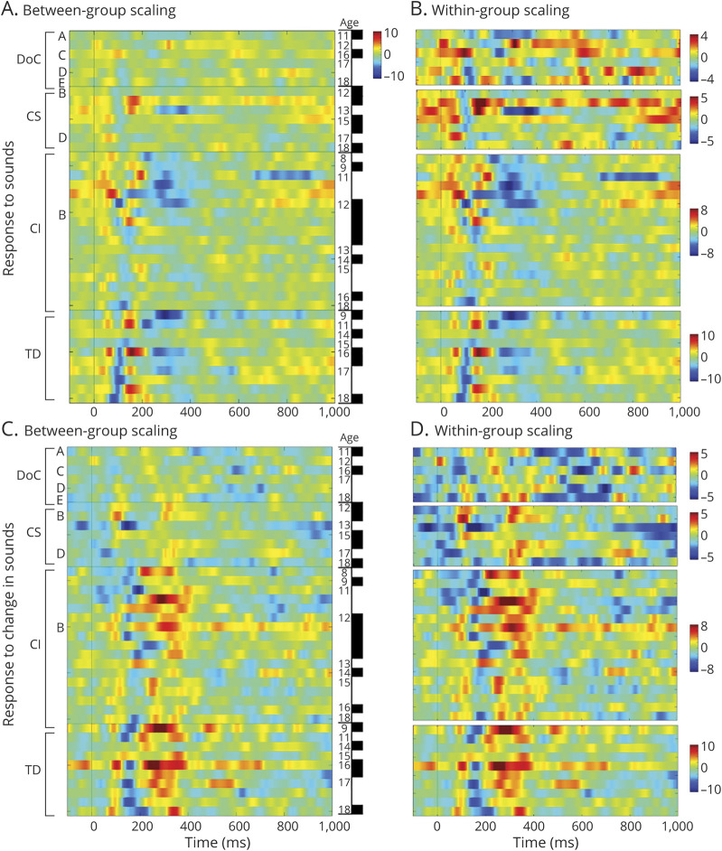
Methods: Participants with and without brain injury were aged 8-18 years, clinically classified according to cognitive recovery state: N = 8 in disorders of consciousness (DoC), N = 7 in confusional state, N = 19 cognitively impaired, and N = 13 typically developing uninjured controls. We prospectively measured electroencephalographic markers of sensory processing and attention in an auditory oddball paradigm, and of covert movement attempts in a command-following paradigm.

Results: In 3 participants with DoC, EEG markers of active attempted command following revealed cognitive function that clinical assessment had failed to detect. These same 3 individuals could also be distinguished from the rest of their group by 2 event-related potentials that correlate with sensory processing and orienting attention in the oddball paradigm. Considered across the whole participant group, magnitudes of these 2 ERP markers significantly increased as cognitive recovery progressed (ANOVA: each p < 0.001); viewed jointly, the 2 ERP markers cleanly delineated the 4 cognitive states.

Discussion: Despite heterogeneity of brain injuries and brain development, our objective EEG markers reflected cognitive recovery independent of motor function. Two of these markers required no active participation. Together, they allowed us to identify 3 individuals who meet the criteria for cognitive-motor dissociation. To diagnose, prognose, and track cognitive recovery accurately, such markers should be used in pediatrics.


Figures

Figure 1
Figure 1. Grand-Averaged Event-Related Potentials (ERPs) in the Auditory Oddball Paradigm
In Panels A--D we display grand-averaged ERPs per cognitive state. Raw amplitudes are shown (not scaled by SD). The SEM (across participants in each group) is shown as a shaded region around each grand-averaged waveform. In panel D, we indicate auditory evoked potential (AEP), N2, and P3 components.
Figure 2
Figure 2. Raster Plot of Individual Event-Related Potentials From the Oddball Paradigm
In panels A and B, individual participants' response to sounds is displayed with between-group and within-group scaling respectively. In panels C and D, individuals' response to change in sounds is displayed with between-group and within-group scaling respectively. Each row corresponds to a different participant (except where participants B and D are marked as reappearing in multiple cognitive states). The horizontal axis shows time relative to the onset of an auditory stimulus. The color scale is a z-score derived from voltage: it indicates electrical potential, measured at the vertex EEG electrode, standardized by its own trial-to-trial variability. Upper panels: response to standard beep stimuli; lower panels: difference wave between deviant and standard stimuli. Left panels: between-group color scaling—all groups are compared on the same scale. Right panels: within-group scaling—each group is presented with a group-specific scale. Within each group, participants are sorted according to their age at testing—each individual's age, in years, is indicated in the column of numbers down the center of the figure. Certain individuals of interest are denoted by letters in the margin: A, C, and E all showed positive EEG responses to attempted movement while in disorders of consciousness; only B and D were measured longitudinally—B appears in the confusional state and cognitively impaired groups, and D appears in disorders of consciousness and confusional state.
Figure 3
Figure 3. Group and Individual Motor Command–Following EEG Results (MCF-EEG)
Panel A displays topographic scalp maps of group results. Within each group, the color scale denotes the number of participants whose EEG response was significant according to the two-group test at each electrode location. Cold colors represent channels where few or no participants responded significantly; hot colors indicate channels where significant responses occurred (greater bandpower in the rest condition than in the attempted hand movement trials). Panel B shows individual statistical test results. Significance values (negative log p values) from permutation tests are shown per participant for attempted left-hand movement on the left side of the panel and attempted right-hand movement on the right side of the panel. Symbol shape and color denote cognitive state, corresponding with Figure 4. Participant numbers and letters on the y axis correspond with the identifiers in Table 1.
Figure 4
Figure 4. Two-Dimensional View of Individuals' Event-Related Potential Magnitudes, Inflected According to Motor Command–Following EEG Results
Each symbol denotes one measurement from a different participant, with the exception of the repeated measurements in the participants marked B and D. (The longitudinal sequence of measurements for each of these participants is connected by faint gray arrows.) Symbol shape and color indicate cognitive state. Letters denote particular individuals of interest, noted in the main text. Filled symbols denote participants for whom there was a positive result in the motor command-following EEG measurement (MCF-EEG). Open symbols denote participants with negative MCF-EEG. Symbols containing crosses denote participants for whom there were no MCF-EEG data. Participants not shown, because of missing event-related potential data, are as follows: 2 disorders of consciousness, 2 cognitively impaired, and 1 typically developing who all had negative MCF-EEG and 2 typically developing who had positive MCF-EEG.

References

    1. Giacino JT, Kalmar K, Whyte J. The JFK Coma Recovery Scale-Revised: measurement characteristics and diagnostic utility. Arch Phys Med Rehabil. 2004;85(12):2020-2029. - PubMed
    1. Edlow BL, Claassen J, Schiff ND, Greer DM. Recovery from disorders of consciousness: mechanisms, prognosis and emerging therapies. Nature reviews. Neurlogy.l 2021;17(3):135-156. - PMC - PubMed
    1. Polich J. Clinical application of the P300 event-related brain potential. Phys Med Rehabil Clin N Am. 2004;15(1):133-161. doi: 10.1016/s1047-9651(03)00109-8. - DOI - PubMed
    1. Meysam A, Kim YK. EEG correlates of cognitive functions and neuropsychiatric disorders: a review of oscillatory activity and neural synchrony abnormalities. Curr Psychiatry Res Rev Former Curr Psychiatry Rev. 2020;16(4):228-243.
    1. Landa L, Krpoun Z, Kolarova M, Kasparek T. Event-related potentials and their applications. Act Nerv Super (Praha). 2014;56(1-2):17-23. doi: 10.1007/BF03379603. - DOI