. 2022 Nov 4;12(49):31711-31728.
doi: 10.1039/d2ra05096j. eCollection 2022 Nov 3.

Nanotechnology meets immunology towards a rapid diagnosis solution: the COVID-19 outbreak challenge

Affiliations

Nanotechnology meets immunology towards a rapid diagnosis solution: the COVID-19 outbreak challenge

Santos B S A S et al. RSC Adv. .

Abstract

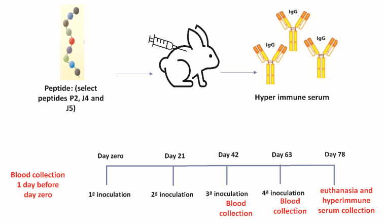
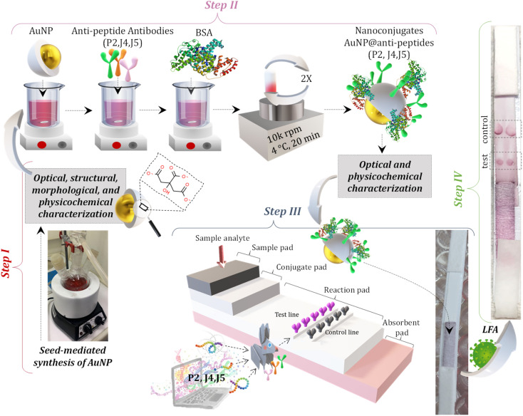
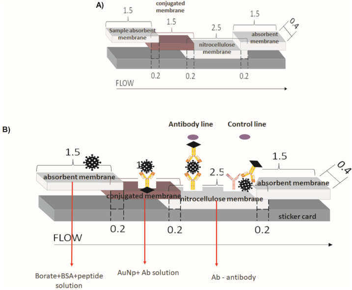
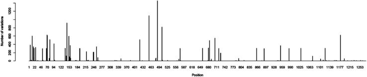
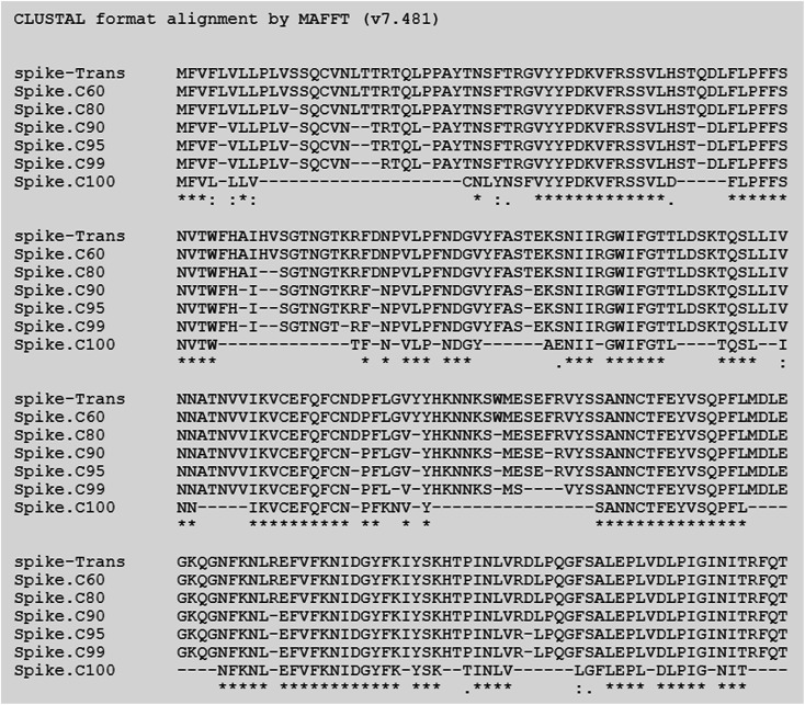
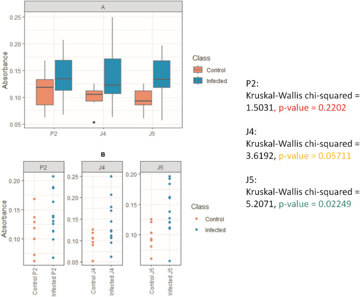
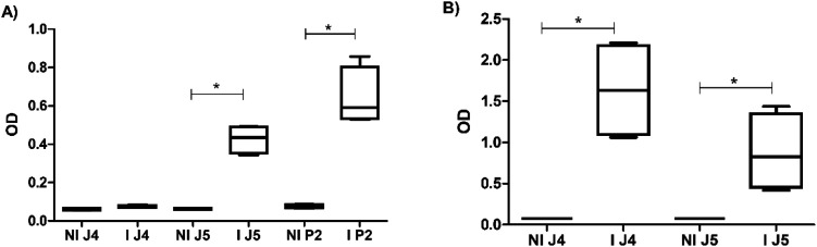
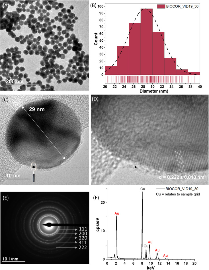
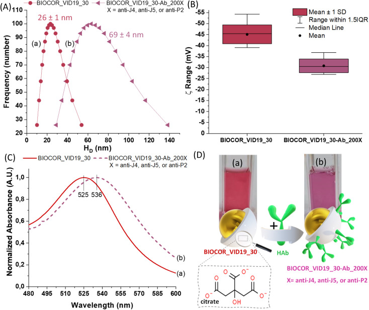
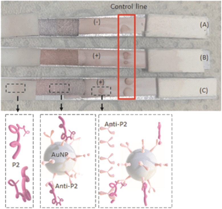
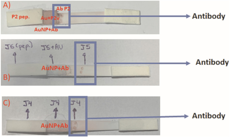
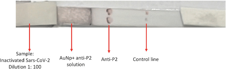
The current COVID-19 pandemic presents one of the greatest challenges in human history. There is a consensus that the rapid and accurate diagnosis of COVID-19 directly affects procedures to avoid dissemination, promote treatments, and favor the prognosis of infected patients. This interdisciplinary study aims at designing new synthetic peptides inspired by the SARS-CoV-2 spike protein (SARS-CoV-2S) to produce rapid detection tests relying on nanomaterial-based colorimetric properties. Hence, in silico analyses of SARS-CoV-2S were performed using advanced bioinformatic simulation tools and algorithms. Five novel peptide sequences were proposed, and three were selected (P2, J4, and J5) based on their prospective reactivity against positive serum from naturally COVID-19-infected humans. Next, hyperimmune sera against the selected peptides were produced in rabbits. Concurrently, gold nanoparticles (AuNP) were synthesized using a green aqueous method under mild conditions through in situ reduction by trisodium citrate salt. They were extensively characterized by their morphological, physicochemical, and optical properties. The AuNPs demonstrated colloidal chemical stability in aqueous media, with an average size of approximately 29 nm (metallic core), and zeta potential before and after bioconjugation of -43 mV and -31 mV, respectively. Moreover, they presented an intense reddish-bluish color due to the surface plasmon resonance (SPR) effect, with maxima at λ = 525 nm and 536 nm, before and after bioconjugation, respectively, evidencing their applicability as colorimetric biomarkers for antigen-antibody immunoassay detection. To develop a rapid COVID-19 diagnosis test using lateral flow assay (LFA), semi-purified anti-SARS-CoV-2S sera against the three selected peptides were bioconjugated to the AuNPs as the highly optically sensitive agents using a considerably low antibody concentration (0.2 mg mL-1). All tested peptide sequences (P2, J4, and J5) induced antibodies capable of identifying the presence of SARS-CoV-2 virus inactivate suspensions (1 : 10, 1 : 100, or 1 : 1000 dilutions). For LFA positive test control, an anti-rabbit antibody was used. In summary, this research comprises several contributions and advances to the broad and multidisciplinary field of nanomaterials-based immunodiagnosis tools, encompassing: (a) the novelty of designing and synthesizing new immunogenic peptides inspired by SARS-CoV-2 virus epitopes using in silico bioinformatics; (b) the peptides induced the immune response in rabbit animal model producing hyperimmune serum; (c) the semi-purified hyperimmune serum rendered effective antibodies to detect SARS-CoV-2 virus in cell suspension; (d) colloidal gold nanoparticles were produced and bioconjugated to the antibodies for qualitative colorimetric detection. As the overall result of this study, it was designed, developed, produced, and validated a new simple, rapid, and sensitive LFA diagnostic test for the SARS-CoV-2 virus using a nanotechnology-based qualitative colorimetric assay, which can be envisioned as promising nanoplatforms for detecting other diseases.


Conflict of interest statement

The authors confirm no competing interests to declare regarding the publication of this article.

Figures

Fig. 1
Fig. 1. .Schematic illustration of the hyperimmune serum production.
Fig. 2
Fig. 2. .Schematic illustration of the complete LFA (Lateral Flow assay). Step I represents the synthesis of AuNPs, and the conjugation stage is depicted in Step II. Step III illustrates the assembly of each pad (sample pad, conjugate pad, reaction pad, and absorbent pad) onto the LFA card; and Step IV shows the LFA test after interaction with an infected sample.
Fig. 3
Fig. 3. .(A) Schematic representation of standard LFA assembly; (B) detailed representation of the complete setup for the LFA-based Immunodiagnostic test for SARS-CoV-2 virus: all components, parts, and conjugates, designed, developed, synthesized, and assembled in this work (dimensions in cm).
Fig. 4
Fig. 4. .Evaluation of SARS-CoV-2 spike protein variant regions. The X-axis corresponds to the positions from the spike protein, and the Y axis is the number of isolates that diverged from the most common amino acid observed in the position, resulting from the alignment of 2850 spike protein sequences from 11 SARS-CoV-2 viral strains.
Fig. 5
Fig. 5. .Representation of the conservation of the initial part of the spike protein. Each line corresponds to a different conservation cut-off value, where “spike-trans” corresponds to the original spike protein reference sequence (YP_009724390.1), and the lines below “spike.C60” to “spikeC100” correspond to the conservation cut-off in 60 to 100% of the isolates. Each column corresponds to a position in the protein. Regions that did not pass a specific cut-off are represented by a “-”. Asterisks at the bottom highlight 100% conserved positions in all evaluated isolates.
Fig. 6
Fig. 6. .Graphs show the differentiation between the peptides tested in positive and negative samples of humans naturally infected with SARS-CoV-2; (A) BoxPlot: boxes represent the median absorbance value, interquartile intervals, and maximum and minimum points; (B) scatterplots where each point corresponds to a sample.
Fig. 7
Fig. 7. .(A) ELISA results for J4 and J5, 15 days after third inoculations (I J4 and I J5), and P2, 15 days after the fourth inoculation (I P2). (B) ELISA results for J4 and J5, 15 days after the fourth inoculation (IJ4 and IJ5). NI – sera before inoculation.
Fig. 8
Fig. 8. .(A) TEM image of uniformly dispersed spherical-like gold nanoparticles (AuNPs). (B) Size distribution of AuNPs measured by TEM image analysis (n = 330; dashed line represents the normal distribution of the data). (C and D) HRTEM images of a single AuNP (zoomed), with typical AuNP diameter (arrow), and the nanocrystallinity by the well-defined interplanar distance. (E) SAED ring patterns of AuNPs. (F) Typical EDS spectrum of AuNPs.
Fig. 9
Fig. 9. .(A) Size distribution, (B) zeta potential (ξ) distribution, (C) UV-vis, and (D) digital images and schematic illustration of BIOCOR_VID19_30 (a) and BIOCOR_VID19_30-Ab_200X (b).
Fig. 10
Fig. 10. .Results of the peptide P2 detection by LFA assay after 10 min of the sample application. Test conditions: (A) control (sample = borate buffer with 2% BSA, pH 8.5), (B) peptide at 0.3 mg mL−1, and (C) peptide at 0.8 mg mL−1 (bottom: sketch of components of each stage).
Fig. 11
Fig. 11. .LFA peptides: (A) sample: P2 peptide; conjugate solution: anti-P2 + AuNP; Antibody membrane: anti-P2. (B) Sample: J5 peptide; conjugate solution: anti-J5 + AuNP; Antibody membrane: anti-J5. (C) Sample: J4 peptide; conjugate solution: anti-J4 + AuNP; antibody membrane: anti-J4. The following were used as samples: 5 μL peptide + 145 μL sol.2% BSA+2 mM borate. The used concentration of each peptide: 20 μg of J5; 20 μg of P2, and 25 μg of J4.
Fig. 12
Fig. 12. .LFA using inactivated SARS-CoV-2 dilution 1 : 100, reacting with AuNP + anti-P2 solution and anti-P2. Control line with commercial anti-rabbit antibody.

References

    1. Anh N. H. et al, Gold nanoparticle-based optical nanosensors for food and health safety monitoring: recent advances and future perspectives. RSC Adv. 2022;12:10950–10988. - PMC - PubMed
    1. Mulder L. et al, A Comparative Study of Nine SARS-CoV-2 IgG Lateral Flow Assays Using Both Post-Infection and Post-Vaccination Samples. J. Clin. Med. 2022;11:2100. - PMC - PubMed
    1. Cucinotta D. Vanelli M. WHO declares COVID-19 a pandemic. Acta Biomedica. 2020;91:157–160. - PMC - PubMed
    1. WHO, World Health Organization, Weekly epidemiological update on COVID-19, 2021, vol. 69, available at: https://www.who.int/publications/m/item/weekly-epidemiological-update-on...
    1. Bruijns B. Folkertsma L. Tiggelaar R. FDA authorized molecular point-of-care SARS-CoV-2 tests: A critical review on principles, systems and clinical performances. Biosens. Bioelectron.: X. 2022;11:100158. - PMC - PubMed