. 2022 Oct 15;15(10):403-411.
eCollection 2022.

Role of macrophages in progression of colorectal cancer: a contrast with the traditional paradigm

Affiliations

Role of macrophages in progression of colorectal cancer: a contrast with the traditional paradigm

Aleksei S Konstantinov et al. Int J Clin Exp Pathol. .

Abstract

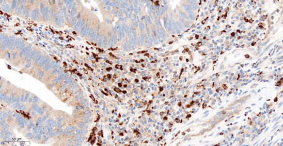
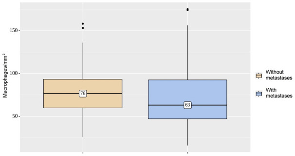
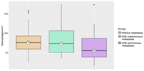
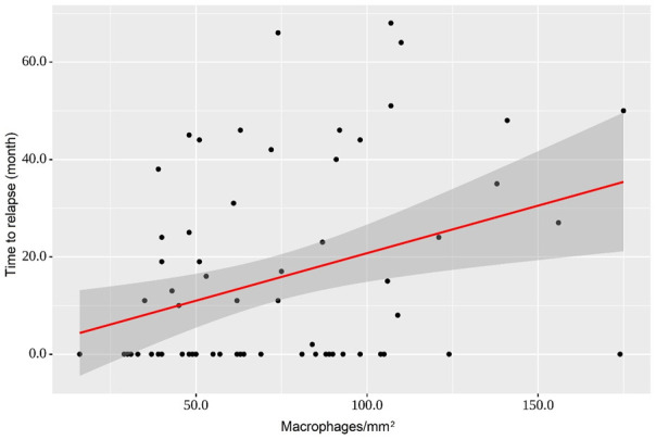
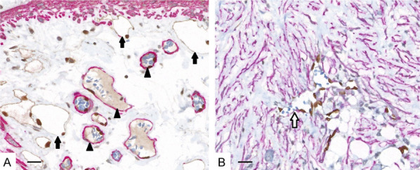
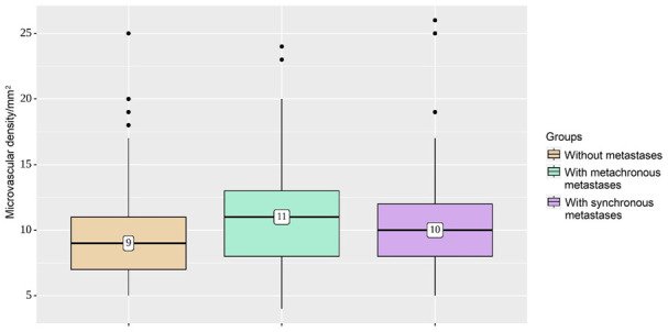
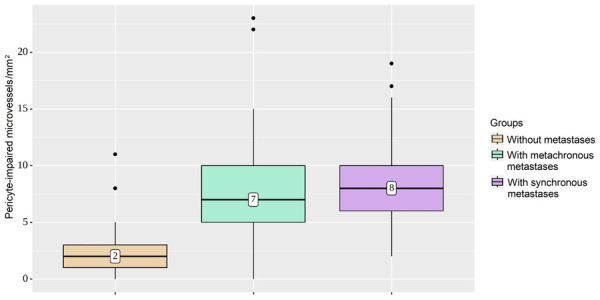
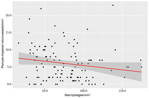
The phenotype of tumor-associated macrophages may be critical for tumor immunity, angiogenesis, and clinical disease outcome. Here, we elucidated the prognostic significance of the neovasculature and macrophages in colorectal cancer. We analyzed the effect of M2 macrophage density on the clinical behavior of 151 primary colorectal carcinomas using CD206 as a marker for type 2 macrophages. Triple immunohistochemical staining (ERG, SMA, and podoplanin) was used for microvessel evaluation. We found that M2 macrophages in colorectal cancer did not have a direct association with metastatic behavior. However, high numbers of CD206 tumor-associated macrophages correlated positively with recurrence-free interval duration (P=0.005). Fewer macrophages in the tumor microenvironment resulted in insufficient coverage of newly formed vessels by pericytes (P=0.011), and a high number of pericyte-impaired microvessels correlated with metastatic behavior (P<0.001). These results suggested that type 2 macrophages had a role in limiting the metastatic process by affecting vascular maturity and normalization. These findings contribute to understanding complex interactions in the tumor microenvironment and the clinical behavior of colorectal cancer.

Keywords: Colorectal cancer; M2; alternatively activated macrophages; angiogenesis; prognosis; tumor microenvironment.


Conflict of interest statement

None.

Figures

Figure 1
Figure 1
Immunohistochemical detection of macrophages in colorectal cancer. Stromal M2 macrophages are clearly indicated by strong cytoplasmic staining with anti-CD206 antibody, 400 x.
Figure 2
Figure 2
Density of macrophage infiltration per square millimeter. Mean numbers of M2 macrophages in the metastasizing and non-metastasizing groups are not different (P=0.144).
Figure 3
Figure 3
Comparison of macrophage infiltration in three groups. The cases with synchronous metastasis have significantly lower M2 macrophage densities, compared to metachronous disease or local colorectal cancer (P=0.029).
Figure 4
Figure 4
Regression curve characterizing the dependence of time to relapse from a stromal density of macrophages. With an increase in macrophage numbers in the tumor microenvironment in CRC by 10/mm2, it is expected that the recurrence-free time will increase by 1.95 months (P=0.005).
Figure 5
Figure 5
Triple immunohistochemical staining of microvessels in normal colon (A) and colorectal cancer (B). Nuclei of endothelial cells of the blood (arrowheads) and lymphatic capillaries (black arrows) are stained brown with ERG. Blood vessel contours appear red due to the presence of pericytes stained with α-SMA. Podoplanin staining defines the brown cytoplasm of endothelial cells of lymphatic vessels. Newly formed malformed blood vessels (white arrow) in the tumor stroma are devoid of pericytes and retain only the outline of their endothelial cells, which are recognizable by nuclei stained with brown anti-ERG. Note the striking desmoplastic change in the stroma in the tumor, with numerous activated myofibroblasts, detected using α-SMA. Scale bar = 200 μm, 400 x.
Figure 6
Figure 6
Microvascular density groups. The average density of microvessels in the group without metastases, compared with metastasizing tumors, is not significantly different (P=0.158).
Figure 7
Figure 7
Characterization of pericyte-impaired microvessel densities in groups. In tumors with metachronous and synchronous metastases, capillaries without pericytes were more common among newly formed vessels, compared with quite low pericyte-impaired microvessels in non-metastasizing tumors (P<0.001).
Figure 8
Figure 8
Regression curve demonstrates the observed variation in pericyte-impaired microvessels with respect to macrophage density. An increase in the number of macrophages of 100/mm2 is accompanied by a decrease in pericyte-impaired microvessels of 2.5/mm2 (P=0.011).

References

    1. Nathan CF, Murray HW, Wiebe ME, Rubin BY. Identification of interferon-gamma as the lymphokine that activates human macrophage oxidative metabolism and antimicrobial activity. J Exp Med. 1983;158:670–689. - PMC - PubMed
    1. Martinez FO, Gordon S. The M1 and M2 paradigm of macrophage activation: time for reassessment. F1000Prime Rep. 2014;6:6–13. - PMC - PubMed
    1. Stein M, Keshav S, Harris N, Gordon S. Interleukin 4 potently enhances murine macrophage mannose receptor activity: a marker of alternative immunologic macrophage activation. J Exp Med. 1992;176:287–292. - PMC - PubMed
    1. Mills CD, Kincaid K, Alt JM, Heilman MJ, Hill AM. M-1/M-2 macrophages and the Th1/Th2 paradigm. J Immunol. 2000;164:6166–6173. - PubMed
    1. Boutilier AJ, Elsawa SF. Macrophage polarization states in the tumor microenvironment. Int J Mol Sci. 2021;22:6995. - PMC - PubMed

LinkOut - more resources