Case Reports
. 2022 Oct 8;14(10):e30081.
doi: 10.7759/cureus.30081. eCollection 2022 Oct.

Bilateral Lupus Chorioretinopathy in a Patient With Active Systemic Lupus Erythematosus

Affiliations
Case Reports

Bilateral Lupus Chorioretinopathy in a Patient With Active Systemic Lupus Erythematosus

Sharoon David et al. Cureus. .

Abstract

Ocular involvement is commonly seen in systemic lupus erythematosus (SLE). However, chorioretinopathy is an easily missed ocular manifestation of SLE. Early recognition and a multidisciplinary treatment approach can play a key role in reducing the ocular and systemic morbidity seen with this condition. This case report describes a patient with active SLE who presented with bilateral lupus chorioretinopathy. The patient demonstrated a significant improvement in ocular symptoms once the systemic disease was controlled.

Keywords: chorioretinopathy; choroidopathy; lupus; ocular manifestations; retinopathy; serous retinal detachment; systemic lupus erythematosus.


Conflict of interest statement

The authors have declared that no competing interests exist.

Figures

Figure 1
Figure 1. Optical coherence tomography OU - initial visit: macular edema with lipid exudates, subretinal fluid (white arrow), and intraretinal fluid (black arrow)
Figure 2
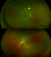
Figure 2. Ultra-wide field fundus photography OU - initial visit: intraretinal deposits (black arrows), intraretinal and subretinal hemorrhages (white arrow), and tortuous veins
Figure 3
Figure 3. Fluorescein angiography OU - initial visit: multiple hypofluorescent spots consistent with intraretinal hemorrhages and hyperfluorescent areas (black arrows) consistent with dye leakage during the full venous phase
Figure 4
Figure 4. Optical coherence tomography OU - follow-up visit: multiple intraretinal deposits (black arrow) and thinning of the outer retina (absence of ellipsoid zone). Some cystic changes were also noted in the right eye (white arrow)
Figure 5
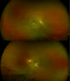
Figure 5. Ultra-wide field fundus photography OU - follow-up visit: multiple intraretinal deposits (white arrows), parafoveal/perifoveal depigmentation with few intraretinal hemorrhages, and tortuosity of veins bilaterally
Figure 6
Figure 6. Fluorescein angiography OU - follow-up visit: enlarged foveal avascular zone (FAZ) and multiple hypofluorescent areas consistent with intraretinal hemorrhages (white arrows). No evidence of leakage was present

References

    1. Systemic lupus erythematosus. Manson JJ, Rahman A. Orphanet J Rare Dis. 2006;1:6. - PMC - PubMed
    1. The diagnosis and treatment of systemic lupus erythematosus. Kuhn A, Bonsmann G, Anders HJ, Herzer P, Tenbrock K, Schneider M. Dtsch Arztebl Int. 2015;112:423–432. - PMC - PubMed
    1. García-Carrasco M, Pinto MC, Poblano JCS, Morales IE, Cervera R, Anaya JM. Autoimmunity: From Bench to Bedside. Vol. 1. Bogota, Colombia: El Rosario University Press; 2013. Systemic lupus erythematosus; p. 25. - PubMed
    1. Ocular manifestations in systemic lupus erythematosus. Silpa-archa S, Lee JJ, Foster CS. Br J Ophthalmol. 2016;100:135–141. - PubMed
    1. Ocular manifestations of systemic lupus erythematosus: a review of the literature. Palejwala NV, Walia HS, Yeh S. Autoimmune Dis. 2012;2012:290898. - PMC - PubMed

Publication types

LinkOut - more resources