. 2022 Oct 26;4(6):fcac272.
doi: 10.1093/braincomms/fcac272. eCollection 2022.

Comprehensive characterization of elevated tau PET signal in the absence of amyloid-beta

Affiliations

Comprehensive characterization of elevated tau PET signal in the absence of amyloid-beta

Alexandra J Weigand et al. Brain Commun. .

Erratum in

Abstract

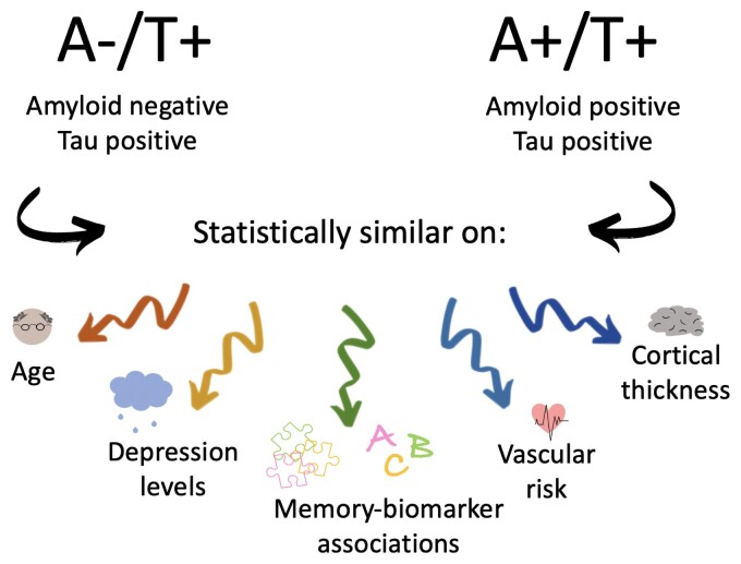
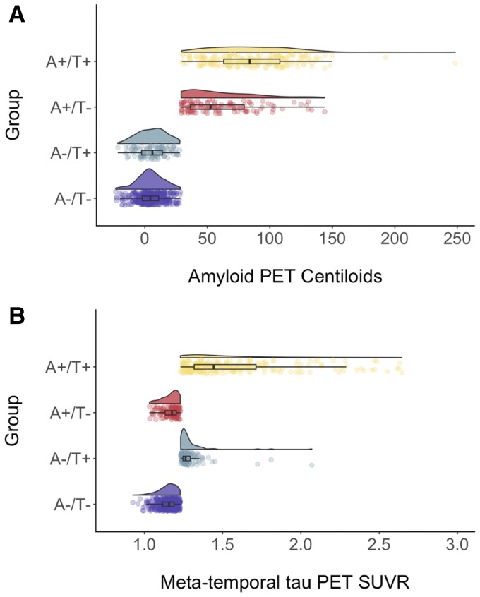
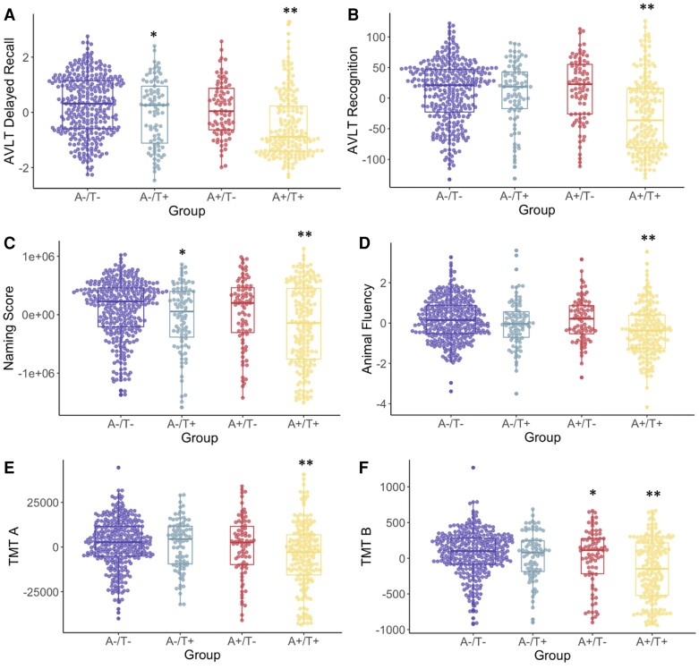
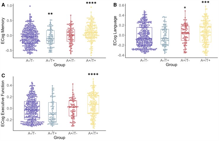
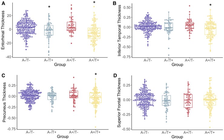
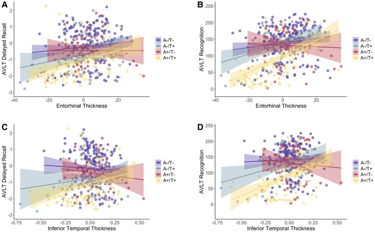
Recently proposed biomarker-only diagnostic frameworks propose that amyloid-beta is necessary for placement on the Alzheimer's disease continuum, whereas tau in the absence of amyloid-beta is considered to be a non-Alzheimer's disease pathologic change. Similarly, the pathologic designation of tau in the absence of amyloid-beta is characterized as primary age-related tauopathy and separable from Alzheimer's disease. Our study sought to identify an early-to-moderate tau stage with minimal amyloid-beta using PET imaging and characterize these individuals in terms of clinical, cognitive and biological features. Seven hundred and three participants from the Alzheimer's Disease Neuroimaging Initiative were classified into one of the four groups (A-/T-, A-/T+, A+/T- and A+/T+) based on PET positivity or negativity for cortical amyloid-beta (A-/A+) and early-to-moderate stage (i.e. meta-temporal) tau (T-/T+). These groups were then compared on demographic and clinical features, vascular risk, multi-domain neuropsychological performance, multi-domain subjective cognitive complaints, apolipoprotein E epsilon-4 carrier status and cortical thickness across Alzheimer's disease-vulnerable regions. The proportion of participants classified in each group was as follows: 47.23% A-/T-, 13.51% A-/T+, 12.23% A+/T- and 27.03% A+/T+. Results indicated that the A-/T+ and A+/T+ groups did not statistically differ on age, sex, depression levels, vascular risk and cortical thickness across temporal and parietal regions. Additionally, both A-/T+ and A+/T+ groups showed significant associations between memory performance and cortical thickness of temporal regions. Despite the different pathologic terminology used for A-/T+ and A+/T+, these groups did not statistically differ on a number of clinical, cognitive and biomarker features. Although it remains unclear whether A-/T+ reflects a pathologic construct separable from Alzheimer's disease, our results provide evidence that this group typically characterized as 'non-Alzheimer's pathologic change' or 'primary age-related tauopathy' should be given increased attention, given some similarities in cognitive and biomarker characteristics to groups traditionally considered to be on the Alzheimer's continuum.

Keywords: Alzheimer’s disease; amyloid-beta; biomarkers; primary age-related tauopathy; tau.


Figures

Graphical abstract
Graphical abstract
Figure 1
Figure 1
A/T group distributions of Aβ PET centiloid and tau PET SUVR levels. Raincloud plots depicting Aβ PET centiloid levels (A) and tau PET SUVR levels (B) across A−/T− (purple, bottom), A−/T + (blue–green, second from bottom), A+/T− (red, second from top) and A+/T+ (yellow, top)) groups. ANCOVAs indicated that groups differed on levels of Aβ PET centiloid levels [F(3,653) = 528.3, P < 0.001] an meta-temporal tau PET SUVR levels [F(3,653) = 153.3, P < 0.001].
Figure 2
Figure 2
A/T group differences in neuropsychological scores. Neuropsychological scores (A, AVLT delayed recall; B, AVLT recognition; C, animal fluency; D, naming score; E, TMT A; F, TMT B) across A/T groups. Transformed and residualized data are depicted. ANCOVAs indicated a significant difference between groups on all measures: AVLT delayed recall [F(3,650) = 19.5, P < 0.001], AVLT recognition [F(3,648) = 25.2, P < 0.001], naming [F(3, 647) = 10.0, P < 0.001], animal fluency [F(3,651) = 11.3, P < 0.001], TMT A [F(3,647) = 8.9, P < 0.001] and TMT B [F(3,638) = 17.8, P < 0.001]. *Statistically different from the A−/T− group. **Significantly different from all other groups.
Figure 3
Figure 3
A/T group differences in ECog questionnaire scores. ECog scores (A, memory; B, language; C, executive function) across A/T groups. Transformed and residualized data are depicted. ANCOVAs indicated a significant difference between groups on all measures: Memory [F(3,646) = 14.0, P < 0.001], language [F(3,642) = 6.1, P < 0.001] and executive function [F(3,634) = 8.8, P < 0.001]. *Significantly different from the A−/T− group. **Significantly different from the A−/T+ group. ***Significantly different from the A−/T− and A−/T+ groups. ****Significantly different from all other groups.
Figure 4
Figure 4
A/T group differences in cortical thickness. Cortical thickness profiles (A, entorhinal; B, inferior temporal; C, precuneus; D, superior frontal) across A/T groups. Transformed and residualized data are depicted. ANCOVAs indicated a significant difference between groups for the entorhinal cortex [F(3,412) = 12.6 P < 0.001], the inferior temporal gyrus [F(3,397) = 5.6, P < 0.001] and the precuneus [F(3,412) = 5.3, P = 0.001]. Groups did not significantly differ for the superior frontal gyrus. *Statistically different from the A−/T− and A+/T− groups.
Figure 5
Figure 5
A/T group associations between cortical thickness and neuropsychological scores. Scatterplots depicting AVLT delayed recall (A, C) and recognition (B, D) associations with entorhinal (A, B) and inferior temporal (C, D) cortical thickness across A/T groups. Transformed and residualized data are depicted. (A) AVLT delayed recall was significantly associated with entorhinal thickness only in the A+/T+ group (t = 3.5, P = 0.001). (B) AVLT recognition was significantly associated with entorhinal thickness in the A−/T− (t = 2.3, P = 0.02), A−/T+ (t = 2.9, P = 0.006) and A+/T+ (t = 6.3, P < 0.001) groups but not the A+/T− group. (C) AVLT delayed recall was significantly associated with inferior temporal thickness only in the A+/T+ group (t = 2.8, P = 0.01). (D) AVLT recognition was significantly associated with inferior temporal thickness in the A−/T+ (t = 2.4, P = 0.02) and A+/T+ (t = 4.8, P < 0.001) groups but not the A−/T− or A+/T− group.

References

    1. Alzheimer A. Über eine eigenartige Erkrankung der Hirnrinde. Allg Zeitschr Psychiatr Psychiatr Gerichtl Med. 1907;64:146–148.
    1. Hardy J. The discovery of Alzheimer-causing mutations in the APP gene and the formulation of the “amyloid cascade hypothesis”. FEBS J. 2017;284(7):1040–1044. - PubMed
    1. Hardy J, Allsop D. Amyloid deposition as the central event in the aetiology of Alzheimer's disease. Trends Pharmacol Sci. 1991;12(10):383–388. - PubMed
    1. Jack CR Jr, Bennett DA, Blennow K, et al. . NIA-AA Research framework: Toward a biological definition of Alzheimer's disease. Alzheimers Dement. 2018;14(4):535–562. - PMC - PubMed
    1. Crary JF, Trojanowski JQ, Schneider JA, et al. . Primary age-related tauopathy (PART): A common pathology associated with human aging. Acta Neuropathol. 2014;128(6):755–766. - PMC - PubMed