. 2022;5(1):1-25.
doi: 10.26502/jbsb.5107030. Epub 2022 Feb 3.

Characterization of Two Novel EF-Hand Proteins Identifies a Clade of Putative Ca2+-Binding Protein Specific to the Ambulacraria

Affiliations

Characterization of Two Novel EF-Hand Proteins Identifies a Clade of Putative Ca2+-Binding Protein Specific to the Ambulacraria

Arisnel Soto-Acabá et al. J Bioinform Syst Biol. 2022.

Abstract

In recent years, transcriptomic databases have become one of the main sources for protein discovery. In our studies of nervous system and digestive tract regeneration in echinoderms, we have identified several transcripts that have attracted our attention. One of these molecules corresponds to a previously unidentified transcript (Orpin) from the sea cucumber Holothuria glaberrima that appeared to be upregulated during intestinal regeneration. We have now identified a second highly similar sequence and analyzed the predicted proteins using bioinformatics tools. Both sequences have EF-hand motifs characteristic of calcium-binding proteins (CaBPs) and N-terminal signal peptides. Sequence comparison analyses such as multiple sequence alignments and phylogenetic analyses only showed significant similarity to sequences from other echinoderms or from hemichordates. Semi-quantitative RT-PCR analyses revealed that transcripts from these sequences are expressed in various tissues including muscle, haemal system, gonads, and mesentery. However, contrary to previous reports, there was no significant differential expression in regenerating tissues. Nonetheless, the identification of unique features in the predicted proteins and their presence in the holothurian draft genome suggest that these might comprise a novel subfamily of EF-hand containing proteins specific to the Ambulacraria clade.


Figures

Figure 1:
Figure 1:
Orpin A and Orpin B are isoforms. Differences between sequences are highlighted. White bars: 5’ UTR and 3’ UTR regions of both sequences; pink bar: predicted signal peptides; orange bar: ORF regions; purple bars: predicted EF-hand motifs; green: conservation level; top sequences: nucleotide and amino acid consensus sequences. Differences between nucleotide sequences are highlighted. It is shown a significant difference, especially between both 3’ UTR sequences. Analysis was done using the MAFFT plugin in Geneious 11.1.5.
Figure 2:
Figure 2:
Primers for sq-RT-PCR of Orpin A. Orpin A UTRs regions (blue boxes) and coding region (green box) of the Orpin A gene. Primer sequences designed to specifically amplify Orpin A (light red letters). Primer sequences used for the identification of the original Orpin sequence in previous reports (green letters). These primers were designed prior to identification Orpin isoform.
Figure 3:
Figure 3:
Primers for sq-RT-PCR of Orpin B. Orpin B UTR sequences (blue boxes) and coding region (green box). Primer sequences designed to specifically amplify Orpin B (blue letters). Sequence comparisons among the two Orpins from H. glaberrima and the two Orpin-like sequences from S. kowalevskii show that the latter shared 46–50% identity and 76–77% similarity with the Orpin A (Fig 4). Similarly, Orpin B translated amino acid sequence shared 46–50% identity and 66–67% similarity with the sequences from S. kowalevskii (Fig 4). Furthermore, we identified three additional putative Orpin homologs from another sea cucumber species, Apostichopus japonicus, one from the starfish Acanthaster planci, and two from the sea urchin Strongylocentrotus purpuratus with expected values (E-value < 0.001 and total scores > 47.8). All Orpin-like sequences contain one domain that is predicted to be a calcium-binding domain composed of two EF-hand motifs at their carboxy-terminal (Figs 5 and 6).
Figure 4:
Figure 4:
Orpin homologs pairwise sequence divergence. Translated amino acid sequences comparison by (A) identity% and (B) similarity%. The alignments were done using Muscle with 50 iterations using Geneious 11.1.5.
Figure 5:
Figure 5:
Orpin homologs alignment. The most conserved residues are indicated by letters in black boxes, green identity regions, and large cartoon letters at the sequence Logo. The exception is PIK49419.1 because 20 amino acid residues from the N-terminal portion are not compared to other sequences. We can see the additional N-terminal regions from A. japonicus sequences ARI48335.1 and PIK49419.1 that did not match to the other homologs. Blue box: signal peptide prediction; red box: EF-Hand motif pair prediction. This alignment was done by Muscle plugin with 50 iterations using Geneious 11.1.5.
Figure 6:
Figure 6:
Orpin homologs EF-hand motifs alignment. The predicted odd EF-hands match with the canonical EF-hand pattern and the predicted even EF-hands were identified as non-canonical motifs (14 residues vs 12 residues) (red boxes). The non-canonical motifs are similar to vertebrates S100s. Predicted calcium coordinating residues from the identified EF-hands patterns are indicated by blue triangles. Holothurian Orpins contain a Cys residue at the −Z−1 position of the predicted calcium-binding loop (left orange box). The characteristic residues from Orpin residues are highlighted by orange boxes. Alignment was done using the MAFFT plugin in Geneious 11.1.5.
Figure 7:
Figure 7:
Orpin A and Orpin B bioinformatics characterization. (A) Orpin A and (B) Orpin B have predicted signal peptides at their transmembrane N-terminal regions including cleavage sites. Also, the two isoforms have predicted EF-hand motifs in their non-cytoplasmic regions. These were predicted by various bioinformatics plugin tools using Geneious 11.1.5.
Figure 8:
Figure 8:
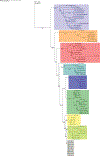
Orpin isoforms are specific to the Ambulacraria clade. EF-hand protein representative sequences from different subfamilies were aligned to build a phylogenetic tree. Orpin homologs were clustered together as a group, separated to the other EF-hand proteins. The tree was made using the PhyML plugin ran through Geneious 11.1.5. The parameters used for this analysis were JTT model of amino acid substitution and 1000 bootstraps. Scale bar: 1. Protein sequences accession numbers are included in S1 Table.
Figure 9:
Figure 9:
Orpin A expression in different tissues. Composite image from RT PCR amplification of Orpin A from H. glaberrima tissues. Orpin A expression (top band) was detected in haemal system (H), muscle (Mu), gonads (G), and mesentery (Me) relative to the expression of NADH. Orpin A was detected neither in the nerve (N) nor in the respiratory tree (RT). The image is a composite from different gels and is divided by a white line.
Figure 10:
Figure 10:
Orpin B expression in different tissues. Composite image from RT PCR amplification of Orpin B from H. glaberrima tissues. Orpin B expression (top band) was detected in haemal system (H), muscle (Mu), gonads (G), and mesentery (Me) relative to expression of NADH. Orpin B was detected neither in the nerve (N) nor in the respiratory tree (RT). The faint band below the NADH band corresponded to primer dimers. The image is a composite from different gels and is divided by a white line.
Figure 11:
Figure 11:
Orpin A expression during intestine regeneration grouped tissues. Semi-quantitative RT-PCR amplification of Orpin A transcripts from mRNA samples from different intestine regenerative days compared to the corresponding expression in samples from normal intestine (NI) and normal mesentery (NM). A statistical high differential expression was found between NM and 7–10 days post evisceration (dpe) (P < .05; P = .002); and between 3–5 dpe and 7–10 dpe (P < .05; P = .03) as indicated in the all pairs Tukey-Kramer HSD test. The large red circle (low number of data points) displays the significant difference between the small grey circles (high number of data points) group means. Red boxes: outlier box plots summarizing the distribution of points at each factor level from the quantiles report. Green diamonds: sample mean and confidence interval ([1 − alpha] x 100). Blue lines: standard mean error. JMP®, Version 12 software was used for the statistical analyses.
Figure 12:
Figure 12:
Orpin B expression during intestine regeneration grouped tissues. Semi-quantitative RT-PCR amplification of Orpin B transcripts from mRNA samples from different intestine regenerative days compared to the corresponding expression in samples from normal intestine (NI) and normal mesentery (NM). A statistical high differential expression was found between NM and 3–5 dpe (P < .05; P = .02), and to 7–10 dpe (P < .001) as indicated in the all pairs Tukey-Kramer HSD test. The large red circle (low number of data points) displays the significant difference between the small grey circles (high number of data points) group means. Red boxes: outlier box plots summarizing the distribution of points at each factor level from the quantiles report. Green diamonds: sample mean and confidence interval ([1 − alpha] × 100). Blue lines: standard mean error. JMP®, Version 12 software was used for the statistical analyses.
Figure 13
Figure 13
(A): Genomic annotation of Orpin A and Orpin B in the draft genome of H. glaberrima. Numbers under each exon represent coordinates in each scaffold. (B) Alignment of exon 3 of both Orpin transcripts obtained from transcriptomic data; both matched the same sequence on scaffold 22267 coordinates 5414–5466.

Similar articles

Cited by

References

    1. García-Arrarás JE, Estrada-Rodgers L, Santiago R, Torres II, Díaz-Miranda L, Torres-Avillán I. Cellular mechanisms of intestine regeneration in the sea cucumber, Holothuria glaberrima Selenka (Holothuroidea:Echinodermata). J Exp Zool 281 (1998): 288–304. - PubMed
    1. García-Arrarás JE, Greenberg MJ. Visceral Regeneration in Holothurians. Microsc Res Tech. 55(2001): 438–451. - PubMed
    1. Rojas-Cartagena C, Ortiz-Pineda P, Ramírez-Gómez F, Suárez-Castillo EC, Matos-Cruz V, Rodríguez C, et al. Distinct profiles of expressed sequence tags during intestinal regeneration in the sea cucumber Holothuria glaberrima. Physiol Genomics 31 (2007): 203–215. - PMC - PubMed
    1. San Miguel-Ruiz JE, García-Arrarás JE. Common cellular events occur during wound healing and organ regeneration in the sea cucumber Holothuria glaberrima. BMC Dev Biol 7 (2007): 115. - PMC - PubMed
    1. Mashanov VS, Zueva OR, García-Arrarás JE. Organization of glial cells in the adult sea cucumber central nervous system. Glia 58(2010): 1581–1593. - PubMed

LinkOut - more resources