. 2022 Nov 30;289(1987):20221747.
doi: 10.1098/rspb.2022.1747. Epub 2022 Nov 16.

The mutational spectrum of SARS-CoV-2 genomic and antigenomic RNA

Affiliations

The mutational spectrum of SARS-CoV-2 genomic and antigenomic RNA

Lele Zhao et al. Proc Biol Sci. .

Abstract

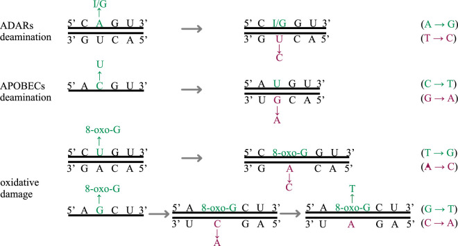
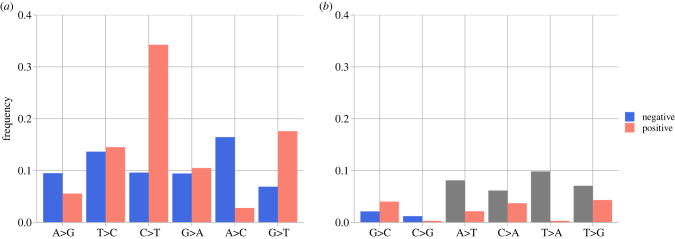
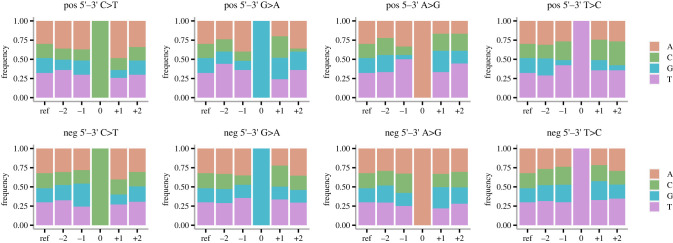
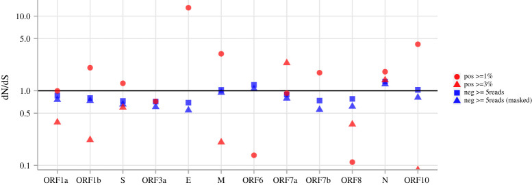
The raw material for viral evolution is provided by intra-host mutations occurring during replication, transcription or post-transcription. Replication and transcription of Coronaviridae proceed through the synthesis of negative-sense 'antigenomes' acting as templates for positive-sense genomic and subgenomic RNA. Hence, mutations in the genomes of SARS-CoV-2 and other coronaviruses can occur during (and after) the synthesis of either negative-sense or positive-sense RNA, with potentially distinct patterns and consequences. We explored for the first time the mutational spectrum of SARS-CoV-2 (sub)genomic and anti(sub)genomic RNA. We use a high-quality deep sequencing dataset produced using a quantitative strand-aware sequencing method, controlled for artefacts and sequencing errors, and scrutinized for accurate detection of within-host diversity. The nucleotide differences between negative- and positive-sense strand consensus vary between patients and do not show dependence on age or sex. Similarities and differences in mutational patterns between within-host minor variants on the two RNA strands suggested strand-specific mutations or editing by host deaminases and oxidative damage. We observe generally neutral and slight negative selection on the negative strand, contrasting with purifying selection in ORF1a, ORF1b and S genes of the positive strand of the genome.

Keywords: RNA editing; SARS-CoV-2; antigenomes; mutational spectrum.


Conflict of interest statement

We declare we have no competing interests.

Figures

Figure 1.
Figure 1.
Example of the three types of mutations. The green-coloured bases show the original mutational changes; the magenta-coloured bases show the subsequent changes in base pairing. (Online version in colour.)
Figure 2.
Figure 2.
Consensus > variant pair frequencies for SARS-CoV-2 positive and complemented negative strands; (a) shows mutational changes attributable to known mechanisms (i.e. ADARs, APOBECs, oxidative damage); (b) shows other mutational changes, with A > T, C > A, T > A, T > G frequencies (grey) on the negative strand not interpreted. (Online version in colour.)
Figure 3.
Figure 3.
Genomic context of candidate sites for RNA editing on the positive (top row) and non-complemented negative (bottom row) strands. The site of change is labelled 0, with the base positions to the 5′ direction labelled −2 and −1, base positions to the 3′ direction labelled + 1 and + 2. The ‘ref’ columns show the genomic content of the SARS-CoV-2 reference sequence [GenBank ID: NC_045512.2]. (Online version in colour.)
Figure 4.
Figure 4.
dN/dS of SARS-CoV-2 coding regions computed with differently filtered variants. Red circles are from positive-strand variant sites with no less than 1% MAF; red triangles are from positive-strand variant sites with no less than 3% MAF. Blue squares are from negative-strand variant sites with at least five reads supporting the minor variant, blue triangles are the same sites but with A > T, C > A, T > A, T > G sites masked. ‘Pos > = 3%’ dN/dS = Inf are not shown for ORF3a, ORF6 and ORF7b. No ‘Pos > = 3%’ variant sites were called within E; therefore, dN/dS is not shown. (Online version in colour.)
Figure 5.
Figure 5.
SARS-CoV-2 polymorphic sites shared by both strands. The x-axis indicates the genomic positions of the sites, the y-axis ([pos] for positive strand, [neg] for negative strand) indicates the consensus base frequencies of the mismatching sites. 'O's mark sites with complementing consensus-variant pairs from the two strands (base frequencies not shown). 'X's mark consensus-variant pair mismatching sites, with positive consensus frequency on the top, negative consensus frequency on the bottom, connected by a vertical line. Each mismatching site is labelled with genomic position and positive consensus > variant in the top half of the plot and complemented negative consensus > variant (in the positive sense) in the lower half of the plot. (Online version in colour.)

References

    1. World Health Organization. 2022. COVID-19 weekly epidemiological update, edition 110, 21 September. https://apps.who.int/iris/bitstream/handle/10665/363125/nCoV-weekly-sitr....
    1. Stern A, Andino R. 2016. Viral evolution: it is all about mutations. In Viral pathogenesis: from basics to systems biology (eds Katze MG, Korth MJ, Law GL, Nathanson N), pp. 233-240. New York, NY: Academic Press.
    1. Eguia RT, Crawford KHD, Stevens-Ayers T, Kelnhofer-Millevolte L, Greninger AL, Englund JA, Boeckh MJ, Bloom JD. 2021. A human coronavirus evolves antigenically to escape antibody immunity. PLoS Pathog. 17, e1009453. (10.1371/journal.ppat.1009453) - DOI - PMC - PubMed
    1. Bordería AV, Stapleford KA, Vignuzzi M. 2011. RNA virus population diversity: implications for inter-species transmission. Curr. Opin. Virol. 1, 643-648. (10.1016/j.coviro.2011.09.012) - DOI - PubMed
    1. Davies NG, et al. 2021. Estimated transmissibility and impact of SARS-CoV-2 lineage B.1.1.7 in England. Science 372, eabg3055. (10.1126/science.abg3055) - DOI - PMC - PubMed

Publication types