Review
. 2023 Mar;293(3):275-292.
doi: 10.1111/joim.13585. Epub 2022 Dec 18.

The role of extracellular histones in COVID-19

Affiliations
Review

The role of extracellular histones in COVID-19

Femke de Vries et al. J Intern Med. 2023 Mar.

Abstract

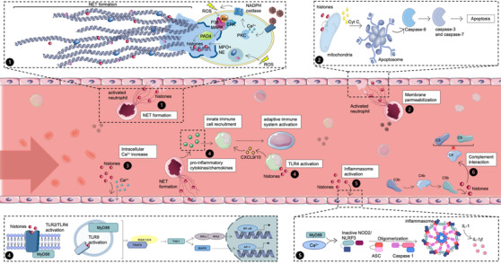
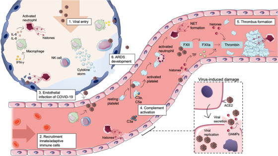
The severe acute respiratory syndrome coronavirus 2 (SARS-CoV-2) had spread from China and, within 2 months, became a global pandemic. The infection from this disease can cause a diversity of symptoms ranging from asymptomatic to severe acute respiratory distress syndrome with an increased risk of vascular hyperpermeability, pulmonary inflammation, extensive lung damage, and thrombosis. One of the host defense systems against coronavirus disease 2019 (COVID-19) is the formation of neutrophil extracellular traps (NETs). Numerous studies on this disease have revealed the presence of elevated levels of NET components, such as cell-free DNA, extracellular histones, neutrophil elastase, and myeloperoxidase, in plasma, serum, and tracheal aspirates of severe COVID-19 patients. Extracellular histones, a major component of NETs, are clinically very relevant as they represent promising biomarkers and drug targets, given that several studies have identified histones as key mediators in the onset and progression of various diseases, including COVID-19. However, the role of extracellular histones in COVID-19 per se remains relatively underexplored. Histones are nuclear proteins that can be released into the extracellular space via apoptosis, necrosis, or NET formation and are then regarded as cytotoxic damage-associated molecular patterns that have the potential to damage tissues and impair organ function. This review will highlight the mechanisms of extracellular histone-mediated cytotoxicity and focus on the role that histones play in COVID-19. Thereby, this paper facilitates a bench-to-bedside view of extracellular histone-mediated cytotoxicity, its role in COVID-19, and histones as potential drug targets and biomarkers for future theranostics in the clinical treatment of COVID-19 patients.

Keywords: COVID-19; DAMPs; NETosis; NETs; extracellular histones; histones.


Conflict of interest statement

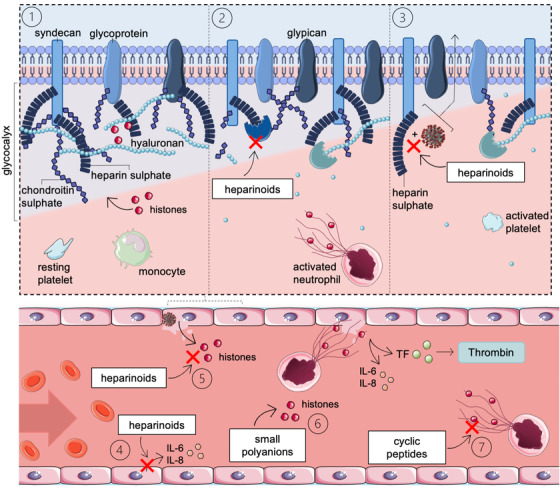
CR and GN are inventors of a patent owned by the Maastricht University on the use of non‐anticoagulant heparin to treat histone‐mediated inflammatory disease. KW and GN are inventors of a patent filed by the Maastricht University to use peptides to target extracellular histones H4 and H2A.

Figures

Fig. 1
Fig. 1
Release and mechanisms of action of extracellular histones. (1.1) Histone release via NETosis. Activated neutrophils can undergo neutrophil extracellular trap (NET) formation to release their DNA and cytotoxic proteins, including histones, thereby killing pathogens. (1.2) Histone‐mediated membrane disruption. The direct effect of histones on plasma and mitochondrial membranes causes permeabilization. Cyt C is released from the mitochondria that lead to the formation of apoptosomes and activation of caspases. The cell will eventually undergo apoptosis. (1.3) Histone‐induced increase of intracellular calcium. Histones can actively bind to nonselective cation channels to rapidly increase calcium concentrations inside the cell leading to a disruption of calcium homeostasis. (1.4) Histone‐mediated pro‐inflammatory cytokine/chemokine release. Activation of Toll‐like receptors (TLR) receptors by histones triggers myD88 and TRAP. This cascade eventually leads to nuclear factor kappaB (NF‐κB) and activator protein 1 (AP‐1) transcription. (1.5) Histone‐mediated NOD2/nucleotide‐binding oligomerization domain‐like receptor protein 3 (NLRP3) inflammasome activation. Extracellular histones can activate nucleotide binding oligomerization domain containing 2 (NOD2) via binding to TLR2/4, whereby the further establishment of the inflammasome is possible. Intracellular Ca2+ from the mitochondria or ER can enhance this pathway. (1.6) Complement‐mediated histone release. Extracellular histones are released when MAC lyses cells infected with the SARS‐CoV‐2 virus. Soluble complement protein C4 can be bound by extracellular histones, inhibiting complement proteins C3 and C5. Akt, protein kinase B; cyt C, cytochrome C; ERK, extracellular signal‐regulated kinases; IKK, IkappaB kinase; IL‐1, interleukin 1; IRAK, interleukin‐1 receptor–associated kinase; MAPK, mitogen‐activated protein kinases; MPO, myeloperoxidase; myD88, myeloid differentiation factor 88; NADPH, nicotinamide adenine dinucleotide phosphate; NE, neutrophil elastase; PAD4, protein arginine deiminase 4; PKC, protein kinase C; ROS, reactive oxygen species; TAK, transforming growth factor beta‐activated kinase‐1; TRAF, TNF receptor–associated factor. Source: Servier Medical Art was used to make these figures: smart.servier.com.
Fig. 2
Fig. 2
The role of extracellular histones during early and severe coronavirus disease 2019 (COVID‐19) infection. Binding of the coronavirus spike protein to the human angiotensin‐converting enzyme 2 (ACE‐2) receptor allows for the entry of the virus into the target host cell. After entering the cytoplasm and uncoating of the viral particle, viral gene expression is ensued. Viral particles, including histones, are released during apoptosis or necrosis. (2.1) When the SARS‐CoV‐2 virus enters the upper respiratory tract, innate immune cells are activated by infected respiratory cells. Alveolar macrophages secrete pro‐inflammatory cytokines to activate neutrophils, which undergo NETosis. In later phases of the infection, the SARS‐CoV‐2 virus infects the lower respiratory tract, and the adaptive immune system is activated. (2.2) Recruitment of innate and adaptive immunes cells to the site of infection. Secretion of pro‐inflammatory cytokines by alveolar macrophages results in the recruitment of other macrophages, invasion of neutrophils and natural killer (NK) cells. (2.3) Due to activation of neutrophils and neutrophil extracellular trap (NET) formation, infected epithelial cells are cleared via secretion of cytotoxic components. However, these components damage the epithelial and endothelial layer of the alveoli and blood vessel wall, leading to a spread of the SARS‐CoV‐2 virus toward endothelial cells. (2.4) Infected endothelial cells initiate the recruitment of platelets and neutrophils to the microvasculature. Activated platelets can trigger neutrophils to form NETs. During a viral infection, platelets can engulf viral particles mediated by TLR7, triggering complement C3‐dependent NET formation. (2.5) Coagulation can be initiated in different ways during COVID‐19 infection. NETs can form a surface for thrombin formation, whereas histones can also directly stimulate the activation of prothrombin by activated factor X (FXa). Furthermore, the coagulation pathway can be triggered directly by viral damage or by tissue factor exposure. (2.6) Newly recruited immune cells secrete high levels of pro‐inflammatory cytokines/chemokines that, when excessive, cause a cytokine storm, leading to epithelial and endothelial damage. Over time, the alveolar permeability barrier will disrupt, and acute respiratory distress syndrome (ARDS) will develop. DAMPs, damage‐associated molecular patterns; FVa, activated factor V; PAMPs, pathogen‐associated molecular patterns; TLR, Toll‐like receptor. Source: Servier Medical Art was used to make these figures: smart.servier.com.
Fig. 3
Fig. 3
Working mechanisms of unfractionated heparin (UFH)/low‐molecular weight heparin (LMWH) to prevent cellular damage in coronavirus disease 2019 (COVID‐19) patients. (3.1) The glycocalyx forms a natural barrier between blood and tissues that can reduce extracellular histone cytotoxicity. Histones are attracted to the glycocalyx through electrostatic interactions. Upon glycocalyx degradation, as occurs in severe disease states, mobilized extracellular histones can damage the underlying endothelium. (3.2) UFH/LMWH inhibits heparinase activity, preventing heparan sulfate degradation. (3.3) UFH/LMWH inhibits cell attachment of spike glycoproteins (SGPs) from viruses via heparin sulfate. (3.4) UFH/LMWH inhibits various pro‐inflammatory pathways. For example, LPS‐stimulated endothelial cells increase interleukin (IL)‐6 and IL‐8, whereas a UFH treatment after LPS‐stimulation reduced the pro‐inflammatory cytokine levels. (3.5, 3.6, 3.7) UFH/LMWH, small polyanions and cyclic peptides can neutralize extracellular histones by binding. TF, tissue factor. Source: Servier Medical Art was used to make these figures: smart.servier.com.

Similar articles

Cited by

References

    1. World Health Organisation 2013 . WHO Coronavirus (COVID‐19) Dashboard . The United Nations, https://covid19.who.int. Accessed 30 November 2022
    1. Gibson PG, Qin L, Puah SH. COVID‐19 acute respiratory distress syndrome (ARDS): clinical features and differences from typical pre‐COVID‐19 ARDS. Med J Aust. 2020;213(2):54–6 e1. - PMC - PubMed
    1. Ng H, Havervall S, Rosell A, Aguilera K, Parv K, von Meijenfeldt FA, et al. Circulating markers of neutrophil extracellular traps are of prognostic value in patients with COVID‐19. Arterioscler Thromb Vasc Biol. 2021;41:988–94 ATVBAHA120315267. - PMC - PubMed
    1. Arcanjo A, Logullo J, Menezes CCB, de Souza Carvalho Giangiarulo TC, Dos Reis MC, et al. The emerging role of neutrophil extracellular traps in severe acute respiratory syndrome coronavirus 2 (COVID‐19). Sci Rep. 2020;10(1):19630. - PMC - PubMed
    1. Castanheira FVS, Kubes P. Neutrophils and NETs in modulating acute and chronic inflammation. Blood. 2019;133(20):2178–85. - PubMed

Publication types