Review
. 2024 Feb;37(1):6-16.
doi: 10.1177/19714009221140540. Epub 2022 Nov 16.

Skull base infections, their complications, and management

Affiliations
Review

Skull base infections, their complications, and management

Phat Tan Nguyen et al. Neuroradiol J. 2024 Feb.

Abstract

Objective: Our review aims to summarize the current literature on skull base infections (SBIs) and retrospectively analyze any such cases encountered at our institution.

Design: A literature search was conducted using online databases PubMed, MEDLINE, and ResearchGate with the terms "skull base osteomyelitis," "temporal bone osteomyelitis," "skull base infections," "necrotizing otitis media," and "SBO". References from the resulting manuscripts were reviewed for relevant articles. A search of our electronic health records using the same key terms was also performed to identify patients with a tissue biopsy-confirmed diagnosis of skull base infections. Patients with an indeterminate diagnosis or inaccessible/poor imaging were excluded.

Setting: A level one trauma and major tertiary academic medical center.

Participants: All patients treated at the University of California Davis Health System with a confirmed diagnosis of skull base infections from January 2005 to November 2020.

Main outcome measures: Imaging results, symptoms, treatment, morbidity, and mortality.

Results: Our literature search yielded 59 articles ranging from 1982 to 2021. A retrospective search of our electronic health records identified two cases of skull base infections.

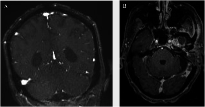
Conclusion: Skull base infections have no pathognomonic findings. A multimodal approach with computed tomography (CT), magnetic resonance imaging (MRI), and nuclear medicine is necessary to characterize the disease process in addition to a biopsy for definitive diagnosis. Other diagnoses can mimic SBI on imaging, such as nasopharyngeal carcinoma and inflammatory pseudotumor. Culture-guided antimicrobial treatment and surgery are mainstay therapies. Other adjuvant strategies currently lack the robust evidence necessary to characterize their risks and benefits.

Keywords: Skull base; infection; malignant; mimics; necrotizing otitis media; osteomyelitis.


Conflict of interest statement

Declaration of conflicting interestsThe author(s) declared no potential conflicts of interest with respect to the research, authorship, and/or publication of this article.

Figures

Figure 1.
Figure 1.
(a) Axial fluid attenuated inversion recovery demonstrates subtle T2 hyperintensity in the left petrous apex extending into the nasopharynx. (b) Axial T1 demonstrates low signal and marrow replacement in the left petrous apex and clivus with blurring of the left superior parapharyngeal space. (c) Diffusion weighted image at the same level demonstrating restricted diffusion in the left petrous apex.
Figure 2.
Figure 2.
(a) Gallium-avid foci in the left skull base in the region of the left petrous apex and clivus extending to the nasopharynx.
Figure 3.
Figure 3.
(a,b) Computed tomography demonstrates erosive changes in the clivus and right occipital condyle. (c) Initial magnetic resonance imaging shows mild osseous enhancement of the basisphenoid and left greater than right enhancing inflammatory tissues in the infratemporal fossa.
Figure 4.
Figure 4.
Computed tomography demonstrates worsening erosions of the left petrous apex and central skull base osteolysis. Left opacification of the mastoids and retroauricular soft tissue swelling and cellulitis is consistent with clinical otomastoiditis.
Figure 5.
Figure 5.
(a,b) Post-contrast fast spin echo T1 sequences demonstrate lack of opacification of the left distal transverse sinus with corresponding T2 weighted images demonstrating lack of the flow void in this same region.
Figure 6.
Figure 6.
(a,b) Magnetic resonance venography demonstrates lack of flow-related enhancement in left distal transverse sinus. Post-contrast fast spoiled gradient echo sequences (right) demonstrate lack of opacification in the left sigmoid sinus.
Figure 7.
Figure 7.
(a) Follow-up magnetic resonance imaging demonstrates increased enhancement of skull base and surrounding tissue, wall thickening and enhancement of the traversing internal carotid arteries, and left coalescent mastoiditis. (b) Uptake on follow-up Gallium-67 single photon emission computed tomography displays the extent and progression of infection. (c) Magnetic resonance angiography demonstrates subtle narrowing of the left petrous internal carotid artery secondary to soft tissue involvement from skull base infection.
Figure 8.
Figure 8.
(a,b,c) Post-contrast images of the nasopharynx and skull base demonstrating increased enhancement and worsening extension of skull base infection. Diffusion weighted images show restricted diffusion in this same region.
Figure 9.
Figure 9.
(a) Post-contrast axial T1 with fat saturation shows abnormal enhancement of the left petrous apex with extension to the nasopharynx. (b) Axial T2 with fat saturation shows opacification of the left mastoid air cells and abnormal signal in the left petrous apex. (c) Diffusion weighted imaging shows abnormal restricted diffusion in the left petrous apex.

References

    1. Johnson AK, Batra PS. Central skull base osteomyelitis: An emerging clinical entity. Laryngoscope 2014; 124: 1083–1087. - PubMed
    1. Albataineh T, Mukherjee S, Donahue JH, et al. Skull Base Infection. Semin Ultrasound CT MRI 2021; 42: 253–265. - PubMed
    1. Khan M, Quadri SQ, Kazmi A, et al. A comprehensive review of skull base osteomyelitis: Diagnostic and therapeutic challenges among various presentations. Asian J Neurosurg 2018; 13: 959. - PMC - PubMed
    1. Chang PC, Fischbein NJ, Holliday RA. Central skull base osteomyelitis in patients without otitis externa: Imaging findings. Am J Neuroradiology 2003; 24: 1310–1316. - PMC - PubMed
    1. Chawdhary G, Hussain S, Corbridge R. Delayed diagnosis of central skull-base osteomyelitis with abscess: Case report and learning points. Ann R Coll Surg Engl 2017; 99: e24–e27. - PMC - PubMed

MeSH terms

LinkOut - more resources