. 2022 Nov 16;18(11):e1010924.
doi: 10.1371/journal.ppat.1010924. eCollection 2022 Nov.

Cryo-EM reveals the conformational epitope of human monoclonal antibody PAM1.4 broadly reacting with polymorphic malarial protein VAR2CSA

Affiliations

Cryo-EM reveals the conformational epitope of human monoclonal antibody PAM1.4 broadly reacting with polymorphic malarial protein VAR2CSA

Sai Sundar Rajan Raghavan et al. PLoS Pathog. .

Abstract

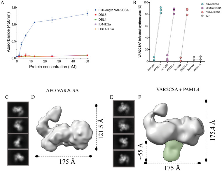
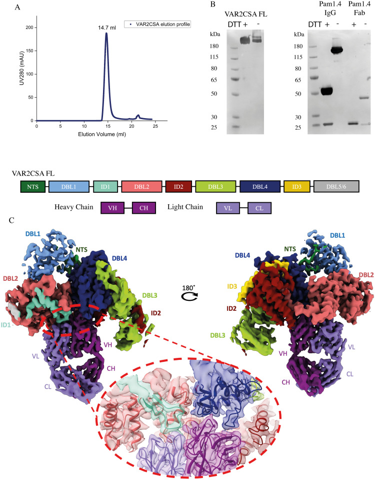
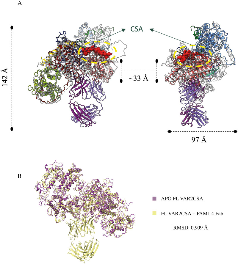
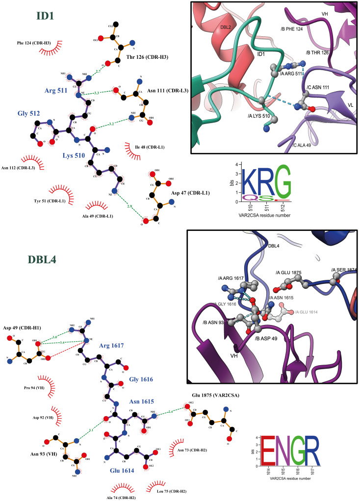
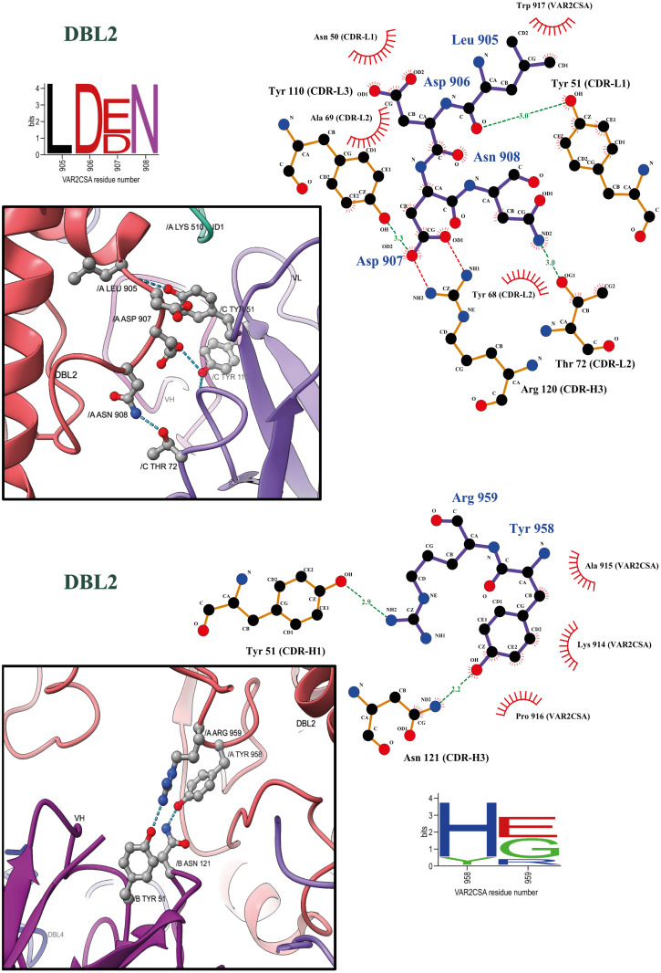
Malaria during pregnancy is a major global health problem caused by infection with Plasmodium falciparum parasites. Severe effects arise from the accumulation of infected erythrocytes in the placenta. Here, erythrocytes infected by late blood-stage parasites adhere to placental chondroitin sulphate A (CS) via VAR2CSA-type P. falciparum erythrocyte membrane protein 1 (PfEMP1) adhesion proteins. Immunity to placental malaria is acquired through exposure and mediated through antibodies to VAR2CSA. Through evolution, the VAR2CSA proteins have diversified in sequence to escape immune recognition but retained their overall macromolecular structure to maintain CS binding affinity. This structural conservation may also have allowed development of broadly reactive antibodies to VAR2CSA in immune women. Here we show the negative stain and cryo-EM structure of the only known broadly reactive human monoclonal antibody, PAM1.4, in complex with VAR2CSA. The data shows how PAM1.4's broad VAR2CSA reactivity is achieved through interactions with multiple conserved residues of different sub-domains forming conformational epitope distant from the CS binding site on the VAR2CSA core structure. Thus, while PAM1.4 may represent a class of antibodies mediating placental malaria immunity by inducing phagocytosis or NK cell-mediated cytotoxicity, it is likely that broadly CS binding-inhibitory antibodies target other epitopes at the CS binding site. Insights on both types of broadly reactive monoclonal antibodies may aid the development of a vaccine against placental malaria.


Conflict of interest statement

We have read the journal’s policy and the authors of this manuscript have the following competing interests: AS, and MAN are listed as co-inventors on a patent family covering the use of VAR2CSA to target and diagnose cancer. AS and is listed as co-inventor on a patent on using VAR2CSA as a prophylactic malaria vaccine during pregnancy. The other authors have no conflicts of interest.

Figures

Fig 1
Fig 1. Reactivity of PAM1.4 to full-length recombinant VAR2CSA and native VAR2CSA.
(a) ELISA analysis showing binding of PAM1.4 Fab to full-length recombinant VAR2CSA and not to single domain constructs ID1-1D2a, DBL1-ID2a, DBL4, DBL5. (b) P. falciparum laboratory-adapted clones FCR3, NF54, and 7G8 expressing VAR2CSA on the infected erythrocyte surface and a non-VAR2CSA 3D7 clone were incubated with 10μg/ml PAM1.4 IgG or IgG isotype control and evaluated by flow cytometry as described to confirm reactivity to the native protein. Independent experiments are shown as individual points. (c) Representative of reference free 2D classification of APO VAR2CSA. (d) Refined 3D models after iterative rounds of 2D and 3D classification. (e) 2D class averages of VAR2CSA complexed with PAM1.4 Fab. (f) Refined 3D model of the complex VAR2CSA and PAM1.4 Fab. Extra density corresponding to the Fab is highlighted in green.
Fig 2
Fig 2. CryoEM reveals conformational epitope binding of PAM1.4 Fab to VAR2CSA.
(a). Gel filtration profile of VAR2CSA full length in 20mM Tris HCl, 150mM KCl, pH. 7.5 buffer. (b) Silver stained SDS-PAGE reduced/ non-reduced of full-length VAR2CSA (307 kDa), PAM1.4 IgG and PAM1.4 Fab after IgG digestion and Fc removal by reverse protein A purification. (c) Overall architecture of VAR2CSA with PAM1.4 Fab, flipped 180°. The domains are colored individually. NTS–N-terminal Sequence, DBL- Duffy Binding Like domain, ID- Inter-domain, VH, CH–Variable and Constant Heavy, VL, CL–Variable and Constant Light. The binding region is magnified and highlighted with 50% transparency to show conformational epitope binding.
Fig 3
Fig 3. Overlay of VAR2CSA PAM 1.4 Fab complex structure with VAR2CSA complexed with CSA (PDB ID: 7JGH) and APO VAR2CSA.
(a) Alignment of the models show no major conformational changes between the structures, generating a RMSD of 1.078Å. CSA is shown in red spherical model and the nearest distance between the epitope of CSA and epitope of PAM 1.4 Fab is measured to be ~33Å showing that the CSA binding site is not affected by PAM 1.4 Fab binding. (b) Overlay of APO VAR2CSA (S5 Fig) and VAR2CSA PAM 1.4 Fab complex structure. Alignment of the models shows no major conformational changes on VAR2CSA upon PAM1.4 Fab binding. RMSD: 0.909 Å.
Fig 4
Fig 4. PAM1.4 Fab interaction analysis to ID1 and DBL4.
Binding plot analysis using Ligplot+ v.2.7. Hydrogen bonds are shown in green dotted lines, electrostatic interactions in red dotted lines and hydrophobic contacts are shown as short spokes radiating from each atom or residue. Sequence conservation of PAM1.4-interacting amino acids in FCR3 VAR2CSA is shown in sequence LOGOs extracted from a previously published sequence alignment LOGO of 3737 VAR2CSA variants found in [13]. FCR3 VAR2CSA sequence positions 510 and 1614 correspond to alignment positions 523 and 1760 respectively. Representation of corresponding Ligplot+ in UCSFChimeraX showing ID1 and DBL4 interactions to Heavy and light chain CDRs of PAM1.4 Fab. Hydrogen bonds are represented in dotted blue lines. Non-CDR residue N93 of heavy chain is shown to form hydrogen bond with N1615 of DBL4 domain in VAR2CSA.
Fig 5
Fig 5. PAM1.4 Fab interaction analysis to DBL2.
Similar to Fig 4, Binding analysis of PAM1.4 Fab to DBL2 is represented using Ligplot+ and UCSF ChimeraX. FCR3 VAR2CSA sequence positions 905 and 958 corresponds to alignment positions 982 and 1073 respectively.

Similar articles

Cited by

References

    1. World Malaria Report 2021. 2021.
    1. Fried M, Duffy PE. Adherence of Plasmodium falciparum to chondroitin sulfate A in the human placenta. Science. 1996;272(5267):1502–4. Epub 1996/06/07. doi: 10.1126/science.272.5267.1502 . - DOI - PubMed
    1. Desai M, ter Kuile FO, Nosten F, McGready R, Asamoa K, Brabin B, et al.. Epidemiology and burden of malaria in pregnancy. Lancet Infect Dis. 2007;7(2):93–104. Epub 2007/01/26. doi: 10.1016/S1473-3099(07)70021-X . - DOI - PubMed
    1. Miller LH, Baruch DI, Marsh K, Doumbo OK. The pathogenic basis of malaria. Nature. 2002;415(6872):673–9. doi: 10.1038/415673a - DOI - PubMed
    1. Hviid L, Jensen AT. PfEMP1—A Parasite Protein Family of Key Importance in Plasmodium falciparum Malaria Immunity and Pathogenesis. Adv Parasitol. 2015;88:51–84. Epub 2015/04/26. doi: 10.1016/bs.apar.2015.02.004 . - DOI - PubMed

Publication types