. 2022 Nov 16;14(671):eabq5931.
doi: 10.1126/scitranslmed.abq5931. Epub 2022 Nov 16.

Protein kinase Cι mediates immunosuppression in lung adenocarcinoma

Affiliations

Protein kinase Cι mediates immunosuppression in lung adenocarcinoma

Ning Yin et al. Sci Transl Med. .

Abstract

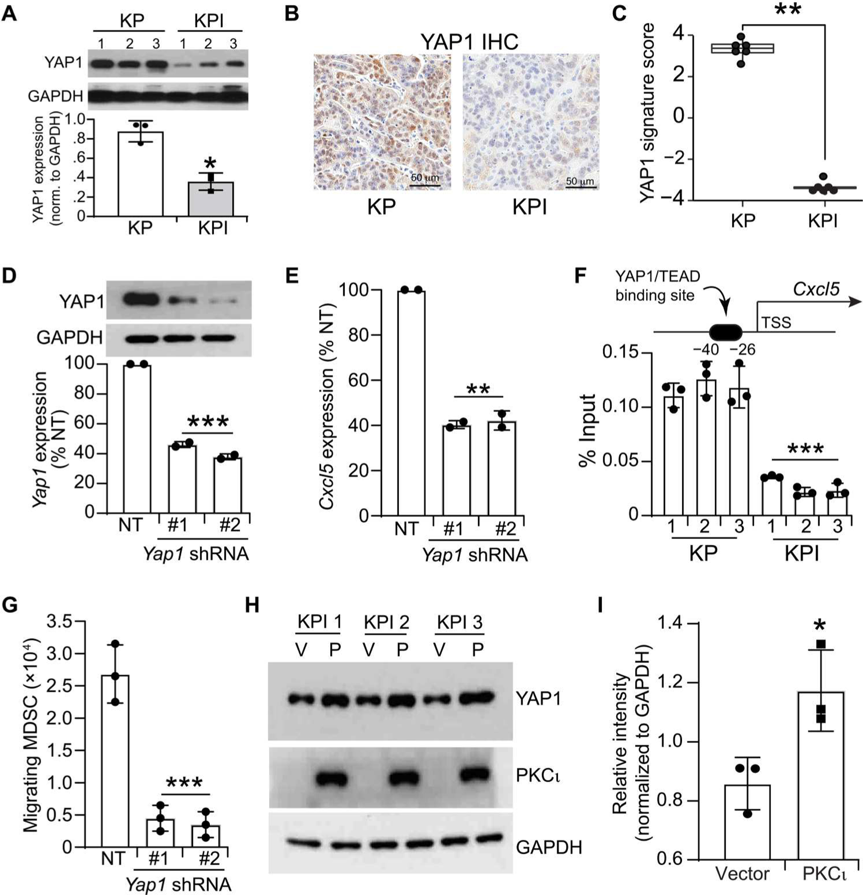
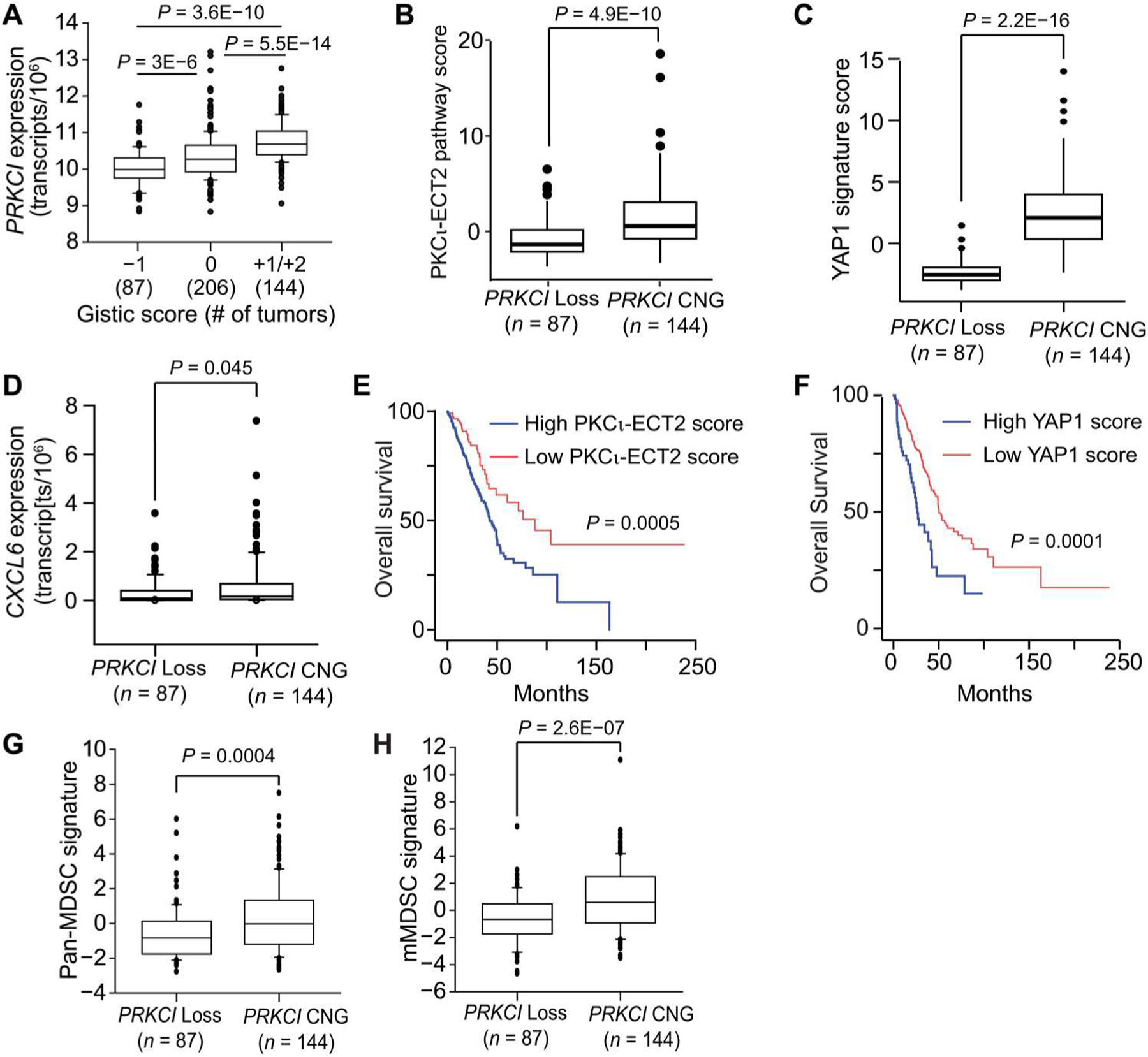
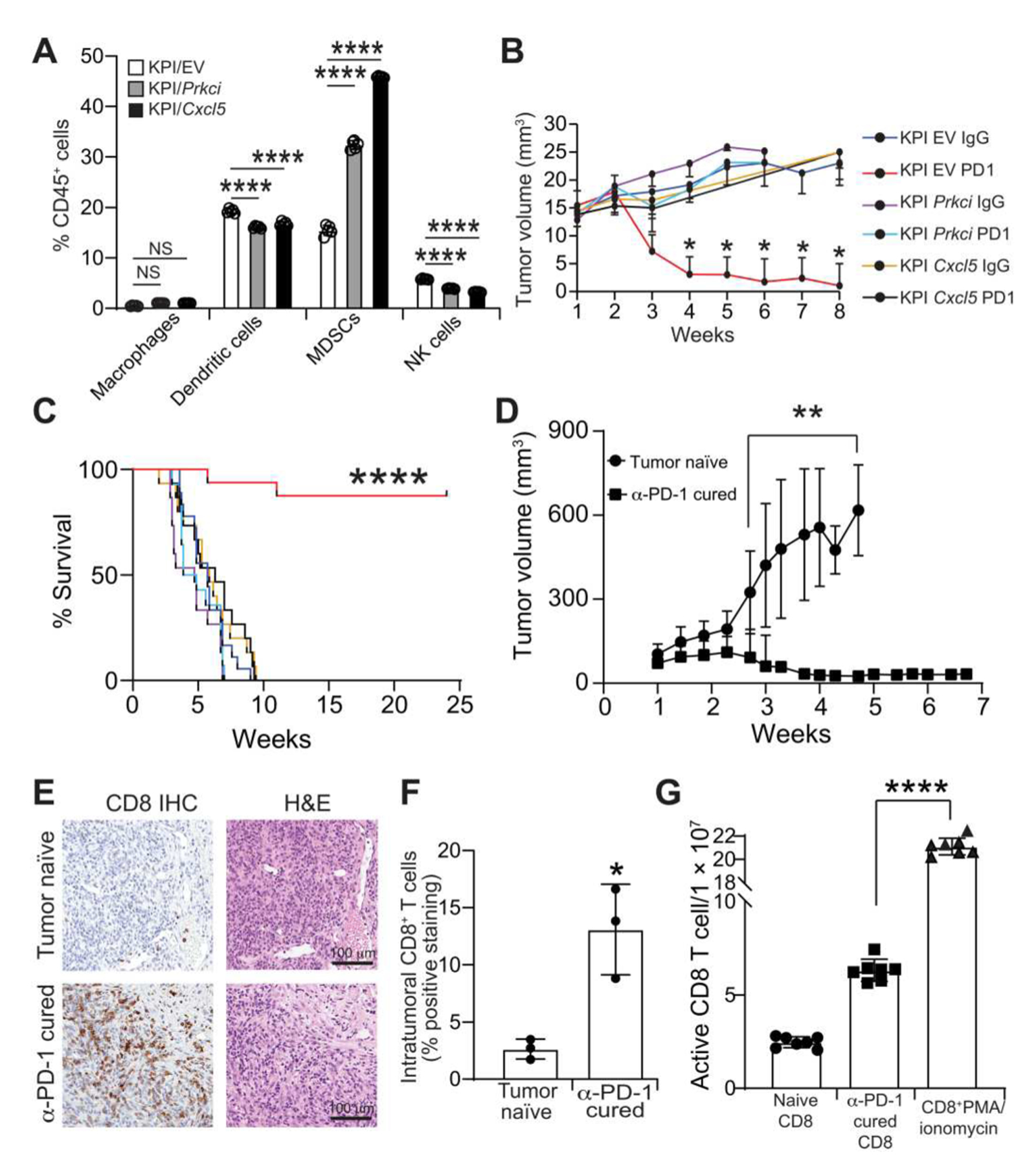
Lung adenocarcinoma (LUAD) is the most prevalent form of non-small cell lung cancer (NSCLC) and a leading cause of cancer death. Immune checkpoint inhibitors (ICIs) of programmed death-1/programmed death-ligand 1 (PD-1/PD-L1) signaling induce tumor regressions in a subset of LUAD, but many LUAD tumors exhibit resistance to ICI therapy. Here, we identified Prkci as a major determinant of response to ICI in a syngeneic mouse model of oncogenic mutant Kras/Trp53 loss (KP)-driven LUAD. Protein kinase Cι (PKCι)-dependent KP tumors exhibited resistance to anti-PD-1 antibody therapy (α-PD-1), whereas KP tumors in which Prkci was genetically deleted (KPI tumors) were highly responsive. Prkci-dependent resistance to α-PD-1 was characterized by enhanced infiltration of myeloid-derived suppressor cells (MDSCs) and decreased infiltration of CD8+ T cells in response to α-PD-1. Mechanistically, Prkci regulated YAP1-dependent expression of Cxcl5, which served to attract MDSCs to KP tumors. The PKCι inhibitor auranofin inhibited KP tumor growth and sensitized these tumors to α-PD-1, whereas expression of either Prkci or its downstream effector Cxcl5 in KPI tumors induced intratumoral infiltration of MDSCs and resistance to α-PD-1. PRKCI expression in tumors of patients with LUAD correlated with genomic signatures indicative of high YAP1-mediated transcription, elevated MDSC infiltration and low CD8+ T cell infiltration, and with elevated CXCL5/6 expression. Last, PKCι-YAP1 signaling was a biomarker associated with poor response to ICI in patients with LUAD. Our data indicate that immunosuppressive PKCι-YAP1-CXCL5 signaling is a key determinant of response to ICI, and pharmacologic inhibition of PKCι may improve therapeutic response to ICI in patients with LUAD.


Conflict of interest statement

Competing interests: The authors declare that they have no competing interests.

Figures

Fig. 1.
Fig. 1.. PKCι stimulates intratumoral infiltration of MDSCs by inducing Cxcl5 expression.
(A) Flow cytometric analysis of innate immune cells in KP and KPI tumors shown as percentage of CD45+ cells. n = 5; data represent mean ± SD. Significance was assessed by ANOVA followed by Tukey’s multiple comparisons test, and P values are shown for each comparison. (B) Quantification of mMDSCs and gMDSCs in KP and KPI tumors. n = 5; data represent mean ± SD. (C) Ability of conditioned medium from KP and KPI tumor cells to stimulate migration of MDSCs in vitro. Data are expressed as number of migrating MDSCs ± SD; n = 4. (D) Effect of silencing PKCι in KP cells on ability of conditioned medium to support MDSC migration. Data are expressed as fold of MDSC migration in KP/NT cells ± SD; N = 4. Inset shows immunoblot for PKCι and glyceraldehyde-3-phosphate dehydrogenase (GAPDH). (E) Effect of reexpressing PKCι in KPI tumor cells on MDSC migration (bottom). Immunoblot of protein extracts from three KP cell lines, KPI/Vector (V) and KPI/PKCι (P) cells for PKCι and GAPDH (top). Data are expressed as fold of MDSC migration in KPI/Vector cells for each cell line pair ± SD; n = 4. (F) Expression of chemokines in KP and KPI LUAD cells. Data are expressed as fold of chemokine RNA in KPI cells ± SD; n = 3. (G) Effect of PKCι KD in KP cells on expression of Cxcl5 RNA. Data expressed as fold NT control ± SD; n = 3. (H) Effect of reexpressing PKCι in KPI LUAD cells on Cxcl5 RNA expression. Data expressed as fold V control ± SD; n = 3. (I) Secretion of CXCL5 from KP and KPI LUAD cells. Data expressed as secreted CXCL5 in pg/ml ± SD; n = 3. (J) Effect of reexpressing PKCι in KPI LUAD cells on secreted CXCL5. Data expressed as secreted CXCL5 in pg/ml ± SD; n = 3. (K) Effect of lentiviral shRNA–mediated KD of Cxcl5 in KP cells on MDSC migration. Data expressed as fold NT control ± SD; n = 3; NT, nontarget. For (B) to (K), *P < 0.05 and **P < 0.01.
Fig. 2.
Fig. 2.. Identification of a PKCι-YAP1-CXCL5 signaling axis in KP cells.
(A) Expression of YAP1 in KP and KPI cells. Immunoblot analysis of cell lysates from three KP and three KPI cell lines for YAP1 and GAPDH (top) and quantification of immunoblot results for YAP1 (bottom). Data are expressed as YAP1 expression normalized to GAPDH ± SD. n = 3. (B) IHC of representative KP and KPI tumors for YAP1. (C) Differential expression of a transcriptional signature of YAP1 activity in KP and KPI cells. n = 6. (D) Characterization of shRNA-mediated Yap1 KD in KP cells. Data represent Yap1 mRNA expressed as % nontarget (NT) control ± SD. n = 2. Immunoblot shows YAP1 protein expression (top). (E) Effect of Yap1 KD on Cxcl5 RNA abundance. Data represent Cxcl5 RNA expressed as %NT control; n = 2. (F) YAP1 binding to the promoter region of the Cxcl5 gene in KP and KPI cells. n = 3. (G) Effect of Yap1 KD on MDSC migration in vitro. Data are expressed as migrating MDSCs ± SD; n = 4. (H) Effect of PKCι reexpression in KPI cells on YAP1 expression. Immunoblot for YAP1, PKCι, and GAPDH in empty vector (V)– and PKCι (P)–overexpressing KPI cells. (I) Quantification of YAP1 immunoblot data in (H). Results are expressed as intensity of YAP1 protein relative to GAPDH ± SD. n = 3. *P < 0.05, **P < 0.01, ***P < 0.001, and ****P < 0.0001.
Fig. 3.
Fig. 3.. Effect of PRKCI CNG or monoallelic loss on PRKCI expression, PKCι signaling activity, and immune cell signatures in human LUAD.
(A) Effect of PRKCI CNG and monoallelic loss on PRKCI expression in LUAD tumors. PRKCI mRNA expression is shown for human LUAD harboring PRKCI CNG (GISTIC score, +1/+2), no copy change (GISTIC score, 0), and monoallelic loss (GISTIC score, −1). Numbers in parentheses indicate the number of tumors in each group. PKCι-ECT2 pathway score (B), YAP1 signature score (C), and CXCL6 expression (D) in human LUAD harboring either PRKCI CNG or PRKCI monoallelic loss. Overall survival of patients with LUAD based on PKCι-ECT2 (E) and YAP1 (F) signature scores. Pan-MDSC signature (G) and mMDSC (H) scores in human LUAD harboring PRKCI CNG or monoallelic loss. P values for comparisons are shown in each panel.
Fig. 4.
Fig. 4.. Response of KP and KPI LUAD tumors to α-PD-1 therapy.
(A) Effect of α-PD-1 on KP and KPI tumor growth. Tumor volume is expressed as mm3; n = 29 (KP/IgG), 24 (KP/αPD-1), 28 (KPI/IgG), and 27 (KPI/αPD-1). (B) Overall survival of KP and KPI tumor–bearing mice in response to α-PD-1 therapy. (C) Representative IHC detection of CD11b+ cells in KP and KPI tumors treated with either α-PD-1 or control IgG. (D) Quantification of CD11b+ cells in KP and KPI tumors. Data are presented as %CD11b+ cells. n = 7 (KP IgG and α-PD-1), n = 11 (KPI control IgG; 4 KPI α-PD-1). NS, not significant. (E) Representative IHC detection of CD8+ cells in KP and KPI tumors treated with either α-PD-1 or control IgG. (F) Quantification of CD8+ cells in KP and KPI tumors. Data are presented as % of total cells. n = 7 KP IgG and α-PD-1, n = 11 KPI control IgG, and n = 4 KPI α-PD-1. *P < 0.05 and ****P < 0.0001.
Fig. 5.
Fig. 5.. Effect of expressing exogenous Prkci and Cxcl5 on response of KPI LUAD tumor to α-PD-1 therapy.
(A) Flow cytometric analysis of innate immune cells in KP and KPI tumors. n = 5; data represent mean ± SD. Significance was assessed by ANOVA followed by Tukey’s multiple comparisons test. (B) Effect of expressing exogenous Prkci and Cxcl5 in KPI cells on orthotopic lung tumor growth and response to α-PD-1 therapy. Data are presented as tumor volume (mm3) and expressed as mean ± SD. Significance between KPI/EV + PD-1 and other groups at each time point is indicated. (C) Effect of expressing exogenous Prkci and Cxcl5 in KPI cells on survival of mice in response to α-PD-1 therapy. (D) Growth of KPI tumors in tumor-naïve and α-PD-1 cured mice. N = 19 (tumor naïve) and 13 (α-PD-1 cured). Each of the indicated time points shows significant difference between groups. (E) IHC detection of CD8+ T cells in xenograft KPI tumors in tumor-naïve and α-PD-1 cured syngeneic mice harvested 2 weeks after inoculation. (F) Quantification of CD8+ T cell staining in (E). Data are expressed as intratumoral CD8+ T cells. n = 3. (G) ELIspot assay for activated CD8+ T cells/100,000 cells from tumor-naïve and α-PD-1 cured mice harvested 2 weeks after inoculation. Activated CD8+ T cells after treatment with PMA and ionomycin (200 ng/ml and 2 μg/ml, respectively) serve as a positive control. Data represent mean ± SD; n = 7. Significance from naïve CD8 is shown. *P < 0.05, **P < 0.01, ***P < 0.001, and ****P < 0.0001.
Fig. 6.
Fig. 6.. Effect of ANF on PKCι-Yap1-Cxcl5 signaling, MDSC migration, and response of KP LUAD tumors to α-PD-1.
KP cells were treated with 1.5 μM ANF for the indicated time, and RNA was subjected to RT-qPCR for Cxcl5 (A) and Yap1 (B) RNA. Data are expressed as fold of 0-hour control. n = 3. (A) Significance compared to 0-hour control is shown. (C) Protein lysates from three independent KP cell lines in (A) were subjected to immunoblot analysis for YAP1, PKCι, and GAPDH. (D) Quantification of YAP1 protein from (C). Results are expressed as mean ± SD and are normalized to GAPDH. n = 3. (E) Immunoblot analysis of cytoplasmic and nuclear fractions of untreated KP cell lines and KP cell lines treated with ANF for 12 hours for YAP1, mitogen-activated protein kinase kinase (MEK) (as a cytoplasmic marker), and lamin A/C (as a nuclear marker). (F) Quantification of YAP1 from (E). Results are expressed as mean ± SD. Cytoplasmic YAP1 is normalized to MEK, and nuclear YAP1 is normalized to lamin A/C. n = 3. (G) Effect of ANF on the ability of KP cells conditioned medium to stimulate MDSC migration in vitro. MDSC migration was assessed in the presence of conditioned medium from either DMSO- or ANF-treated KP cells. Either DMSO or ANF was added directly to the migration assay as indicated to distinguish the effect of ANF on KP cell conditioned medium from possible direct effects on MDSC migration. n = 5. Effect of ANF, α-PD-1, and combined ANF/α-PD-1 treatment on orthotopic lung KP tumor growth (H) and survival (I). n = 15. *P < 0.05, **P < 0.01, and ****P < 0.0001.
Fig. 7.
Fig. 7.. Association of tumor PKCι-YAP1 signaling biomarkers with LUAD patient response to ICI.
Plots of PKCι histoscore (A), YAP1 positivity score (B), and PKCι-YAP1 histoscore (C) in patients with LUAD that either responded or did not respond to ICI. n = 9 (nonresponders), n = 8 (responders). (D) Representative IHC images of YAP1 and PKCι in responder and nonresponder LUAD. Plots of PKCι histoscore (E), YAP1 positivity score (F), and PKCι-YAP1 histoscore (G) in PD-L1+ human LUAD tumors that either responded or did not respond to ICI. n = 4 (nonresponders), n = 6 (responders). *P < 0.05 and **P < 0.01.

References

    1. Siegel RL, Miller KD, Fuchs HE, Jemal A, Cancer statistics, 2021. CA Cancer J. Clin 71, 7–33 (2021). - PubMed
    1. Goldstraw P, Chansky K, Crowley J, Rami-Porta R, Asamura H, Eberhardt WE, Nicholson AG, Groome P, Mitchell A, Bolejack V; International Association for the Study of Lung Cancer Staging and Prognostic Factors Committee, Advisory Boards, and Participating Institutions; International Association for the Study of Lung Cancer Staging and Prognostic Factors Committee Advisory Boards and Participating Institutions, The IASLC lung cancer staging project: Proposals for revision of the TNM stage groupings in the forthcoming (Eighth) Edition of the TNM Classification for Lung Cancer. J. Thorac. Oncol 11, 39–51 (2016). - PubMed
    1. Dafni U, Tsourti Z, Vervita K, Peters S, Immune checkpoint inhibitors, alone or in combination with chemotherapy, as first-line treatment for advanced non-small cell lung cancer. A systematic review and network meta-analysis. Lung Cancer 134, 127–140 (2019). - PubMed
    1. Shukuya T, Carbone DP, Predictive Markers for the efficacy of anti-PD-1/PD-L1 antibodies in lung cancer. J. Thorac. Oncol 11, 976–988 (2016). - PMC - PubMed
    1. Yamauchi Y, Safi S, Blattner C, Rathinasamy A, Umansky L, Juenger S, Warth A, Eichhorn M, Muley T, Herth FJF, Dienemann H, Platten M, Beckhove P, Utikal J, Hoffmann H, Umansky V, Circulating and tumor myeloid-derived suppressor cells in resectable non-small cell lung cancer. Am. J. Respir. Crit. Care Med 198, 777–787 (2018). - PubMed

Publication types