. 2022 Dec 13;55(12):2271-2284.e7.
doi: 10.1016/j.immuni.2022.10.021. Epub 2022 Nov 15.

IKKβ primes inflammasome formation by recruiting NLRP3 to the trans-Golgi network

Affiliations

IKKβ primes inflammasome formation by recruiting NLRP3 to the trans-Golgi network

Niklas A Schmacke et al. Immunity. .

Abstract

The NLRP3 inflammasome plays a central role in antimicrobial defense as well as in the context of sterile inflammatory conditions. NLRP3 activity is governed by two independent signals: the first signal primes NLRP3, rendering it responsive to the second signal, which then triggers inflammasome formation. Our understanding of how NLRP3 priming contributes to inflammasome activation remains limited. Here, we show that IKKβ, a kinase activated during priming, induces recruitment of NLRP3 to phosphatidylinositol-4-phosphate (PI4P), a phospholipid enriched on the trans-Golgi network. NEK7, a mitotic spindle kinase that had previously been thought to be indispensable for NLRP3 activation, was redundant for inflammasome formation when IKKβ recruited NLRP3 to PI4P. Studying iPSC-derived human macrophages revealed that the IKKβ-mediated NEK7-independent pathway constitutes the predominant NLRP3 priming mechanism in human myeloid cells. Our results suggest that PI4P binding represents a primed state into which NLRP3 is brought by IKKβ activity.

Keywords: NEK7; NLRP3; iPSCs; inflammasome; innate immunity; macrophages; monocytes; pattern recognition.


Conflict of interest statement

Declaration of interests The authors declare no competing interests.

Figures

Figure 1
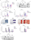
Figure 1. Human iPSC-derived macrophages and human myeloid cell lines activate the NLRP3 inflammasome independently of NEK7
(A) Four clones per indicated genotype of human iPSCs were differentiated into macrophages (hiPS-Macs), primed with LPS for 4 h and then treated with the inflammasome activators Nigericin (NLRP3) or Needle Tox (NAIP-NLRC4) in the presence of the NLRP3 inhibitor MCC950 as indicated before release of LDH (left), IL-1β (middle), and IL-18 (right) was measured. Dots represent separately differentiated iPS cell clones of the indicated genotypes. (B and C) BLaER1 monocytes of the indicated genotypes were primed with LPS for 4 h and subsequently stimulated with Nigericin or Needle Tox. LDH release (B) of one or two clones per genotype is depicted. (C) One representative immunoblot of three independent experiments is shown. (D) Three clones of THP-1 cells of the indicated genotypes were primed with Pam3CSK4 for 4 h and subsequently stimulated with Nigericin for 2 h before release of LDH (left) and TNF (right) were measured. Two different sgRNAs against NEK7 were used (#1 and #2). Dots represent individual clones. (E) THP-1 cells of the indicated genotypes were primed with Pam3CSK4 for 4 h and subsequently stimulated with Nigericin for 2 h before immunoblotting. One representative immunoblot of three independent experiments is shown. (F) NLRP3-/- BLaER1 cells expressing the indicated NLRP3 orthologs under the control of a doxycycline-inducible promoter were treated with doxycycline for the last 24 h of differentiation, primed with LPS for 4 h and subsequently stimulated with Nigericin (left) or Needle Tox (right) for 2 h. The same vector expressing mCherry instead of NLRP3 was used as a mock control. (G) Western blot of cells treated as in (F), one representative of three independent experiments is shown. Data are represented as mean ± SEM with dots representing biological replicates conducted on separate days unless indicated otherwise (one outlier in B is not depicted #). ***p < 0.001, **p < 0.01, *p < 0.05, ns p ≥ 0.05 calculated by two-way ANOVA followed by Tukey’s test (A, B, and D: TNF) or Šidák’s test (D: LDH). See also Figures S1 and S2.
Figure 2
Figure 2. Priming activates IKKβ to bypass NEK7 via a translation-independent mechanism in mouse cells
(A–C) Mouse macrophages constitutively expressing mmNlrp3 (mmMacs) of the indicated genotypes were stimulated with LPS + Nigericin simultaneously for 4 h, with DNA for 28 h or with LPS + ATP for 2 h. (B) One immunoblot representative of two clones from two independent experiments is shown. (D) mmMacs were pretreated with cycloheximide (CHX) for 30 min and stimulated as in (C). (E) Two mmMacs clones per genotype were stimulated as indicated. Release of IP-10 (left), TNF (middle), and LDH (right) of two clones (sub-columns) from three independent experiments (sub-rows) are depicted as heatmaps. (F and G) mmMacs of the indicated genotypes were stimulated as in (A) before the release of LDH (F) and TNF (G) was measured. (H) mmMacs of the indicated genotypes were stimulated as in (A) for 2 h. One representative of three independent biological replicates is shown. Data are represented as mean ± SEM with dots representing biological replicates conducted on separate days. ***p < 0.001, **p < 0.01, *p < 0.05, ns p ≥ 0.05 calculated by two-way ANOVA followed by Tukey’s test (A, C, and F), Šidák’s test (D), or Dunnett’s test (G). See also Figures S3 and S4 and Table S1.
Figure 3
Figure 3. K+ efflux works synergistically with IKKβ to bypass NEK7
(A) mmMacs of the indicated genotypes were stimulated with the NLRP3 inflammasome inducers Nigericin or Imiquimod in the presence of LPS as indicated for 4 h (Nigericin) or 6 h (Imiquimod). (B and C) mmMacs of the indicated genotypes were stimulated as indicated and imaged every 30 min (C) for 3 h in Hank’s balanced salt solution with or without potassium + 10% FCS and 5 mg/mL propidium iodide before LDH release was measured (B). Data are represented as mean ± SEM with dots representing biological replicates conducted on separate days unless indicated otherwise. ***p < 0.001, **p < 0.01, *p < 0.05, ns p ≥ 0.05 calculated by two-way ANOVA followed by Tukey’s test.
Figure 4
Figure 4. NLRP3 priming through IKKβ is required for inflammasome activation in human myeloid cell lines
(A) LDH release from BLaER1 clones of the indicated genotypes primed with LPS for 2 h and subsequently treated with Nigericin or Needle Tox for 2 h. (B) One representative of three immunoblots from cells treated as in (A). The asterisk denotes an unspecific band. (C) Immunoblot of IKBKB-/- BLaER1 cells expressing wild-type IKKβ or kinase-dead IKKβ-K44M under the control of a doxycycline-inducible promoter treated with doxycycline during the last 8 h of differentiation. (D) LDH release from BLaER1 cells as in (C) primed with LPS for 2 h and subsequently treated with Nigericin or Needle Tox as indicated. (E) LDH release from BLaER1 monocytes primed with LPS for 2 h before stimulation with Nigericin or Needle Tox. TPCA-1 was added at different time points as indicated. Data are represented as mean ± SEM with dots representing biological replicates conducted on separate days. ***p < 0.001, **p < 0.01, *p < 0.05, ns p ≥ 0.05 calculated by two-way ANOVA followed by Dunnett’s test (A and E) or Šidák’s test (D). See also Figure S5.
Figure 5
Figure 5. NEK7 accelerates NLRP3 activation at early priming time points in iPSC-derived human macrophages
(A) THP-1 cells were primed with Pam3CSK4 for 4 h and then stimulated with Nigericin for 30 min before lysates were immunoprecipitated with anti-NEK7 antibody or isotype control. One representative immunoblot of three independent experiments is shown. (B) NEK7-/- HEK293T cells were transiently transfected with plasmids driving expression of an ASC-RFP fusion protein and mouse or human orthologs of NLRP3 and NEK7 as indicated. ASC-RFP specks were imaged 24 h after transfection. Dots represent technical replicates from one representative of three independent experiments. (C and D) Four clones per genotype of NEK7-/- or wild-type human iPS cells were differentiated into hiPS-Macs and treated with Nigericin or LPS + Nigericin for 4 h or LPS + Nigericin for 1 h in the presence of the NLRP3 inhibitor MCC950 as indicated. Dots represent individual clones. ***p < 0.001, **p < 0.01, *p < 0.05, ns p ≥ 0.05 calculated by two-way ANOVA followed by Dunnett’s test (B) or Šidák’s test (C and D). See also Figure S6.
Figure 6
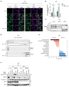
Figure 6. IKKβ-mediated recruitment of NLRP3 to PI4P enables NEK7-independent inflammasome formation
(A) Nlrp3-/- × Pycard-/- J774 cells of the indicated Nek7 genotypes expressing mCherry tethered to phosphatidylinositol-4-phosphate (PI4P) via the PH domain of OSBP (OSBP(PH)-mCherry) and doxycycline-inducible mVenus-mmNlrp3 were treated with doxycycline for 24 h and TPCA-1 for 1 h before stimulation with LPS for 30 min. Scale bars represent 10 μm. (B) Quantification of at least 10 randomly chosen fields of view per experimental condition from three independent experiments described in (A). Data are represented as mean ± SEM with dots representing biological replicates conducted on separate days. (C) Lysates of J774 cells pretreated with TPCA-1 for 1 h and then stimulated with LPS for 30 min were depleted of nuclei (5 min 1,000 × g), and the supernatant was then centrifuged at 5,000 × g for 10 min (pellet P5) followed by 100,000 × g for 20 min (pellet P100, supernatant S100) before immunoblotting. One representative of three independent experiments is shown. (D) P100 fractions from (C) were further fractionated across a linear sucrose gradient (20%–60%) into 12 fractions which were then immunoblotted. One representative of three independent biological replicates is shown. (E) Enrichment of organelle-specific protein sets identified via mass spectrometry analysis of the protein content of fractions #2 and #11. p-values for set enrichment were calculated based on proteins differing between the two fractions (FC R 1.5, FDR < 0.05) using Fisher’s exact test with Benjamini-Hochberg correction. (F) Nlrp3-/- J774 cells of the indicated Nek7 genotypes expressing doxycycline-inducible variants of Nlrp3 as indicated were treated with doxycycline for 18 h followed by 2 h of Nigericin before immunoblotting. ***p < 0.001, **p < 0.01, *p < 0.05, ns ≥ 0.05 calculated by two-way ANOVA followed by Tukey’s test unless indicated otherwise. See also Figure S6 and Table S2.

Comment in

  • Humans pIKK-up NLRP3 to skip NEK7.
    Malireddi RKS, Kanneganti TD. Malireddi RKS, et al. Trends Immunol. 2022 Dec;43(12):947-949. doi: 10.1016/j.it.2022.10.007. Epub 2022 Nov 17. Trends Immunol. 2022. PMID: 36404209
  • IKKβ ignites the inflammasome.
    Flemming A. Flemming A. Nat Rev Immunol. 2023 Jan;23(1):6. doi: 10.1038/s41577-022-00818-w. Nat Rev Immunol. 2023. PMID: 36470972 No abstract available.

References

    1. Broz P, Dixit VM. Inflammasomes: mechanism of assembly, regulation and signalling. Nat Rev Immunol. 2016;16:407–420. doi: 10.1038/nri.2016.58. - DOI - PubMed
    1. Dinarello CA. Overview of the IL-1 family in innate inflammation and acquired immunity. Immunol Rev. 2018;281:8–27. doi: 10.1111/imr.12621. - DOI - PMC - PubMed
    1. Vande Walle L, Lamkanfi M. Pyroptosis. Curr Biol. 2016;26:R568–R572. doi: 10.1016/j.cub.2016.02.019. - DOI - PubMed
    1. Patel MN, Carroll RG, Galván-Peña S, Mills EL, Olden R, Triantafilou M, Wolf AI, Bryant CE, Triantafilou K, Masters SL. Inflammasome priming in sterile inflammatory disease. Trends Mol Med. 2017;23:165–180. doi: 10.1016/j.molmed.2016.12.007. - DOI - PubMed
    1. Swanson KV, Deng M, Ting JP-Y. The NLRP3 inflammasome: molecular activation and regulation to therapeutics. Nat Rev Immunol. 2019;19:477–489. doi: 10.1038/s41577-019-0165-0. - DOI - PMC - PubMed

Publication types