. 2022 Dec 1;29(12):1653-1668.e8.
doi: 10.1016/j.stem.2022.10.009. Epub 2022 Nov 15.

Elevated CD47 is a hallmark of dysfunctional aged muscle stem cells that can be targeted to augment regeneration

Affiliations

Elevated CD47 is a hallmark of dysfunctional aged muscle stem cells that can be targeted to augment regeneration

Ermelinda Porpiglia et al. Cell Stem Cell. .

Abstract

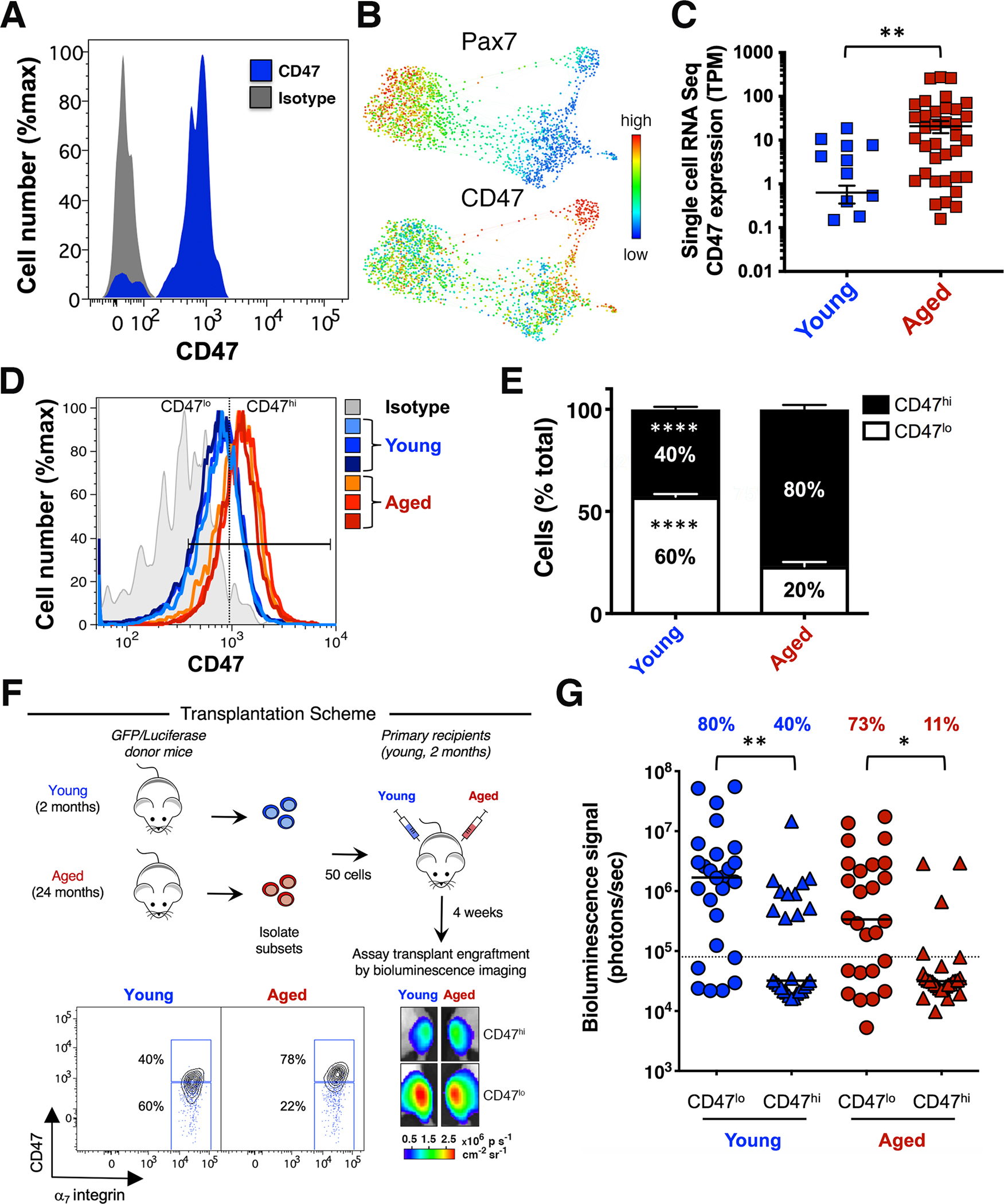
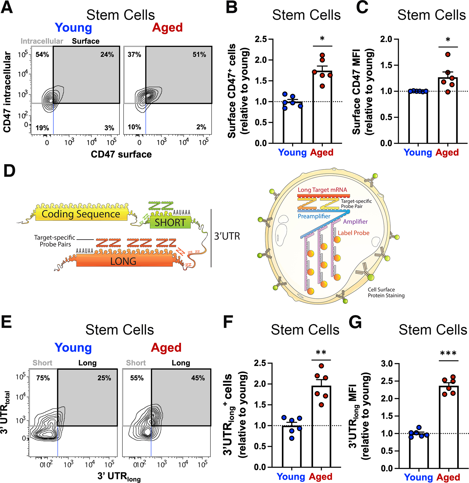
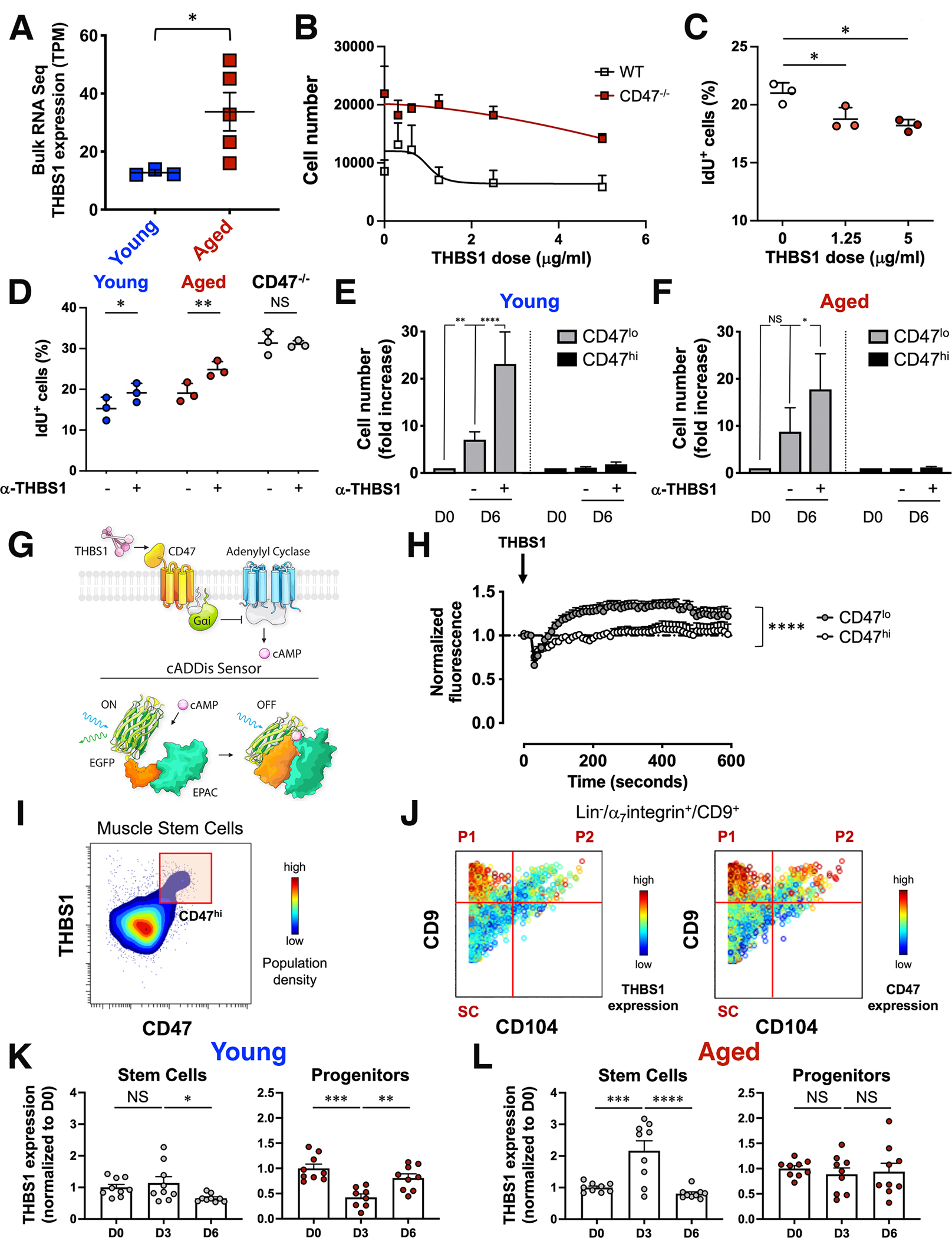
In aging, skeletal muscle strength and regenerative capacity decline, due in part to functional impairment of muscle stem cells (MuSCs), yet the underlying mechanisms remain elusive. Here, we capitalize on mass cytometry to identify high CD47 expression as a hallmark of dysfunctional MuSCs (CD47hi) with impaired regenerative capacity that predominate with aging. The prevalent CD47hi MuSC subset suppresses the residual functional CD47lo MuSC subset through a paracrine signaling loop, leading to impaired proliferation. We uncover that elevated CD47 levels on aged MuSCs result from increased U1 snRNA expression, which disrupts alternative polyadenylation. The deficit in aged MuSC function in regeneration can be overcome either by morpholino-mediated blockade of CD47 alternative polyadenylation or antibody blockade of thrombospondin-1/CD47 signaling, leading to improved regeneration in aged mice, with therapeutic implications. Our findings highlight a previously unrecognized age-dependent alteration in CD47 levels and function in MuSCs, which underlies reduced muscle repair in aging.

Keywords: CyTOF; U1 snRNA; aging; alternative polyadenylation; antisense morpholino oligonucleotide; in vivo antibody blockade; muscle stem cells; paracrine signaling; regeneration; thrombospondin-1/CD47 signaling.


Conflict of interest statement

Declaration of interests H.M.B. is cofounder of Rejuvenation Technologies Inc. and Epirium Bio. H.M.B. and E.P. are named inventors on patent application no. PCT/US2021/038549 held by Stanford University.

Figures

Figure 1.
Figure 1.. CD47 expression levels distinguish functionally and molecularly distinct aged muscle stem cell subsets.
(A) Cell surface marker analysis of MuSCs from Pax7-ZsGreen reporter mice. Histogram overlay shows CD47 expression in ZsGreen+ cells (blue) and the CD47 isotype control (gray). (B) CyTOF analysis of the MuSC population in TA and GA muscle isolated from young mice. Gated Live/Lineage7integrin+/CD34+ MuSCs were analyzed with the X-shift algorithm (K=30) yielding 3 clusters that were visualized using single-cell force-directed layout. Expression levels of Pax7 and CD47 distinguish unique MuSC subsets (representative experiment, n= 3 mice; 4 independent experiments). (C) Scatter plot shows CD47 expression in MuSCs from young and aged mice, measured by single-cell RNA-seq analysis (mean ± SEM). Two-tailed unpaired t-test analysis with Welch’s correction. (D) Histogram overlay shows CD47 expression in MuSCs from young (blue) and aged (red) mice and the FMO + CD47 isotype control (gray) (representative experiment, n= 3 young and 3 aged mice). (E) Stacked columns indicate the relative proportion of CD47lo and CD47hi MuSC subsets in young and aged mice (mean ± SEM from n=9 mice, 3 independent experiments). Two-way ANOVA analysis with Sidak correction for multiple comparisons. (F) Scheme depicting the in vivo assay of regenerative capacity. Overlaid blue dots in the biaxial plot indicate the FMO + CD47 isotype control. Representative BLI images at 4 weeks post-transplant are shown (lower right panel). (G) Scatter plot shows the percentage of transplants from each condition that engrafted above threshold (dashed line) into recipient tissue and the BLI signal intensity (y axis). Line represents the median BLI signal (n= 26 mice, 3 independent experiments). Kruskal Wallis test with significance determined by Dunn’s multiple comparisons test.
Figure 2.
Figure 2.. Alternative polyadenylation regulates CD47 expression and is altered in aged muscle stem cells.
(A, B) Surface and intracellular CD47 protein expression was measured by flow cytometry in young and aged muscle cells. (A) Contour plot of intracellular CD47-PECy7 (y axis) by surface CD47-BV605 (x axis) in young and aged MuSCs (representative sample, n=3 mice). (B) Bar graph indicates the abundance of aged stem cells expressing surface CD47 relative to young (mean ± SEM, n=6 mice, 2 independent experiments). Two-tailed paired t-test analysis. (C) Bar graph shows the expression level of CD47 protein on the surface of aged MuSCs, quantified relative to young MuSCs (mean ± SEM from n=6 mice, 2 independent experiments). Two-tailed paired t-test analysis. (D) Scheme depicting the CD47 coding sequence followed by the 3’ untranslated region (UTR) (left) and the PrimeFlow RNA assay (right). (E, F) CD47 mRNA isoforms distribution was measured by PrimeFlow RNA assay in young and aged muscle. (E) Contour plot of CD47 mRNA-3’UTRtotal (y axis) by CD47 mRNA-3’UTRlong (x axis) shows the distribution of cells expressing the short (upper left quadrant) or the long CD47 mRNA isoform (upper right quadrant) as a fraction of total CD47 mRNA (upper left and right quadrants) in young (left panels) and aged (right panels) MuSCs (representative sample). (F) Bar graph indicates the abundance of aged MuSCs expressing the long CD47 mRNA isoform, quantified relative to young MuSCs (mean ± SEM, n=6 mice, 2 independent experiments). Two-tailed paired t-test analysis. (G) Bar graph shows the expression level of the long CD47 mRNA isoform in aged MuSCs, quantified relative to young MuSCs (mean ± SEM, n=6 mice, 2 independent experiments).
Figure 3.
Figure 3.. U1 snRNA drives alternative polyadenylation and skews the balance toward long CD47 mRNA isoforms in aged muscle stem cells.
(A) CD47 mRNA sequences upstream of polyadenylation site 1 (PAS1) from indicated species were aligned. A highly conserved U1 snRNA binding site (blue box) was identified upstream of PAS1 (red box). (B) Scatter plot shows the expression levels of U1 snRNA in sorted aged MuSCs relative to young MuSCs, measured by q-RT-PCR (mean ± SEM, n= 4 young and 4 aged mice, 2 independent experiments). Two-tailed paired t test. (C) Bar graph shows the relative abundance of CD47 mRNA with long 3’ UTR, as a fraction of the total CD47 mRNA, in young and aged MuSCs treated in vitro with AMOs to PAS1, U1 snRNA binding site on CD47 transcript (CD47 U1), and control (ctr) (mean ± SEM, 4 independent experiments). Two-way ANOVA analysis. (D, E) Surface CD47 protein expression was measured by flow cytometry in young and aged MuSCs treated in culture with AMOs to PAS1, CD47 U1 and control AMO. (D) Bar graph shows the proportion of young and aged MuSCs expressing surface CD47 upon treatment with PAS1 (red) or CD47 U1 (blue) AMO, relative to ctr AMO treated MuSCs (mean ± SEM, 3 independent experiments). Two-way ANOVA. (E) Bar graph shows the expression level of surface CD47 protein on young (left) and aged (right) MuSCs treated as described above, measured as MFI and quantified relative to control AMO treated MuSCs (mean ± SEM, 3 independent experiments). Two-way ANOVA. (F) MuSCs sorted from young and aged GFP/Luciferase mice were treated overnight with AMOs to CD47 U1 or control (ctr) AMO and transplanted (100 cells/injection) into the TA muscle of hindlimb-irradiated NOD/SCID mice. To monitor MuSC engraftment, mice were imaged weekly by BLI. Scatter plot shows the BLI signal intensity of the engrafted transplants, 3 weeks after transplantation. Line represents the median BLI signal with interquartile range (n= 14 mice, 2 independent experiments). Wilcoxon test. (G) Model for post-transcriptional regulation of CD47 in aged MuSCs. U1 snRNA, which is upregulated in aged MuSCs, binds to its site on CD47 3’UTR upstream of PAS1, blocks usage of CD47 PAS1 site, and shifts the balance toward the long isoform, leading to increased surface CD47 expression. Treatment of aged MuSCs with the CD47U1 AMO (intervention), which competes with U1 snRNA for its binding site, prevents alternative polyadenylation, leads to increased production of the short CD47 mRNA isoform and decreased surface CD47 expression, and improves MuSC engraftment to levels similar to those of young MuSCs. The speed dial shows that increasing levels of CD47U1 AMO shift the balance towards the short CD47 isoform and a more youthful molecular phenotype.
Figure 4.
Figure 4.. Aberrant thrombospondin-1 signaling via CD47 inhibits the proliferative capacity of aged muscle stem cells.
(A) Scatter plot shows thrombospondin-1 (THBS1) mRNA levels in bulk RNAseq data from young and aged MuSCs (mean ± SEM, n=3 young and 5 aged mice). Two-tailed unpaired t-test analysis with Welch’s correction. (B) Sorted MuSCs from young wild type and CD47−/− mice were treated in vitro with increasing doses of recombinant THBS1 and proliferation was measured by cell count (mean ± SEM, n=14 wild type and 4 CD47−/− replicates, 4 independent experiments). (C) Scatter plot shows the fraction of young IdU+ MuSCs in response to a 6-day in vitro treatment with increasing doses of recombinant THBS1 (mean ± SD, n= 3 young mice). One-way ANOVA analysis with Tukey’s correction for multiple comparisons. (D) Scatter plot shows the fraction of IdU+ MuSCs in response to a 6-day treatment of sorted wild type (young, aged) and CD47−/− aged MuSCs with a blocking antibody to THBS1 (α-THBS1) (mean ± SD, n= 3 mice per genotype). Two-way ANOVA with Sidak’s correction for multiple comparisons. (E, F) CD47lo and CD47hi MuSCs subsets, sorted from young and aged mice, were treated in vitro on biomimetic hydrogels in the presence (+) or absence (−) of a blocking antibody to THBS1 (α-THBS1), and cell number was quantified by cell count at day 0 (D0) or at day 6 (D6) after treatment. Bar graph shows cell number normalized to D0 for each subset (mean ± SEM from n= 7 young and 3 aged replicates). Two-way ANOVA analysis with Tukey’s correction for multiple comparisons. (G) (Top scheme) THBS1–CD47–cAMP signaling axis. THBS1–CD47 signals through Guanine nucleotide-binding protein Gi’s alpha subunit which inhibits adenylyl cyclases to reduce cAMP levels. (Bottom scheme) The cADDis downward sensor detects changes in cAMP concentration in living cells. cAMP binding to the cADDis downward sensor reduces GFP fluorescence. (H) Sorted CD47lo and CD47hi young MuSCs, transduced with a baculovirus encoding the cAMP sensor, were treated with THBS1 and individually imaged with a confocal microscope for 5 minutes at 10 sec intervals. The graph shows the average normalized GFP fluorescence of CD47lo and CD47hi MuSCs over time (mean ± SEM, 4 mice, 2 independent experiments). Two-tailed paired t-test analysis. (I, J) CyTOF analysis of young hindlimb muscle. (I) Representative contour plot of THBS1 (y axis) by CD47 (x axis) colored by population density shows a CD47hi MuSC subset that expresses high levels of THBS1. (J) Representative biaxial dot plots of CD9 (y axis) by CD104 (x axis) colored by channel show the expression of THBS1 (left) and CD47 (right) in stem (SC) and progenitor cells (P1, P2). (K, L) Intracellular THBS1 protein measurement by CyTOF during an injury time course. Bar graphs show THBS1 levels at day 3 (D3) and day 6 (D6) post injury, normalized to day 0 (D0), in SC (left) and P1 (right) population from young (K) and aged (L) mice (mean ± SEM, n=9 mice from 2 independent experiments). One-way ANOVA analysis with Tukey’s correction for multiple comparisons.
Figure 5.
Figure 5.. Thrombospondin-1 blockade in vivo activates aged muscle stem cells and increases muscle strength in absence of injury.
(A) Experimental scheme (upper panel). Endogenous MuSC expansion was assayed by BLI in Pax7Cre-ERT2;Rosa26LSL-Luc mice upon intramuscular injection in the TA and GA muscles (three times, at two-days interval) with a blocking antibody to THBS1 (α-THBS1) or a control IgG (contralateral leg). Graph shows BLI signal intensity (y axis) over time for each treatment group (mean ± SEM, n = 7 mice per condition, 2 independent experiments). Multiple t-test with Holm-Sidak correction for multiple comparisons. (B) Experimental scheme (upper panel). Young and aged wild type mice were treated with α-THBS1 as in (A) and muscle tissue was collected for CyTOF analysis at the end of treatment. (Left) Representative biaxial dot plots of CD98 (y axis) by CD44 (x axis) colored by channel show IdU incorporation in activated MuSCs (upper right quadrant). (Upper right) Scatter plot indicates the proportion of CD98+/CD44+ activated MuSCs (mean ± SEM, n = 6 mice, 2 independent experiments). Two-tailed paired t-test. (Lower right) Scatter plot shows the proportion of IdU+ cells in the MuSC population (mean ± SEM, n = 6 mice, 2 independent experiments). Two-tailed paired t-test. (C) Experimental scheme. Wild type young and aged mice were treated with α-THBS1 as in (A) and hindlimb muscle tissue was collected for CyTOF analysis, 6 days after the last antibody injection. (D) Gated Live/Lineage7integrin+/CD9+ cells were clustered using the X-shift algorithm (K=50) yielding 61 clusters that were visualized using single-cell force-directed layout. The expression levels of the myogenic transcription factor Pax7 and the surface marker CD47 were overlaid and are shown in the panel composite (n= 3 per condition). (E) Bar graph shows the change in the proportion of each MuSC subset in aged mice treated with α-THBS1, relative to IgG control (n= 3 mice per condition). One-tailed paired t-test. (F) Bar graph shows the fraction of Pax7+ cells in the Live/Lineage7integrin+/CD9+ cell population for each condition (n= 3 mice per condition). One-tailed unpaired t-test (young and aged IgG ctr); one-tailed paired t-test (aged IgG ctr and α-THBS1). (G, H) Young and aged mice were treated with α-THBS1 as in (A) and hindlimb muscle tissue was collected for histology 10 days after the end of treatment. (G) Scatter plot shows the fraction of Pax7+ cells in IgG treated and α-THBS1 treated TA muscles 10 days after the end of treatment (n=3 per condition). Paired t-test. (H) Myofiber cross sectional area (CSA) was quantified in IgG treated and α-THBS1 treated TA muscles and curve fitting was performed (n=3 per condition). Chi-square test. (I, J) Wild type young and aged mice, and young CD47−/− mice were treated with α-THBS1 as in (A) and grip strength (I) and tetanic force (J) were measured 10 days after the end of treatment. (I) Scatter plot shows grip strength measurements in α-THBS1 treated muscles, normalized to IgG treated contralateral muscles (mean ± SEM, n=9 young wild type; n=8 aged wild type and young CD47−/−, 3 independent experiments). Two-tailed paired t-test. (J) Scatter plot shows tetanic force measurements in α-THBS1 treated muscles, normalized to IgG treated contralateral muscles (mean ± SEM, n=9 young wild type; n=7 aged wild type and young CD47−/−, 3 independent experiments). One-tailed paired t-test.
Figure 6.
Figure 6.. Thrombospondin-1 blockade in vivo enhances the regenerative response of aged muscle leading to increased strength.
(A) Experimental scheme. Wild type young and aged mice were injected intramuscularly in the TA and GA muscles with a blocking antibody to THBS1 or IgG control prior to injury and muscle tissue was collected for CyTOF analysis 3- or 6-days post injury. (B) Scatter plot shows the proportion of MuSCs within the myogenic compartment at day 3 post injury for each condition (mean ± SEM, n=6 per condition, 2 independent experiments). Two-way ANOVA with Sidak’s correction for multiple comparisons. (C) (Left) Representative biaxial dot plots of CD98 by CD44 colored by channel, show IdU incorporation in activated MuSCs (upper right quadrant). (Upper right) Scatter plot shows the proportion of CD98+/CD44+ activated MuSCs (mean ± SEM, n=6 per condition, 2 independent experiments). One-tailed paired t-test. (Lower right) Scatter plot shows the proportion of IdU+ cells in the MuSC population (mean ± SEM, n=6 per condition; 2 independent experiments). One-tailed paired t-test. (D, E) Wild type young and aged mice, and young CD47−/− mice were treated with α-THBS1 as in (A) and grip strength (D) and tetanic force (E) were measured 10 days post injury. (D) Scatter plot shows grip strength measurements in α-THBS1 treated muscles, normalized to IgG treated contralateral muscles (mean ± SEM, n=8 for each group; 3 independent experiments). Two-tailed paired t-test. (E) Scatter plot shows tetanic force measurements in α-THBS1 treated muscles, normalized to IgG treated contralateral muscles (mean ± SEM, n=9 young wild type and CD47−/−, n=6 aged wild type; 3 independent experiments). One-tailed paired t-test. (F) Model. Skeletal muscle injury leads to MuSC activation. In young muscle during regeneration, progenitor cells participate in a negative feedback loop whereby they produce THBS1 to limit MuSC proliferation, and promote MuSC return to quiescence, therefore preventing MuSC exhaustion. In aged muscle accumulation of a dysfunctional CD47hi MuSC subset, which precociously secretes THBS1, creates a dysregulated microenvironment that inhibits the proliferation and function of the CD47lo MuSC subset, impairing muscle regeneration.

Comment in

Similar articles

Cited by

References

    1. Blau HM, Cosgrove BD, and Ho ATV (2015). The central role of muscle stem cells in regenerative failure with aging. Nat Med 21, 854–862. 10.1038/nm.3918. - DOI - PMC - PubMed
    1. Feige P, Brun CE, Ritso M, and Rudnicki MA (2018). Orienting Muscle Stem Cells for Regeneration in Homeostasis, Aging, and Disease. Cell Stem Cell 23, 653–664. 10.1016/j.stem.2018.10.006. - DOI - PMC - PubMed
    1. MAURO A (1961). Satellite cell of skeletal muscle fibers. J Biophys Biochem Cytol 9, 493–495. 10.1083/jcb.9.2.493. - DOI - PMC - PubMed
    1. Schmidt M, Schüler SC, Hüttner SS, von Eyss B, and von Maltzahn J (2019). Adult stem cells at work: regenerating skeletal muscle. Cellular and Molecular Life Sciences 76, 2559–2570. 10.1007/s00018-019-03093-6. - DOI - PMC - PubMed
    1. Sousa-Victor P, and Muñoz-Cánoves P (2016). Regenerative decline of stem cells in sarcopenia. Mol Aspects Med 50, 109–117. 10.1016/j.mam.2016.02.002. - DOI - PubMed

Publication types