. 2022 Dec 31;17(1):2129291.
doi: 10.1080/15592324.2022.2129291.

Building bridges: mycelium-mediated plant-plant electrophysiological communication

Affiliations

Building bridges: mycelium-mediated plant-plant electrophysiological communication

Matthew Adam Thomas et al. Plant Signal Behav. .

Abstract

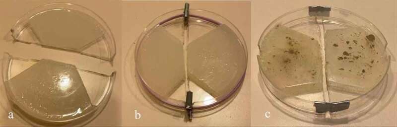
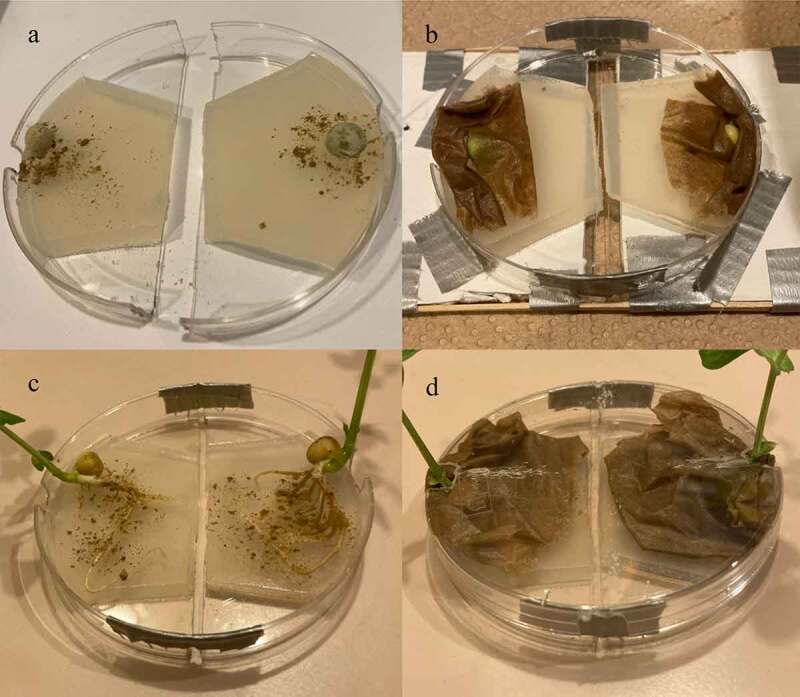
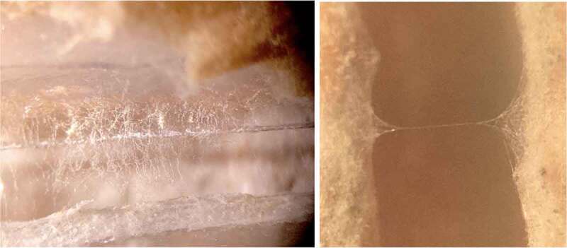
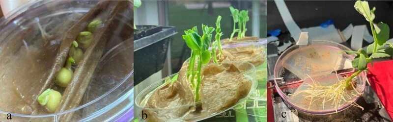
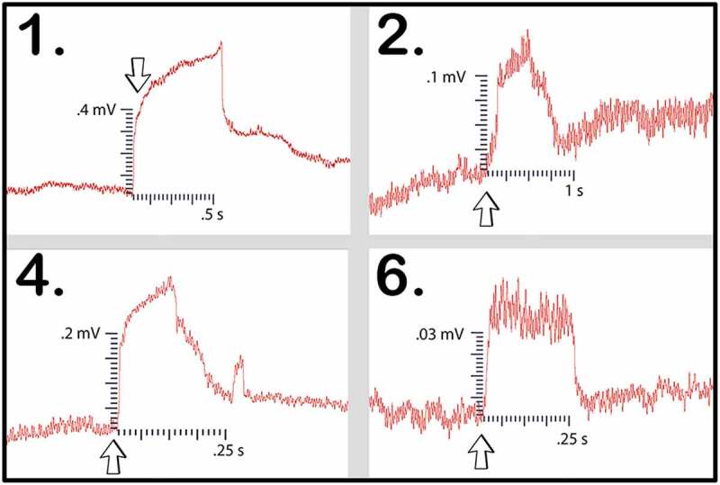
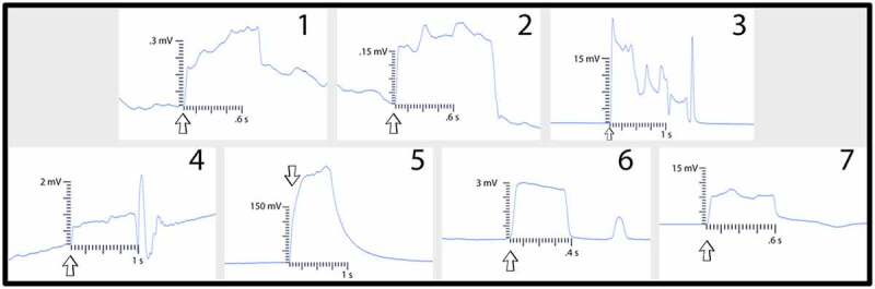
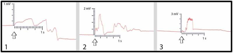
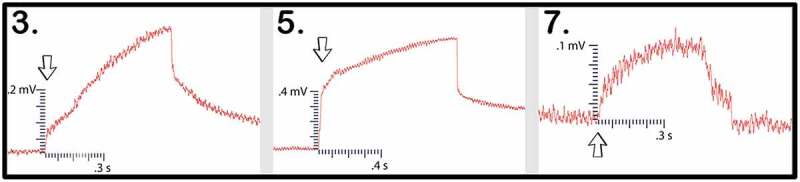
Whether through root secretions or by emitting volatile organic compounds, plant communication has been well-documented. While electrical activity has been documented in plants and mycorrhizal bodies on the individual and ramet, electrical propagation as a means of communication between plants has been hypothesized but understudied. This study aimed to test the hypothesis that plants can communicate with one another electrically via conductively isolated mycelial pathways. We created a bio-electric circuit linking two plants using a mycelial network grown from a blend of mycorrhizal fungi which was directly inoculated onto potato dextrose agar, or onto the host plants placed on the agar. The mycelium that grew was forced to cross, or "bridge," an air gap between the two islands of agar - thus forming the isolated conductive pathway between plants. Using this plant-fungal biocircuit we assessed electrical propagation between Pisum sativum and Cucumis sativus. We found that electrical signals were reliably conducted across the mycelial bridges from one plant to another upon the induction of a wound response. Our findings provide evidence that mechanical input can be communicated between plant species and opens the door to testing how this information can affect plant and fungal physiology.

Keywords: Plant; action; electric; electrophysiology; fungal; graded; mycelium; mycorrhizal; networks; potential; signaling.

Plain language summary

Most plants form underground relationships with fungi. These relationships are mutually beneficial. The plants and fungi share, trade, and distribute resources between themselves, their neighbors, and their offspring. Plants employ diverse methods to detect and respond to their environment and the production of electric signals is one of these methods. It would be favorable to a plant’s survival and the survival of their neighbors, if this plant could transmit and share the information these electrical signals contain. Possible avenues of transmission exist in the roots, and the fungi these roots are in contact with. If a fungal mass is in contact with the roots of multiple plants, it could propagate electrical signals throughout the plant network. We found that electric signals were reliably transmitted from one plant to another via fungal pathways upon the induction of a wound response. Our findings provide evidence that mechanical input can be communicated between plant species and opens the door to testing how this information can affect plant and fungal physiology.


Conflict of interest statement

No potential conflict of interest was reported by the author(s).

Figures

Figure 1.
Figure 1.
Petri dish design. Figure 1a illustrates the Petri dish cut in half with the agar on each side cut into the design to guide the mycelium across the agar gap in the center of the dish. Figure 1b depicts the Petri dish reassembled with spacers and rubber bands in place. Figure 1c represents the second method for reassembling the Petri dish using duct tape. Figure 1c also illustrates how the MycoGrow inoculant appeared on the agar after application.
Figure 2.
Figure 2.
Mycelial bridging across the two islands of agar.
Figure 3.
Figure 3.
Setup testing platform This figure illustrates the testing platform used to aid in the isolation of the signal. The gap in the center of the platform kept the Petri dish off the surface of the table. This ensured the dish did not touch the testing table and further suspended the mycelial bridge in midair.
Figure 4.
Figure 4.
The mycelial bridge setups with direct inoculation of pea seeds and developed pea roots. Figure 4a depicts the pea seeds just after being coated with the Endo/Ecto MycoGrow Inoculant. Figure 4b depicts the same pea seeds, now covered with a damp paper towel to maintain moisture, the dish is now fastened together with tape at the ends while maintaining the necessary gap. The dish has also been fastened to the testing platform for stabilization. Figure 4c depicts direct inoculation of the pea’s root systems. Figure 4d depicts these same pea plants with the roots now covered by a damp paper towel to maintain moisture. This setup would also have been fastened to a testing platform, as seen in 4B.
Figure 5.
Figure 5.
Examples of mycelial bridges when viewed under a microscope. This figure depicts the presence of mycelial bridges that the authors would visually confirm under the microscope prior to testing the setup.
Figure 6.
Figure 6.
Pea growing process.This figure depicts the process of growing pea plants from sprouting to testing. Figure 6a depicts the pea plants just beginning to sprout in a moist paper towel enclosure. Figure 6b depicts the same pea plants further along in the growth cycle with the above-mentioned angled setup. At this stage the pea plants are ready for testing. Figure 6c depicts the pea plant removed from the moist paper towel with the roots placed on one of the agar islands.
Figure 7.
Figure 7.
Experimental setup.We denote the side of the dish with the glass electrode and ground wire (6 and 7) as “Side A.” The plant and agar island on the side of the setup not being recorded is denoted “Side B.” The demarcation between side A and B is denoted with the dashed line – representing the gap between the two islands of agar. Each plant is fixed in position with tape and a beaker for stem support. Wax is used to secure the dish to the testing surface which rests on an air table. The numbers indicate where different controls are performed:.
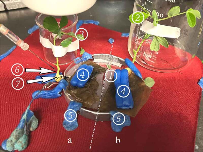
  1. A patch of agar suitable for performing an agar touch on the side B.

  2. A leaf suitable for a leaf nudge and leaf snip on Side B.

  3. A leaf suitable for a leaf nudge and leaf snip on Side A.

  4. Surgical wax is used as weights to hold down the roots under the paper towel, and to keep the roots in good contact with the agar they rest on.

  5. Surgical wax used to secure the setup to the testing pad.

  6. Glass electrode inserted into the plant stem on Side A.

  7. Reference electrode adhered with silver paste to the taproot of the plant

Figure 8.
Figure 8.
Control examples. Figure 8a illustrates an agar touch performed on Side A. Figure 8b provides an example of an agar touch on Side B. Figure 8c is an example of not receiving a signal after a leaf bend. The arrows indicate the moment of induction.
Figure 9.
Figure 9.
Wound responses conducted across the bridge for cucumber trials 1 and 2.This figure depicts the electrical responses that were conducted from one cucumber plant to another via rhyzoelectric pathways for the six setups. The scale values on the x-axis are in seconds and the scale values on the y-axis are in millivolts. The arrows indicate the moment of induction.
Figure 10.
Figure 10.
Pea trial 1 signal propagations. This figure depicts the 5 electrical responses that were recorded from one pea plant to another via rhyzoelectric pathways. The scale values on the x-axis are in seconds and the scale values on the y-axis are in millivolts. In setup 4, the change in electric potential and duration of smaller response was used in the table as this was the initial response. The arrows indicate the moment of induction.
Figure 11.
Figure 11.
Pea trial 2 signal propagations. This figure depicts the electrical responses from the four setups in which a successful signal conduction occurred from one plant to another. The scale values on the x-axis are in seconds and the scale values on the y-axis are in millivolts. The arrows indicate the moment of induction.
Figure 12.
Figure 12.
Pea trial 3 signal propagations. This figure depicts the electrical responses from seven setups which all produced successful signal conductions from one plant to another. The scale values on the x-axis are in seconds and the scale values on the y-axis are in millivolts. The arrows indicate the moment of induction.In the second trial using these direct inoculation methods, all three of the setups successfully conducted electric signals across their respective mycelial bridges. Upon cutting the bridges, the signals could not be conducted from the plant on Side B to the recording electrode in the plant on (Figure 13) and the magnitude and duration of these responses are listed in Table 5.
Figure 13.
Figure 13.
Pea trial 4 signal propagations. This figure depicts the electrical responses from three setups that all produced successful signal conductions from one plant to another. The scale values on the x-axis are in seconds and the scale values on the y-axis are in millivolts. The arrows indicate the moment of induction.
Figure 14.
Figure 14.
Suture thread electric potentials. This figure depicts the three responses conducted from one plant to another with the moist suture thread bridging the gap. The arrows indicate the moment of signal induction.

References

    1. Chen Y, Zhao D-J, Wang Z-Y, Wang Z-Y, Tang G, Huang L.. Plant electrical signal classification based on waveform similarity. Algorithms. 2016;9:70. doi:10.3390/a9040070. - DOI
    1. Smith SE, Read DJ eds. Mycorrhizal symbiosis. 2nd ed. Cambridge, MA: Academic Press; 2016.
    1. Babikova Z, Johnson D, Bruce T, Pickett J, Gilbert L.. How rapid is aphid-induced signal transfer between plants via common mycelial networks? Comm. Integr Biol. 2013;6(6):1. - PMC - PubMed
    1. Song YY, Zeng RS, Zu JF, Li J, Shen X, Yihdego WG. Interplant communication of tomato plants through underground common mycorrhizal networks. Plos One. 2013. doi:10.1371/journal.pone.0013324. - DOI - PMC - PubMed
    1. Akhtar MS, Siddiqui Z. Arbuscular mycorrhizal fungi as potential bioprotectants against plant pathogens (Dordrecht, Nl: ). 2008. p. 6,61. doi:10.1007/978-1-4020-8770-7_3. - DOI

Publication types

LinkOut - more resources