Case Reports
. 2022 Nov 16;22(1):440.
doi: 10.1186/s12886-022-02661-y.

Von Hipple-Lindau disease complicated with central retinal vein occlusion: a case report

Affiliations
Case Reports

Von Hipple-Lindau disease complicated with central retinal vein occlusion: a case report

Xingwang Chen et al. BMC Ophthalmol. .

Abstract

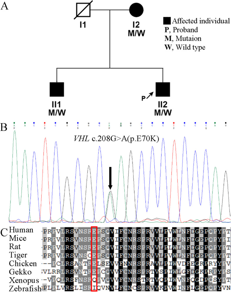
Background: Central Retinal Vein Occlusion (CRVO) is a rare complication of von Hipple-Lindau (VHL) disease. This report presents the first case of VHL disease complicated with CRVO caused by VHL c.208G > A mutation.

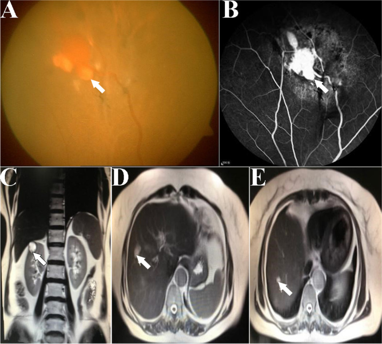
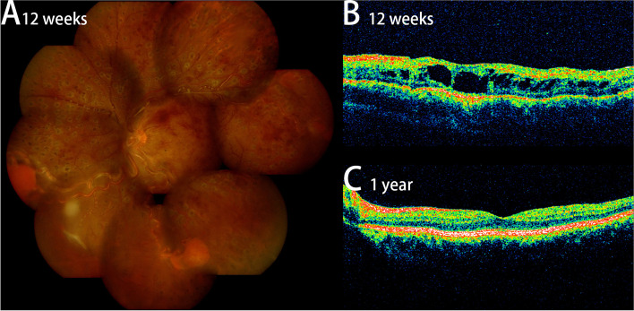
Case presentation: A 20 s man whose left eye visual acuity gradually declined for half a year. The visual acuity of the left eye is counting fingers. Fundus examination revealed that retinal hemangioblastoma was also found in addition to typical CRVO signs such as tortuous expansion of retinal veins and flame-shaped hemorrhage of the retina. Liver tumor, cerebral infarction and erythrocytosis were found during systemic examination, and the diagnosis of polycythemia was confirmed by bone marrow smear. Furthermore, both family history and genetic analysis indicated that the patient had VHL disease caused by VHL c.208G > A. In this patient, a large number of bone marrow erythrocytes proliferated due to VHL disease, which led to the increase of blood viscosity and erythrocyte vascular adhesion, resulting in the obstruction of central retinal vein blood flow, and finally CRVO. For CRVO and its pathogenic factor polycythemia, patient received laser retinal photocoagulation and phlebotomies. After a 1-year follow-up, the vision in the left eye improved to 0.2 logMAR.

Conclusions: This is a rare case of polycythemia complicated by CRVO in patient with VHL disease. It reminds us that the systemic disease factors should be fully considered in the diagnosis of young patients with CRVO, and that treatment requires a coordinated effort of physicians.

Keywords: Case report; Central retinal vein occlusion; Polycythemia; VHL gene mutation; von Hipple-Lindau disease.


Conflict of interest statement

The authors declare that they have no competing interests.

Figures

Fig. 1
Fig. 1
Clinical examination results of proband. A Color fundus image of proband's left eye. Diffuse patchy hemorrhages in the retina, and obvious earthworm-like tortuosity of the veins. Three retinal hemangioblastomas (white arrows) were found in the peripheral retina, all about 1 PD in size. B Color fundus image of proband's right eye. There is no obvious abnormality. C FFA image of proband's left eye. A high fluorescence lesion (white arrow) with the size of 1.5 PD were found. A thick and tortuous nourishing blood vessel is connected to it. Hemorrhage on the lower side of the lesion obscured fluorescence. Fluorescent leakage and staining of retinal veins. D FFA image of proband's left eye. Fluorescent leakage and staining of retinal veins. A high fluorescence lesion on the left side of the image. Non-perfusion areas were fund in the retina on the right and lower sides of the image (white arrows). E FFA late phase image. FFA of the left eye showed tortuous dilation of retinal vein, fluorescein staining of optic disc and retinal vein vessels, and flower-petal appearance of the leakage at the macula. F OCT image of proband's left eye. The retinal thickness in the macular area increased, and dark fluid-filled cyst inside the retina. G MRI image of proband's abdomen. A huge tumor (white arrow) was found in the right anterior lobe of liver with a size of 15.6 cm*11.4 cm, and the boundary with surrounding normal liver tissue was unclear (the white dotted line marks its approximate range). Snowflake enhancement was found in the tumor under enhanced scanning, and the spleen was obviously enlarged. H MRI image of proband's brain. A low signal area was found in the right temporal lobe with a size of 3 cm*2 cm (the white dotted line marks its approximate range, white arrow). I Bone marrow smear showed that nucleated cells proliferated actively, granulocyte: erythroid = 4.24:1, granulocyte: lobulated nuclear granulocyte ratio increased significantly, erythroid: proliferation was dominated by middle and late young erythrocytes, mature erythrocytes were distributed in accumulation, lymphocyte: ratio decreased, cell morphology was not significantly abnormal, combined with blood routine results: wbc12.99 × 109/L、RBC9.55 × 1012 / L, that is consistent with the diagnosis of polycythemia
Fig. 2
Fig. 2
Clinical examination results of proband’s mother. A Color fundus image of I2's left eye. A yellow-white irregular lesion with a size of about 1/4 PD (white arrow) were found. B FFA image of patient I2's left eye. A high fluorescence lesion was found at the lesion corresponding to the Fig. 2A, and fluorescein leakage (white arrow) were found around the lesion. C T2 weighted image of I2's abdomen MRI showed a cystic lesion in the upper right kidney (red arrow). D and E T2 weighted images of I2's abdomen MRI showed two cystic lesions in the right anterior lobe of liver (white arrows)
Fig. 3
Fig. 3
DNA analysis of the patients and family members. A The pedigree of the family with VHL c.208G > A. This family presents a co-segregation of genotypic phenotypes associated with VHL gene heterozygous mutation. B Sanger sequencing electropherogram showing compound heterozygous variant (black arrow) in VHL. C Conservation of E70 in VHL in different mammal, bird, and reptile species. The protein sequences of VHL orthologs at positions 59–100 are aligned. The red box indicates the position of E70
Fig. 4
Fig. 4
Results of proband after treatment. A Color fundus image of proband's left eye after 12 weeks. Most retinal hemorrhages were absorbed, and the tortuosity of retinal veins was partially relieved. Laser spots are widely distributed in the peripheral retina (B) OCT image of proband's left eye after 12 weeks. The retinal thickness in the macular region was not significantly reduced compared with that at the initial visit, and dark fluid-filled cyst inside the retina. C OCT image of proband's left eye after 1 year. The retinal thickness in the macular region was significantly reduced compared with that before, and macular edema was completely absorbed

References

    1. Glasker S, Vergauwen E, Koch CA, Kutikov A, Vortmeyer AO. Von Hippel-Lindau Disease: Current Challenges and Future Prospects. Onco Targets Ther. 2020;13:5669–5690. doi: 10.2147/OTT.S190753. - DOI - PMC - PubMed
    1. Qiu J, Zhang K, Ma K, et al. The Genotype-Phenotype Association of Von Hipple Lindau Disease Based on Mutation Locations: A Retrospective Study of 577 Cases in a Chinese Population. Front Genet. 2020;11:532588. doi: 10.3389/fgene.2020.532588. - DOI - PMC - PubMed
    1. Haddad NM, Cavallerano JD, Silva PS. Von hippel-lindau disease: a genetic and clinical review. Semin Ophthalmol. 2013;28(5–6):377–386. doi: 10.3109/08820538.2013.825281. - DOI - PubMed
    1. Hong B, Ma K, Zhou J, et al. Frequent Mutations of VHL Gene and the Clinical Phenotypes in the Largest Chinese Cohort With Von Hippel-Lindau Disease. Front Genet. 2019;10:867. doi: 10.3389/fgene.2019.00867. - DOI - PMC - PubMed
    1. van Rooijen E, Voest EE, Logister I, et al. Zebrafish mutants in the von Hippel-Lindau tumor suppressor display a hypoxic response and recapitulate key aspects of Chuvash polycythemia. Blood. 2009;113(25):6449–6460. doi: 10.1182/blood-2008-07-167890. - DOI - PubMed

Publication types

MeSH terms