. 2022 Nov 16;19(1):29.
doi: 10.1186/s12983-022-00474-9.

Nematocyst sequestration within the family Fionidae (Gastropoda: Nudibranchia) considering ecological properties and evolution

Affiliations

Nematocyst sequestration within the family Fionidae (Gastropoda: Nudibranchia) considering ecological properties and evolution

Irina A Ekimova et al. Front Zool. .

Abstract

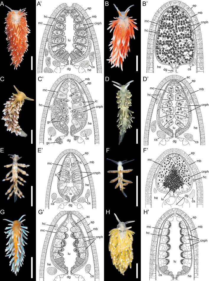
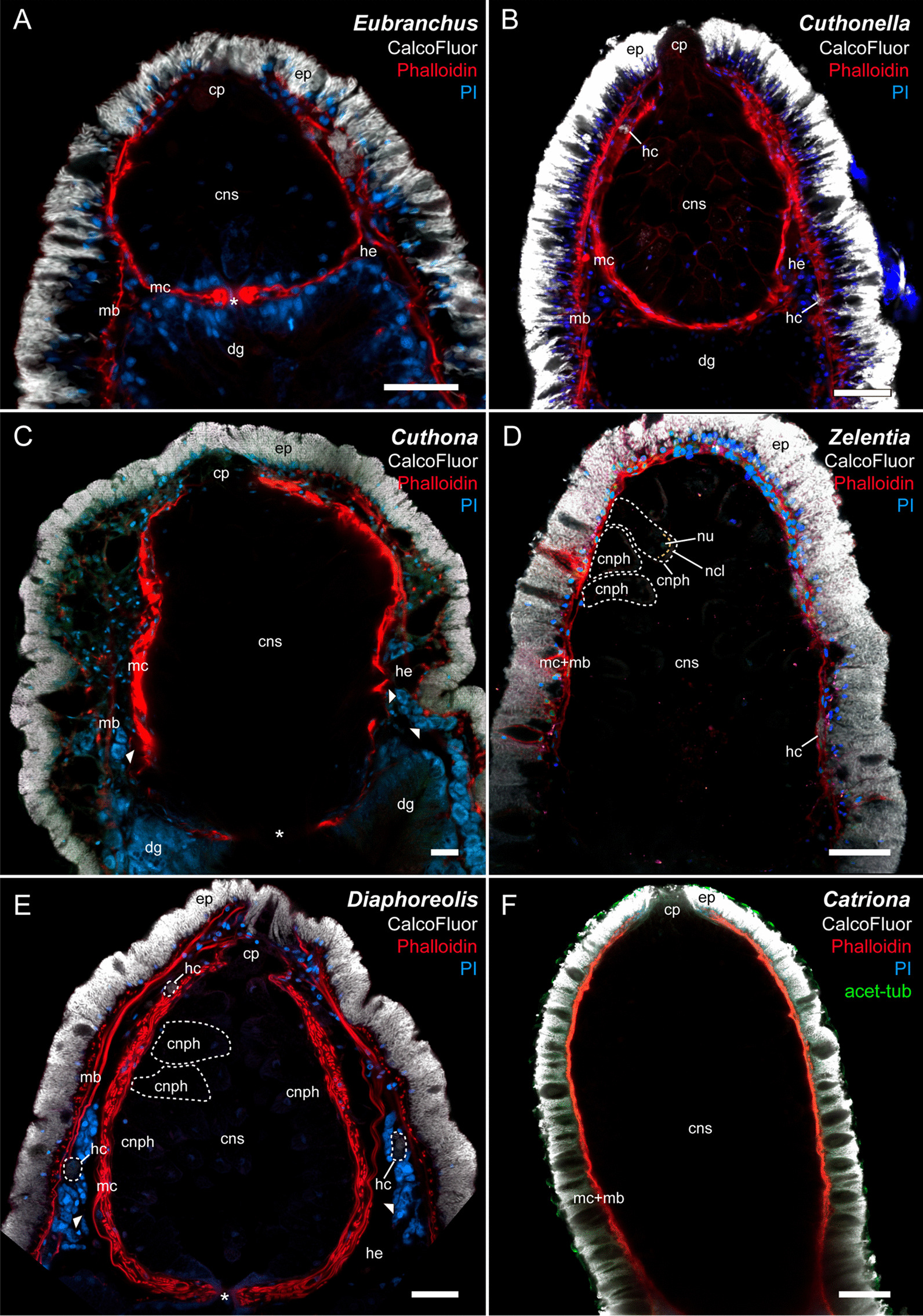
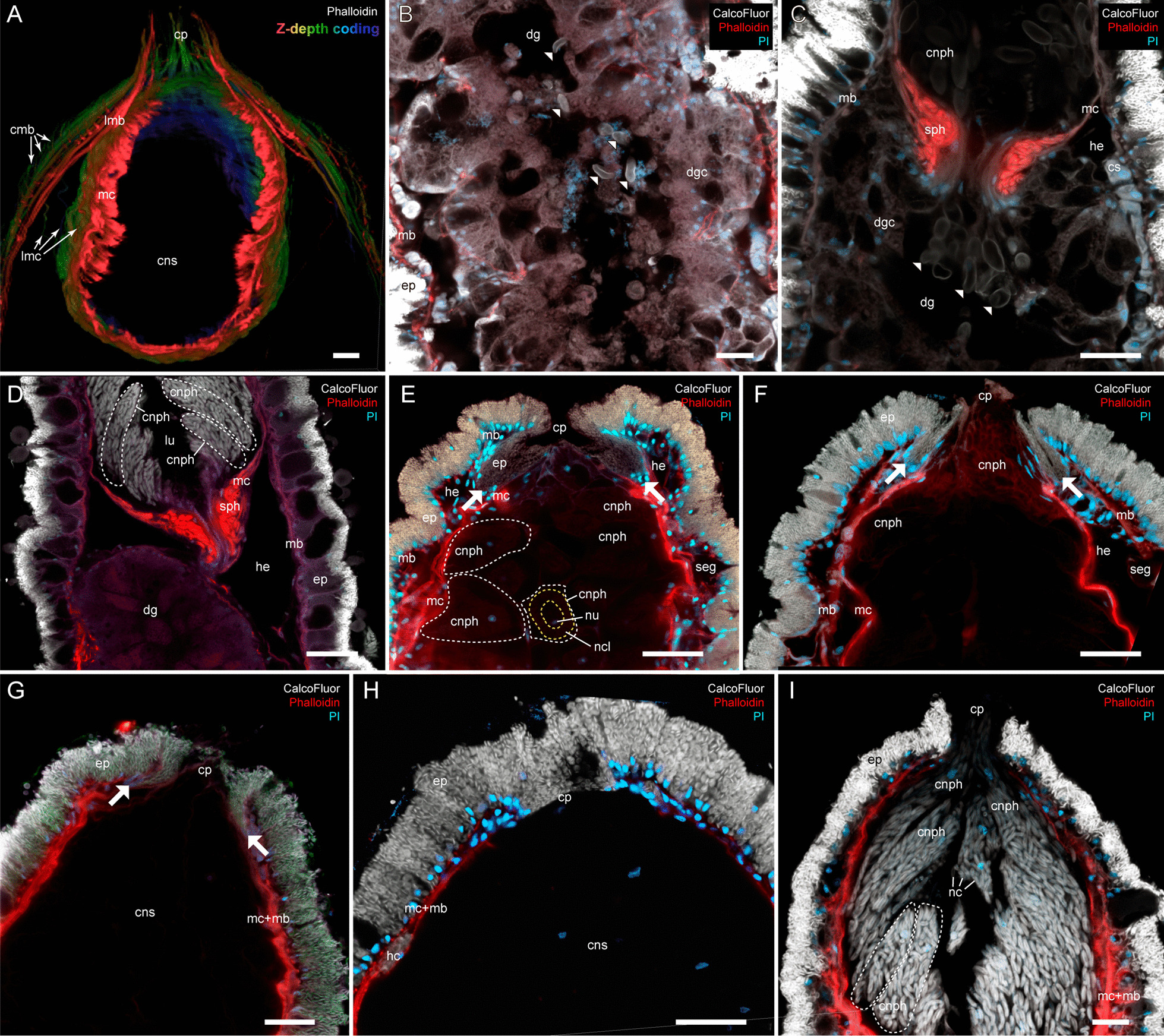
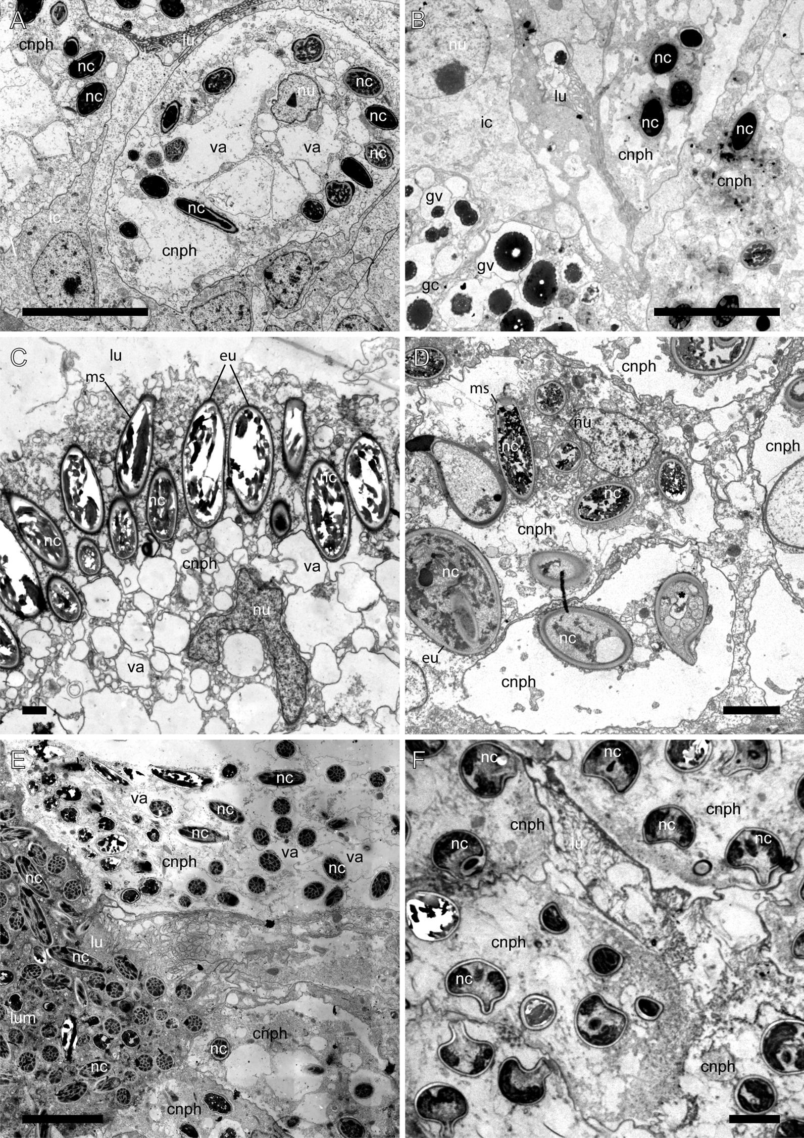
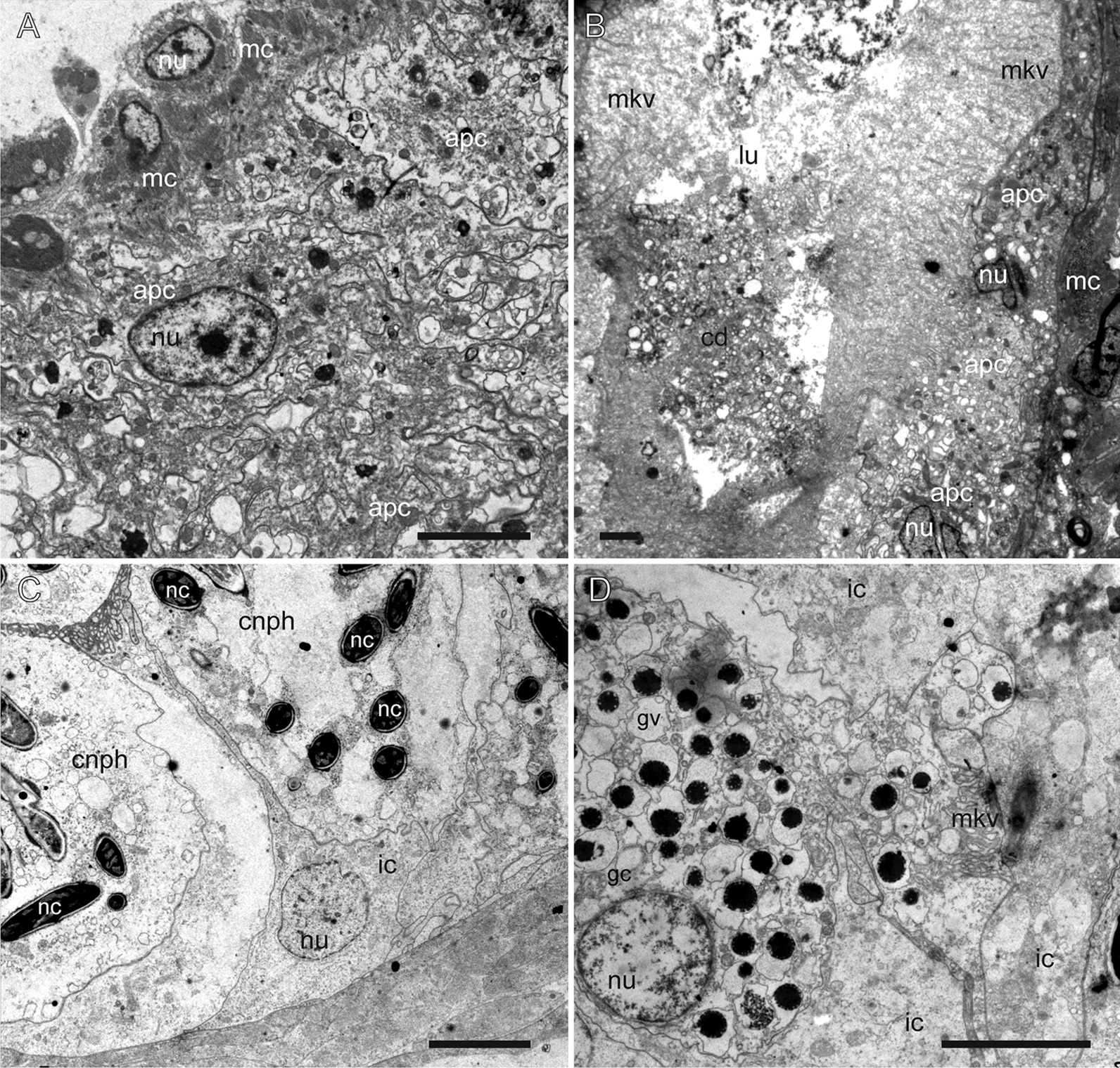
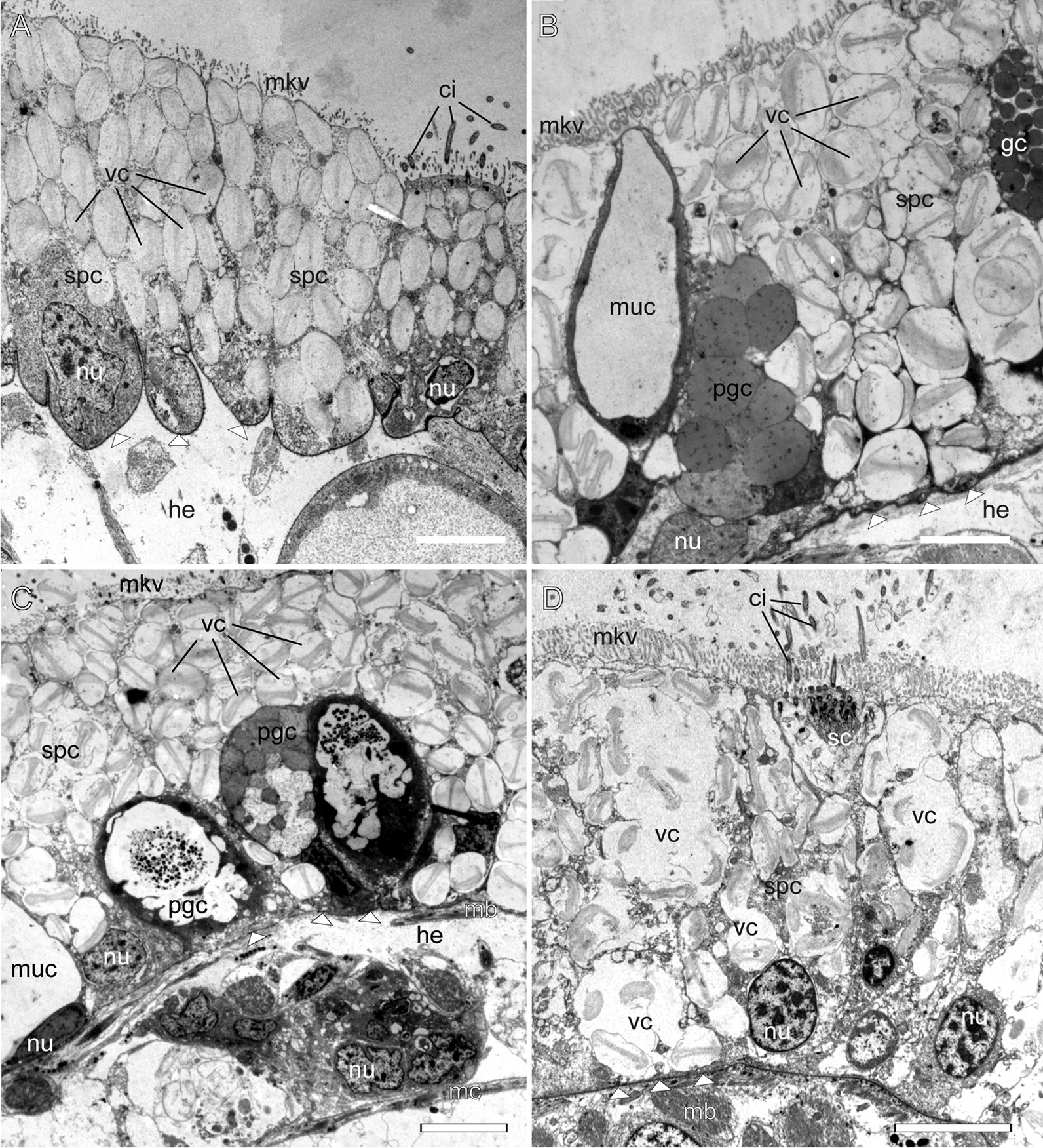
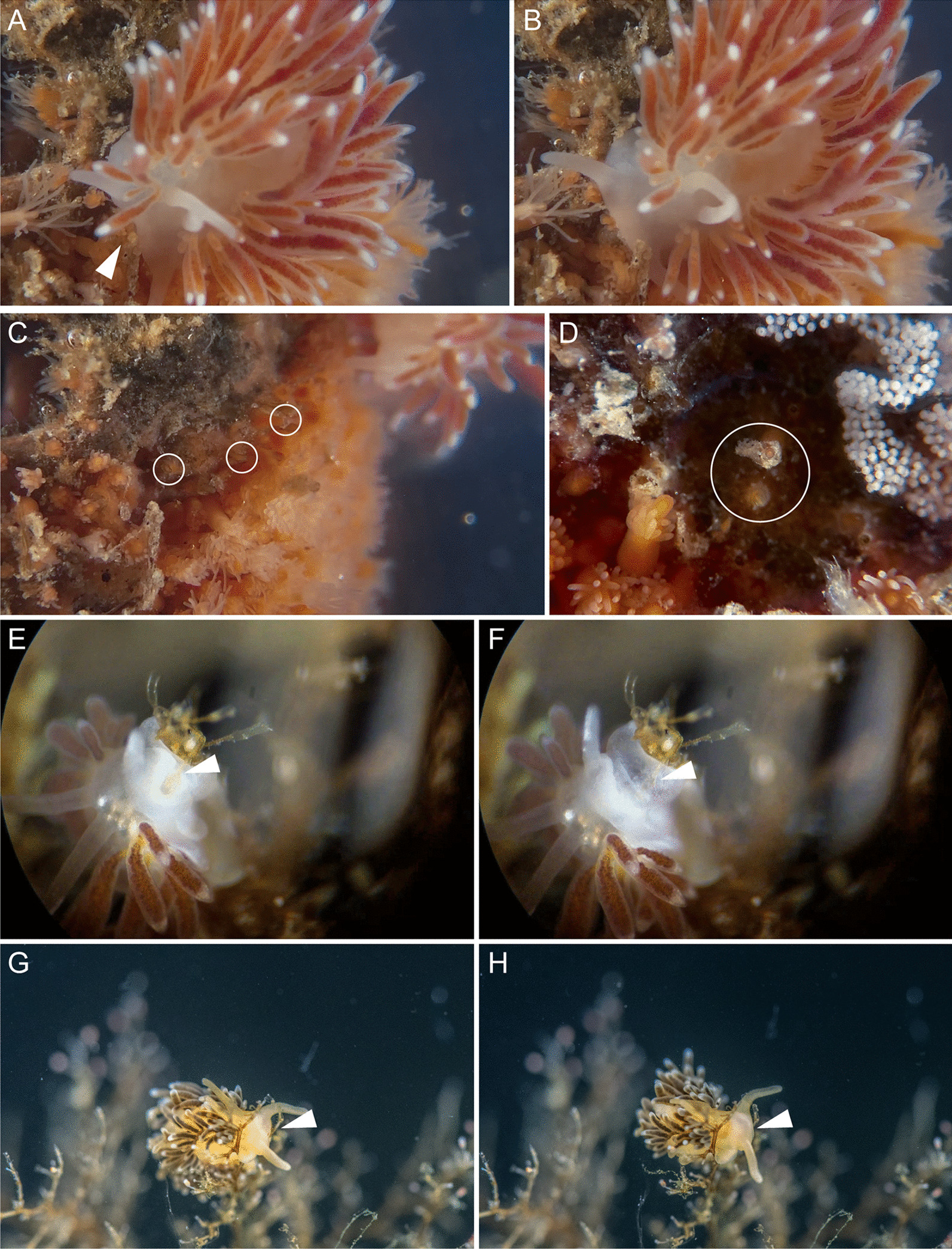
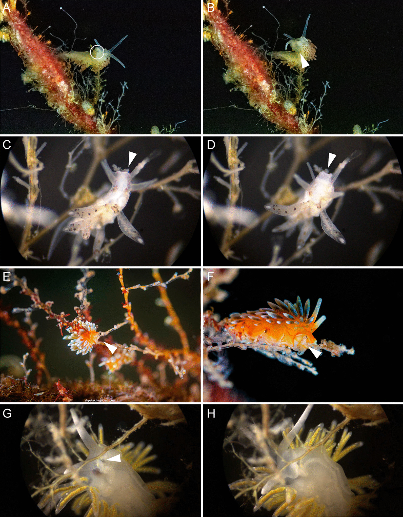
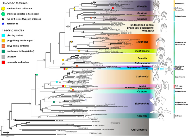
Aeolid nudibranchs are well-known for their ability to incorporate cnidarian nematocysts and use them for defense; this process is tightly linked with the feeding preferences of molluscs. As many nudibranch groups show signs of ecology-based adaptive radiation, studies of prey-based defensive mechanisms can provide valuable insight into details of nudibranch evolutionary history. The main goal of this study is to test the correlation of ecological traits, feeding mechanisms, and prey preferences with cnidosac fine morphology and to pinpoint the phylogenetic value of these traits. We study the cnidosac morphology in thirteen species-representatives of the main lineages within the family Fionidae s.l. The morphological analysis includes histological sections, transmission electron microscopy, confocal laser scanning microscopy, and scanning electron microscopy. For phylogenetic study, available molecular data from public repositories were used, and phylogenetic trees were produced based on Bayesian Inference and Maximum likelihood analysis for a concatenated dataset of three molecular markers (COI, 16S, H3). In general, fionid cnidosacs fit the common aeolid pattern, but among different species we detected a high variation in type of obtained nematocysts, their arrangement within cnidophages, and in number of cell types within cnidosacs. We report on presence of cellules speciale in the haemocoel of all studied species, and for the first time, we report on cells with chitinous spindles in the haemocoel of all fionids except Eubranchus. The function of both these cell types remains unknown. The loss of functional cnidosacs occurred at least three times within Fionidae, and in case of the genera Phestilla, Calma, and Fiona, this loss is linked to their non-cnidarian diet. The diversity of cnidosac fine structure within Fionidae s.l. correlates with that of the radular morphology and feeding preferences of each species. Prey shifts between cnidarian and non-cnidarian prey (both through evolutionary shifts and individual variation) rarely occur within Fionidae s.l.; however, microevolutionary shifts between different hydrozoan species within a single genus are more common. Cnidosac morphology demonstrates considerable resulting changes even when switching between similar hydrozoan species, or changing the feeding site on same prey species. These data indicate that cnidosac morphology likely follows microevolutionary prey shifts-in other words, it is affected by switches in prey species and changes in feeding sites with a single prey species. Thus, the cnidosac morphology may be a useful indicator when studying ecological features of particular species.

Keywords: Adaptive radiation; Character evolution; Chitin; Feeding modes; Functional morphology; Kleptocnidae; Phylogeny.


Conflict of interest statement

The authors declare that they no competing interests.

Figures

Fig. 1
Fig. 1
External morphology of studied nudibranch species and generalized scheme of cnidosac structure in respective species (indicated with apostrophe). A Cuthona nana. B Catriona columbiana. C Cuthonella hiemalis. D Diaphoreolis viridis. E Eubranchus rupium. F Tergipes tergipes. G Trinchesia ornata. H Zelentia pustulata. ac cells without NCs in cnidopore zone, cnph cnidophage, cs cellules speciale, dg digestive gland, ep epithelium, gc cells with granular compound, hc cells with chitinous spindles, he haemocoel, ic interstitial cells, lu lumen, mb body musculature, mc cnidosac musculature. Scale bars: 5 mm. Photo credits: all except C: Tatiana Antokhina, C: Alexander Semenov
Fig. 2
Fig. 2
Longitudinal optical section of cnidosac in different Fionidae species (CLSM). A Eubranchus rupium; B Cuthonella hiemalis; C Cuthona nana; D Zelentia pustulata, proximal end of cnidosac is not seen due to its large size, white dotted lines indicate cnidophages, yellow dotted line indicates NCs layer; E Diaphoreolis viridis, white dotted lines in cnidosac indicate cnidophages, white dotted lines in haemocoel indicates cells with chitinous elements; F Catriona columbiana, proximal end of cnidosac is not seen due to its large size. cnph cnidophage, cns cnidosac, cp cnidopore, dg digestive gland, ep epithelium, hc haemocoel cells with chitinous spindles, he haemocoel, mb body musculature, mc cnidosac musculature. White arrowheads indicate cellules speciale, star—cnidosac entrance (where applicable). Scale bars: 20 µm
Fig. 3
Fig. 3
Musculature, digestive gland diverticula, cnidosac entrance and cnidopore in different Fionidae species (CLSM). A Diaphoreolis viridis, 3D-reconstruction of musculature of ceratal distal part. B Cuthonella concinna, optical longitudinal section, digestive gland diverticula showing intact nematocysts in digestive gland lumen (white arrowheads), the brightness/contrast is enhanced to make nematocysts visible among mollusc tissues. C Cuthonella concinna, optical longitudinal section, cnidosac entrance showing intact nematocysts in digestive gland lumen (white arrowheads), the brightness/contrast is excessive to make nematocysts visible among mollusc tissues. D Eubranchus odhneri, optical longitudinal section, cnidosac entrance, white dotted lines indicate cnidophages with NCs. E Cuthona nana, optical longitudinal section, cnidopore with invagination of epidermal layer closely adjacent to cnidophages (borders are indicated with white arrows), white dotted lines indicate cnidophages, yellow dotted lines indicate NCs layer. F Cuthona nana, optical longitudinal section, discharged cnidosac, cnidopore with ejected cnidophages containing nematocysts (borders are indicated with white arrows). G Catriona columbiana, optical longitudinal section, cnidopore with invagination of epidermal layer closely adjacent to cnidophages (borders are indicated with white arrows). H Zelentia pustulata, optical longitudinal section, cnidopore. I Eubranchus odhneri, optical longitudinal section, cnidopore with ejected cnidophages containing nematocysts, white dotted lines indicate cnidophages with NCs. cmb circular musculature of body, cnph cnidophage, cns cnidosac, cp cnidopore, cs cellules speciale, dg digestive gland, dgc digestive gland cells, ep epithelium, hc haemocoel cell with chitinous spindles, he haemocoel, lmb longitudinal musculature of body, lmc longitudinal musculature of cnidosac, lu lumen, mb body musculature, mc cnidosac musculature, nc NCs, ncl NCs layer within cnidophage, nu nucleus, seg subepidermal mucus gland, sph muscular sphincter of cnidosac. Scale bars: 20 µm
Fig. 4
Fig. 4
Different arrangement of nematocysts within cnidophages of different Fionidae species (TEM). A Mastigophores in Eubranchus rupium. B Mastigophores in Cuthonella hiemalis. C Euryteles and mastigophores in Zelentia pustulata. D Euryteles and mastigophores in Diaphoreolis viridis. E Mastigophores Tergipes tergipes. F Stenoteles in Catriona columbiana. cnph cnidophage, eu euryteles, gv vacuoles with unidentified granular content, ic interstitial cell, lu lumen, ms mastigophores, nc nematocyst, nu nucleus, va vacuole. Scale bars: A, B, E, F—10 µm, C—2 µm, D—5 µm
Fig. 5
Fig. 5
Additional cell types in cnidosacs of different Fionidae species (TEM). A Diaphoreolis viridis. B Trinchesia ornata. C Eubranchus rupium. D Cuthonella hiemalis. apc degraded cells, cd cell debris, cnph cnidophage, gc cell with granular content, gv vacuoles with unidentified granular content, ic interstitial cell, lu lumen, mc cnidosac musculature, mkv microvilli, nc nematocyst, nu nucleus. Scale bars: A, C, D—5 µm, B—2 µm
Fig. 6
Fig. 6
Haemocoel cells (TEM). A, B Zelentia pustulata, cellules speciale. C Cuthonella hiemalis, cellule speciale. D Cuthonella hiemalis, cellules speciale and cell with chitinous spindles. cs cellule speciale, ger granular reticulum, er reticulum (unidentified), he haemocoel, hc haemocoel cell with granular chitin, mc cnidosac musculature, mt mitochondria, nu nucleus, vc vacuoles with chitinous spindles. Scale bars: A 3 µm, B, C 1 µm, D 2 µm
Fig. 7
Fig. 7
Epidermis in different Fionidae species (TEM). A Eubranchus rupium. B Catriona columbiana. C Zelentia pustulata. D Diaphoreolis viridis. ci cilia, gc cell with granular compound, he haemocoel, mb body musculature, mc cnidosac musculature, muc mucous cell, mkv microvilli, nu nucleus, pgc pigment cell, sc sensory cell, spc supportive cells, vc vacuoles with chitinous spindles. White triangles indicate epidermal basal lamina. Scale bars: 5 µm
Fig. 8
Fig. 8
Radular morphology in different Fionidae species (SEM). A Catriona columbiana. B Cuthona nana. C Cuthonella hiemalis. D Diaphoreolis viridis. E Eubranchus odhneri. F Eubranchus rupium. G Tergipes tergipes. H Zelentia pustulata. I Trinchesia ornata. Scale bar: A, C, D, F 30 µm, B 40 µm, E 50 µm, GI 20 µm
Fig. 9
Fig. 9
Feeding processes of several different Fionidae species. A Cuthona nana, discovering prey. B Cuthona nana, consuming prey. C, D Hydractinia echinata colony after C. nana feeding. Circles indicate stalks remaining after polyps consumed. E Cuthonella concinna, discovering prey. F Cuthonella concinna, end of feeding process, the hydrotheca of prey polyp is empty. G Cuthonella hiemalis, discovering prey. H Cuthonella hiemalis, consuming prey polyp. White arrowheads indicate prey polyp
Fig. 10
Fig. 10
Feeding processes of several different Fionidae species. A Diaphoreolis viridis, discovering prey polyp, indicated by white circle. B Diaphoreolis viridis, end of the feeding process, the prey polyp hydrotheca is empty. C Tergipes tergipes, discovering prey polyp bud. D Tergipes tergipes, consuming prey polyp bud. E Trinchesia ornata, discovering prey polyp. Photo by A. Shpatak. F Trinchesia ornata, consuming prey polyp. Photo by A. Shpatak. G Zelentia pustulata, swallowing branch of Halecium sp. colony. H Zelentia pustulata, end of feeding process, branch of the Halecium sp. colony is fully consumed. White arrowheads indicate prey
Fig. 11
Fig. 11
Maximum likelihood phylogenetic tree of the family Fionidae s.l. based on the concatenated dataset of three molecular markers (COI, 16S, H3). Species-level clades and outgroups are collapsed to a single branch. In four cases, two or three distinct species were collapsed to a single branch due to non-monophyly of its representatives (Cuthona nana/divae, Cuthonella osyoro/soboli, Eubranchus viriola/andra, Eubranchus odhneri/malakhovi). Numbers above branches indicate posterior probabilities from Bayesian Inference, numbers below branches—bootstrap support from Maximum Likelihood. Species studied in this work are highlighted in bold font. Several cnidosac features are mapped on respective branches or species as colored circles or stars, the feeding mechanisms of molluscs are mapped on respective branches as squares. For each genus the radular morphology and feeding objects (at high taxonomic level) are indicated

References

    1. Cimino G, Ghiselin MT. Chemical defense and evolutionary trends in biosynthetic capacity among dorid nudibranchs (Mollusca: Gastropoda: Opisthobranchia) Chemoecology. 1999;9(4):187–207.
    1. Wägele H. Potential key characters in Opisthobranchia (Gastropoda, Mollusca) enhancing adaptive radiation. Org Divers Evol. 2004;4(3):175–188.
    1. Burghardt I, Stemmer K, Wägele H. Symbiosis between Symbiodinium (Dinophyceae) and various taxa of Nudibranchia (Mollusca: Gastropoda), with analyses of long-term retention. Org Divers Evol. 2008;8(1):66–76.
    1. Burghardt I, Schrödl M, Wägele H. Three new solar-powered species of the genus Phyllodesmium Ehrenberg, 1831 (Mollusca: Nudibranchia: Aeolidioidea) from the tropical Indo-Pacific, with analysis of their photosynthetic activity and notes on biology. J Mollus Stud. 2008;74(3):277–292.
    1. Greenwood PG. Acquisition and use of nematocysts by cnidarian predators. Toxicon. 2009;54(8):1065–1070. - PMC - PubMed