. 2022 Nov 17;24(1):55.
doi: 10.1186/s12968-022-00895-9.

Non-contrast free-breathing 3D cardiovascular magnetic resonance angiography using REACT (relaxation-enhanced angiography without contrast) compared to contrast-enhanced steady-state magnetic resonance angiography in complex pediatric congenital heart disease at 3T

Affiliations

Non-contrast free-breathing 3D cardiovascular magnetic resonance angiography using REACT (relaxation-enhanced angiography without contrast) compared to contrast-enhanced steady-state magnetic resonance angiography in complex pediatric congenital heart disease at 3T

Alexander Isaak et al. J Cardiovasc Magn Reson. .

Abstract

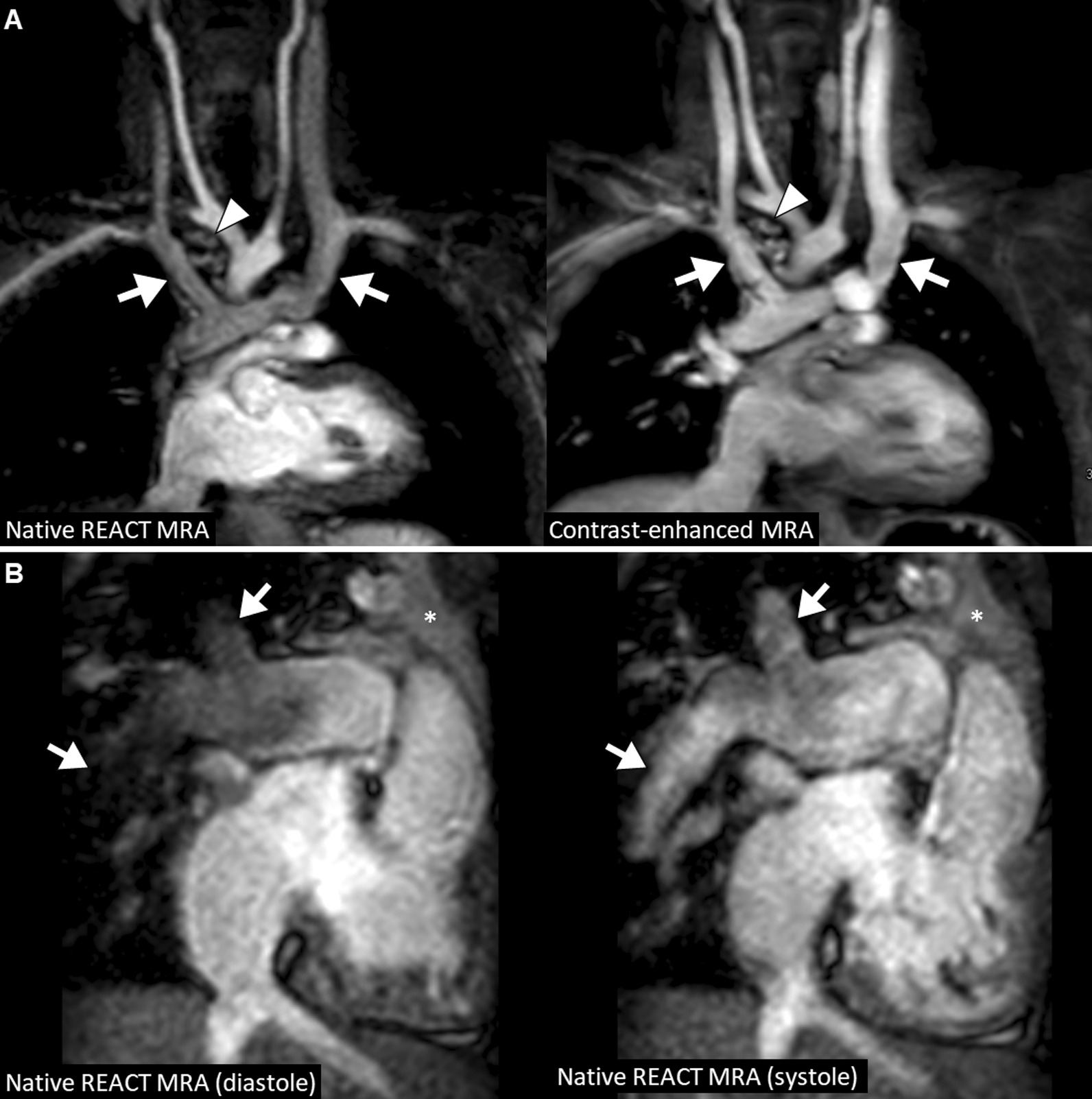
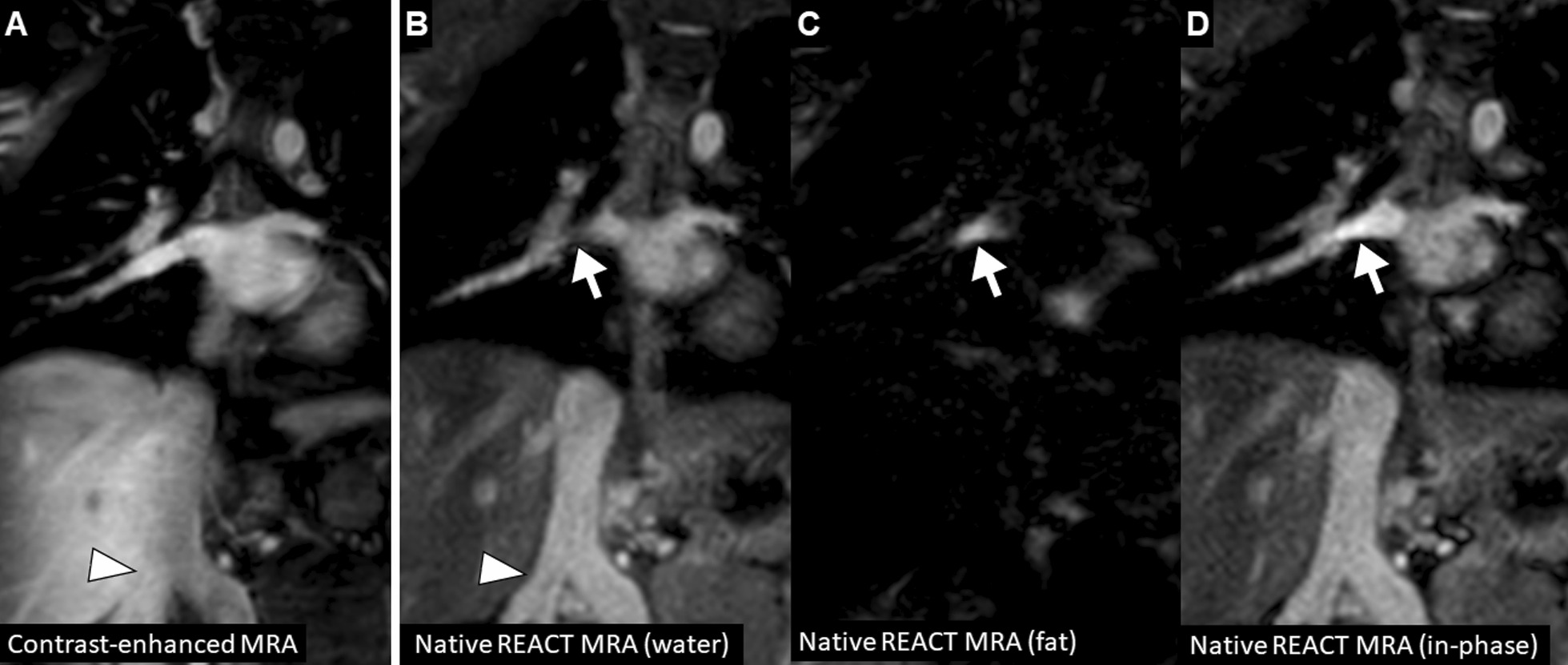
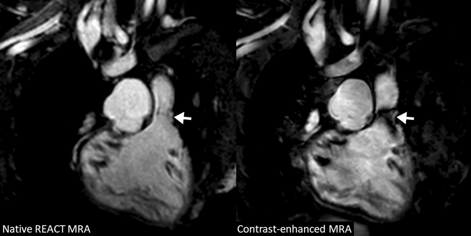
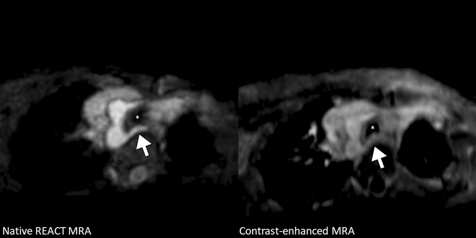
Background: To evaluate the great vessels in young children with complex congenital heart disease (CHD) using non-contrast cardiovascular magnetic resonance angiography (CMRA) based on three-dimensional relaxation-enhanced angiography without contrast (REACT) in comparison to contrast-enhanced steady-state CMRA.

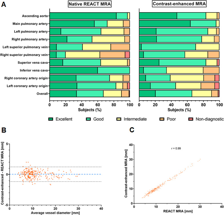
Methods: In this retrospective study from April to July 2021, respiratory- and electrocardiogram-gated native REACT CMRA was compared to contrast-enhanced single-phase steady-state CMRA in children with CHD who underwent CMRA at 3T under deep sedation. Vascular assessment included image quality (1 = non-diagnostic, 5 = excellent), vessel diameter, and diagnostic findings. For statistical analysis, paired t-test, Pearson correlation, Bland-Altman analysis, Wilcoxon test, and intraclass correlation coefficients (ICC) were applied.

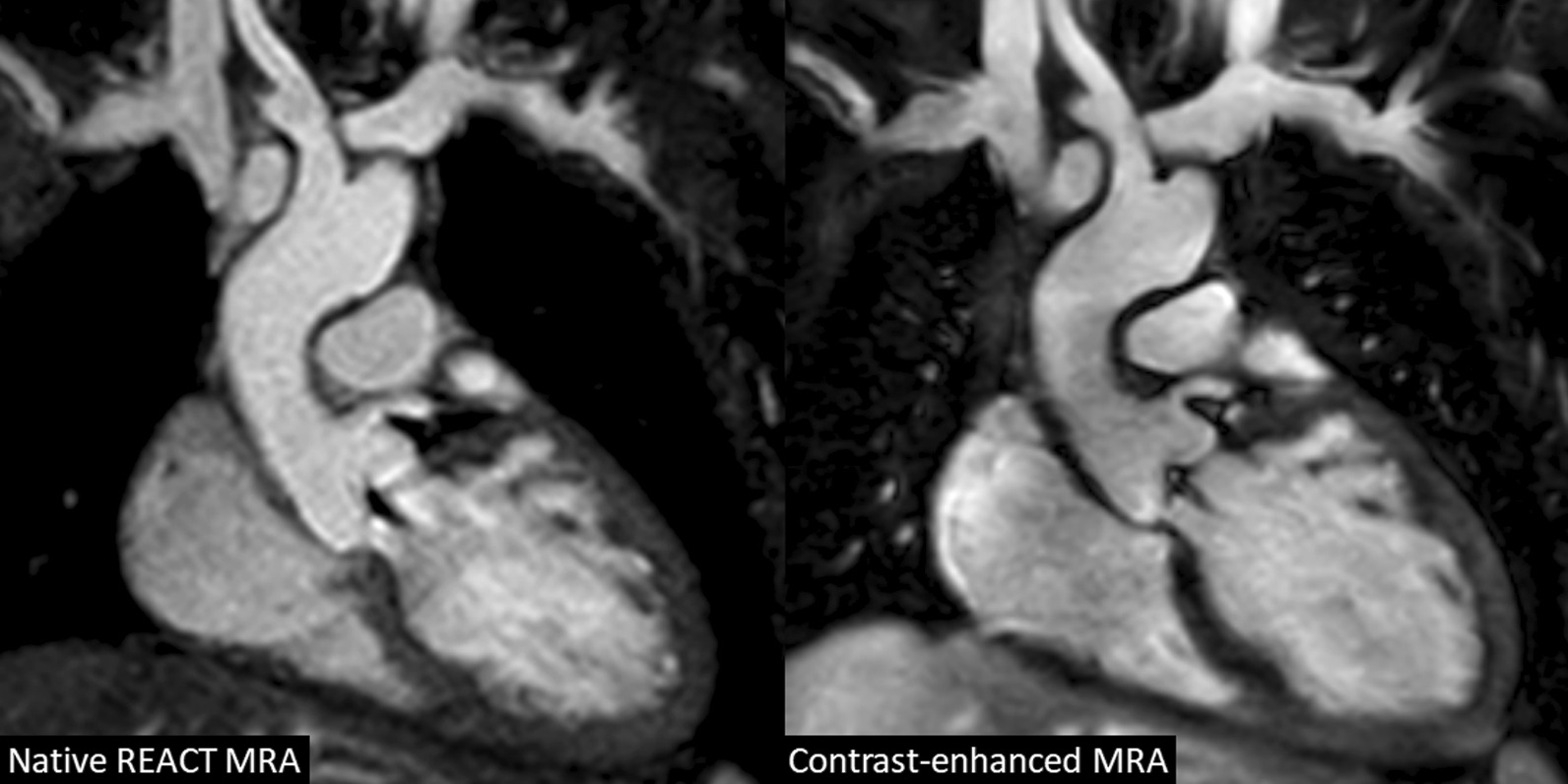
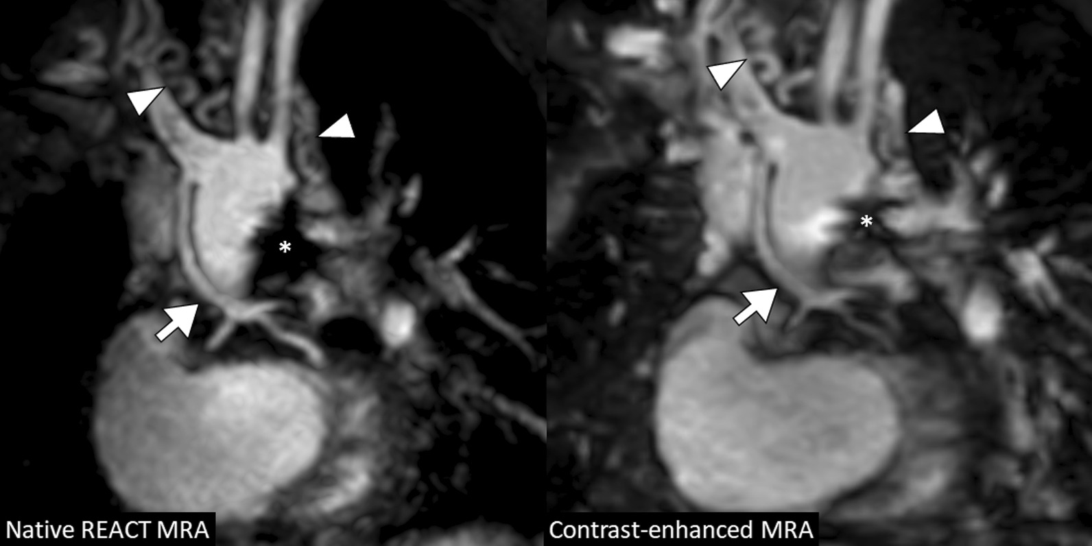
Results: Thirty-six young children with complex CHD (median 4 years, interquartile range, 2-5; 20 males) were included. Native REACT CMRA was obtained successfully in all patients (mean scan time: 4:22 ± 1:44 min). For all vessels assessed, diameters correlated strongly between both methods (Pearson r = 0.99; bias = 0.04 ± 0.61 mm) with high interobserver reproducibility (ICC: 0.99 for both CMRAs). Native REACT CMRA demonstrated comparable overall image quality to contrast-enhanced CMRA (3.9 ± 1.0 vs. 3.8 ± 0.9, P = 0.018). With REACT CMRA, better image quality was obtained at the ascending aorta (4.8 ± 0.5 vs. 4.3 ± 0.8, P < 0.001), coronary roots (e.g., left: 4.1 ± 1.0 vs. 3.3 ± 1.1, P = 0.001), and inferior vena cava (4.6 ± 0.5 vs. 3.2 ± 0.8, P < 0.001). In all patients, additional vascular findings were assessed equally with native REACT CMRA and the contrast-enhanced reference standard (n = 6).

Conclusion: In young children with complex CHD, REACT CMRA can provide gadolinium-free high image quality, accurate vascular measurements, and equivalent diagnostic quality compared to standard contrast-enhanced CMRA.

Keywords: Cardiovascular magnetic resonance angiography; Gadolinium-free; Non-contrast; Pediatric congenital heart disease; Relaxation-enhanced angiography without contrast.


Conflict of interest statement

S.Z., C.K., and O.W. (employees of Philips Healthcare) provided on-site technical support in setting up and adjusting the proposed native CMRA sequence. Nonemployee authors (A.I., J.A.L.) had full control of the data and information submitted for publication. There was no financial support from the industry for this study.

Figures

Fig. 1
Fig. 1
Study flow chart
Fig. 2
Fig. 2
A 7-year-old boy with congenital aortic valve dysplasia (non-reformatted water-images in coronal view). The example demonstrates comparable image quality between native relaxation-enhanced angiography without contrast (REACT) and contrast-enhanced cardiovascular magnetic resonance angiography (CMRA) with excellent fat suppression, high contrast and good vessel border delineation. The left coronary origin is clearly delineated. The left atrial appendage lateral to the main pulmonary artery is partially covered. Note the aortic regurgitant jet due to diastolic acquisition
Fig. 3
Fig. 3
Image quality and vessel diameter measurements results. A Bar plots show image quality scores for native and contrast-enhanced CMRA. B Bland–Altman plot and, C scatter plot show correlation of all vessel diameter measurements between native and contrast-enhanced CMRA. Pearson’s correlation coefficient is given. REACT relaxation-enhanced angiography without contrast
Fig. 4
Fig. 4
A 5-year-old boy with hypoplastic left heart syndrome (atresia of the mitral and aortic valves) and past medical history of several cardiac surgeries including Glenn procedure (multiplanar reformatted water-images in coronal view). Native and contrast-enhanced CMRA images demonstrate severely hypoplastic native ascending aorta that is connected to the brachiocephalic artery and shows distal trifurcation (arrow; note that the proximal coronary arteries are more clearly delineated on native images). Multiple small aorto-pulmonary collaterals with tortuous mediastinal course are visible (arrowheads). Susceptibility artifacts are present due to surgical stent material (asterisk). REACT relaxation-enhanced angiography without contrast
Fig. 5
Fig. 5
A 3-year-old boy with unbalanced atrioventricular septal defect (non-reformatted water-images in coronal view). Images shows lower blood signal intensity in the bilateral Glenn circulation on native compared to contrast-enhanced CMRA (arrows). These are presumably related to turbulent flow and differences in T1 an T2 relaxation times between venous and arterial blood. Small aorto-pulmonary collaterals are visible on both sequences (arrowhead). B A 5-year-old boy with congenitally corrected transposition of the great arteries and pulmonary trunk stenosis with dilatation of the right and left pulmonary arteries (multiplanar reformatted images in coronal-oblique view; out-of-phase images). Additionally performed systolic acquisition of native CMRA provides substantially reduced flow artifacts in the peripheral pulmonary arteries (arrows). Adjacent thymus (asterisk). REACT relaxation-enhanced angiography without contrast
Fig. 6
Fig. 6
A 4-year-old boy with truncus arteriosus communis, ventricular septal defect, and dysplastic tricuspid valve (multiplanar reformatted images in coronal view show different image reconstructions derived from modified Dixon sequence). A Compared to the contrast-enhanced CMRA, a fat-water swapping artifact is seen on native REACT CMRA. B A hypointense signal is observed within the right inferior pulmonary vein (arrow) on native CMRA water-only image. C Fat-only image shows high signal in the same area indicating a fat-water signal swapping (arrow). D In this situation, the in- and out-of-phase images should also be generated, because they are usually not affected by this Dixon-specific artifact. Note the double inferior vena cava variant with better vessel delineation from the adjacent liver on native CMRA (arrowheads)
Fig. 7
Fig. 7
A 4-year-old boy with tricuspid atresia and pulmonary stenosis after Glenn procedure (multiplanar reformatted water images in coronal view). Native and contrast-enhanced CMRA images show dilated right bronchial artery originating from the descending aorta (arrow) indicating competing systemic supply to the lung by major aortopulmonary collateral arteries (MAPCAs). REACT relaxation-enhanced angiography without contrast
Fig. 8
Fig. 8
A 9-year-old girl with interrupted aortic arch and post operative situation (non-reformatted water images in coronal view). Native CMRA has less susceptibility artifacts at the pulmonary valve level (arrow) than contrast-enhanced CMRA and allows for accurate assessment of the right ventricular outflow tract dimension. REACT relaxation-enhanced angiography without contrast
Fig. 9
Fig. 9
A 1-year-old girl with sinus venosus atrial septal defect (multiplanar reformatted water-only images in transversal view). Images show right-sided aortic arch with aberrant left subclavian artery without associated Kommerell diverticulum passing the trachea (asterisk) and the compressed esophagus posteriorly. Native CMRA provides better vessel contrast and delineation from adjacent structures, such as the anterolaterally located thymus, compared to contrast-enhanced CMRA.

Similar articles

Cited by

References

    1. Triedman JK, Newburger JW. Trends in congenital heart disease: the next decade. Circulation. 2016;133:2716–2733. doi: 10.1161/CIRCULATIONAHA.116.023544. - DOI - PubMed
    1. Baumgartner H, de Backer J, Babu-Narayan SV, Budts W, Chessa M, Diller G-P, et al. 2020 ESC guidelines for the management of adult congenital heart disease. Eur Heart J. 2021;42:563–645. doi: 10.1093/eurheartj/ehaa554. - DOI - PubMed
    1. Sachdeva R, Valente AM, Armstrong AK, Cook SC, Han BK, Lopez L, et al. ACC/AHA/ASE/HRS/ISACHD/SCAI/SCCT/SCMR/SOPE 2020 appropriate use criteria for multimodality imaging during the follow-up care of patients with congenital heart disease: a report of the American College of Cardiology Solution Set Oversight Committee and Appropriate Use Criteria Task Force, American Heart Association, American Society of Echocardiography, Heart Rhythm Society, International Society for Adult Congenital Heart Disease, Society for Cardiovascular Angiography and Interventions, Society of Cardiovascular Computed Tomography, Society for Cardiovascular Magnetic Resonance, and Society of Pediatric Echocardiography. J Am Coll Cardiol. 2020;75:657–703. doi: 10.1016/j.jacc.2019.10.002. - DOI - PubMed
    1. Fratz S, Chung T, Greil GF, Samyn MM, Taylor AM, Valsangiacomo Buechel ER, et al. Guidelines and protocols for cardiovascular magnetic resonance in children and adults with congenital heart disease: SCMR expert consensus group on congenital heart disease. J Cardiovasc Magn Reson. 2013;15:51. doi: 10.1186/1532-429X-15-51. - DOI - PMC - PubMed
    1. Fenchel M, Saleh R, Dinh H, Lee MH, Nael K, Krishnam M, et al. Juvenile and adult congenital heart disease: time-resolved 3D contrast-enhanced MR angiography. Radiology. 2007;244:399–410. doi: 10.1148/radiol.2442061045. - DOI - PubMed

Publication types