. 2022 Nov 16;12(1):19720.
doi: 10.1038/s41598-022-22895-6.

Placental vascular alterations are associated with early neurodevelopmental and pulmonary impairment in the rabbit fetal growth restriction model

Affiliations

Placental vascular alterations are associated with early neurodevelopmental and pulmonary impairment in the rabbit fetal growth restriction model

Ignacio Valenzuela et al. Sci Rep. .

Abstract

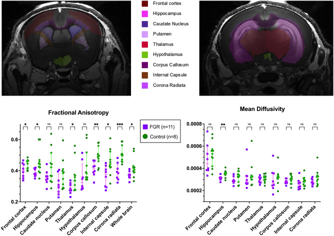
Fetal growth restriction is one of the leading causes of perinatal mortality and morbidity and has consequences that extend well beyond the neonatal period. Current management relies on timely delivery rather than improving placental function. Several prenatal strategies have failed to show benefit in clinical trials after promising results in animal models. Most of these animal models have important developmental and structural differences compared to the human and/or are insufficiently characterized. We aimed to describe placental function and structure in an FGR rabbit model, and to characterize the early brain and lung developmental morbidity using a multimodal approach. FGR was induced in time-mated rabbits at gestational day 25 by partial uteroplacental vessel ligation in one horn. Umbilical artery Doppler was measured before caesarean delivery at gestational day 30, and placentas were harvested for computed microtomography and histology. Neonates underwent neurobehavioral or pulmonary functional assessment the day after delivery, followed by brain or lung harvesting, respectively. Neuropathological assessment included multiregional quantification of neuron density, apoptosis, astrogliosis, cellular proliferation, and oligodendrocyte progenitors. Brain region volumes and diffusion metrics were obtained from ex-vivo brain magnetic resonance imaging. Lung assessment included biomechanical tests and pulmonary histology. Fetal growth restriction was associated with labyrinth alterations in the placenta, driven by fetal capillary reduction, and overall reduced vessels volume. FGR caused altered neurobehavior paralleled by regional neuropathological deficits and reduced fractional anisotropy in the cortex, white matter, and hippocampus. In addition, FGR kittens presented functional alterations in the peripheral lung and structurally underdeveloped alveoli. In conclusion, in a uteroplacental insufficiency FGR rabbit model, placental vascular alterations coincide with neurodevelopmental and pulmonary disruption.


Conflict of interest statement

The authors declare no competing interests.

Figures

Figure 1
Figure 1
Placental histology and microcomputed tomography. (A) Placentas with cytokeratin/lectin double staining, divided by placental zones (left images). Within the labyrinth (zoomed, right), fetal capillaries (arrow), maternal blood spaces (arrowhead) and trophoblast (star) were grid counted to calculate relative and absolute volume densities. For placental volumes, 51 FGR and 59 control placentas were used from 20 litters. For labyrinth structures, 10 FGR and 10 control placentas were used from 10 litters (1 placenta per horn, per litter). (B) Representative image of a placental microcomputed tomography after color coding according to vessel diameter. Data were analysed using a linear mixed-effects model and displayed as mean ± SD with significance as *0.05 ≥ p > 0.01; **0.01 ≥ p > 0.001.; ***0.001 > p > 0.0001; ****p < 0.0001.
Figure 2
Figure 2
Brain assessment in postnatal day 1 rabbits. (A) Neurobehavioral tests (left) and grouped scores (right) from 43 FGR and 61 control subjects from 16 litters. (B) Neuron density was assessed in cresyl violet stained slides in the depicted areas (top images). Bottom images show neuron density in the frontal cortex of FGR and control brains. To the right, neuron density from 27 FGR and 29 control subjects from 16 litters. (C) Representative images of apoptosis (TUNEL), and astrogliosis (GFAP) in hippocampus and corpus callosum, respectively. Data from 14 FGR and 19 control subjects from 11 litters. CA1 cornu ammonis 1, CA3 cornu ammonis 3, DG dentate gyrus, AVTN anteroventral thalamic nuclei. Data were analysed using a linear mixed-effects model and displayed as mean ± SD with significance as *0.05 ≥ p > 0.01; **0.01 ≥ p > 0.001.; ***0.001 > p > 0.0001; ****p < 0.0001.
Figure 3
Figure 3
Ex vivo brain diffusion tensor metrics at postnatal day 1 from 11 FGR and 8 control brains, from 6 litters. Data were analysed using a linear mixed-effects model and displayed as mean ± SD with significance as *0.05 ≥ p > 0.01; **0.01 ≥ p > 0.001.; ***0.001 > p > 0.0001; ****p < 0.0001.
Figure 4
Figure 4
Pulmonary assessment in postnatal day 1. (A, B) Pulmonary function tests results from 19 FGR and 21 control subjects from 6 litters. (C) Representative images of histological H&E-stained slides (left) and alveolar architecture data (right) from 16 FGR and 21 control subjects from 6 litters. Data were analysed using a linear mixed-effects model and displayed as mean ± SD with significance as *0.05 ≥ p > 0.01; **0.01 ≥ p > 0.001.; ***0.001 > p > 0.0001; ****p < 0.0001.

Similar articles

Cited by

References

    1. Resnik R. Intrauterine growth restriction. Obstet. Gynecol. 2002;99:490–496. doi: 10.1016/s0029-7844(01)01780-x. - DOI - PubMed
    1. Flenady V, et al. Major risk factors for stillbirth in high-income countries: A systematic review and meta-analysis. Lancet. 2011;377:1331–1340. doi: 10.1016/s0140-6736(10)62233-7. - DOI - PubMed
    1. Lawn JE, et al. Stillbirths: Rates, risk factors, and acceleration towards 2030. Lancet. 2016;387:587–603. doi: 10.1016/s0140-6736(15)00837-5. - DOI - PubMed
    1. Allanson ER, et al. The WHO application of ICD-10 to deaths during the perinatal period (ICD-PM): Results from pilot database testing in South Africa and United Kingdom. BJOG. 2016;123:2019–2028. doi: 10.1111/1471-0528.14244. - DOI - PubMed
    1. von Beckerath AK, et al. Perinatal complications and long-term neurodevelopmental outcome of infants with intrauterine growth restriction. Am. J. Obstet. Gynecol. 2013;208(130):e131–136. doi: 10.1016/j.ajog.2012.11.014. - DOI - PubMed

Publication types