. 2022 Nov 16;12(1):19630.
doi: 10.1038/s41598-022-24152-2.

Ferroptosis inhibition by deferiprone, attenuates myelin damage and promotes neuroprotection in demyelinated optic nerve

Affiliations

Ferroptosis inhibition by deferiprone, attenuates myelin damage and promotes neuroprotection in demyelinated optic nerve

Atefeh Rayatpour et al. Sci Rep. .

Abstract

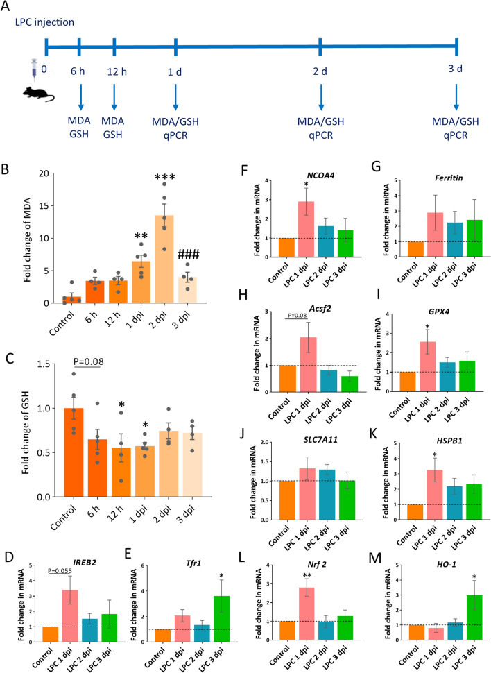
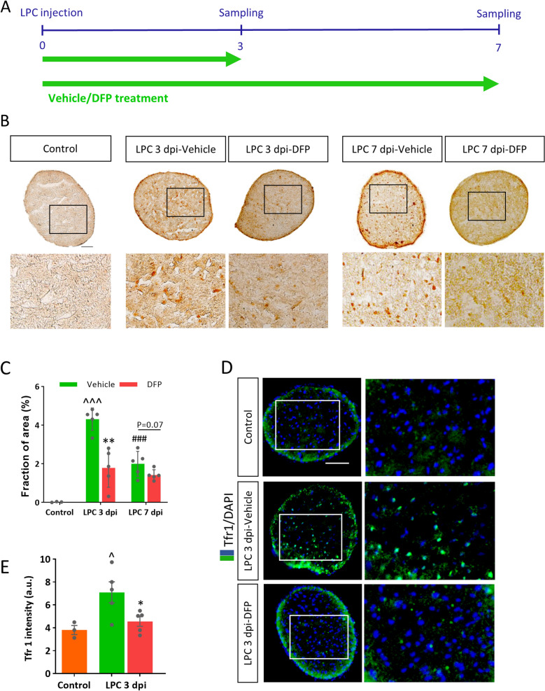
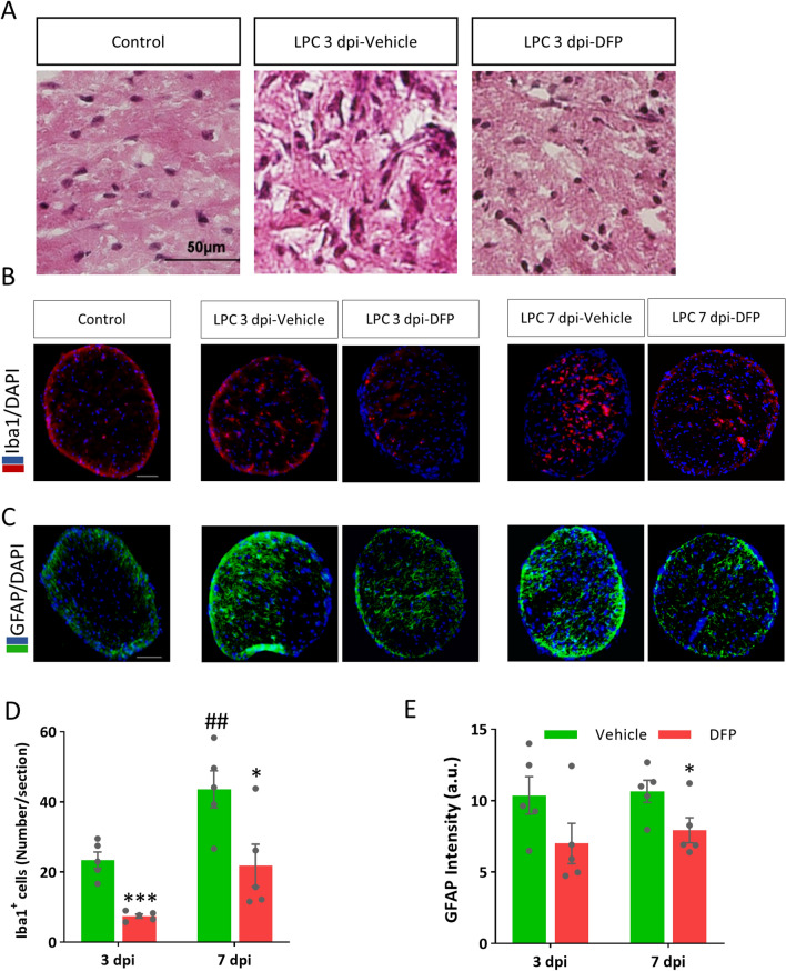
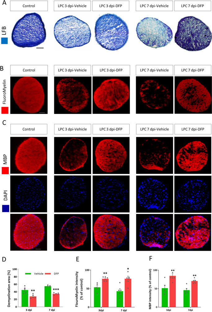
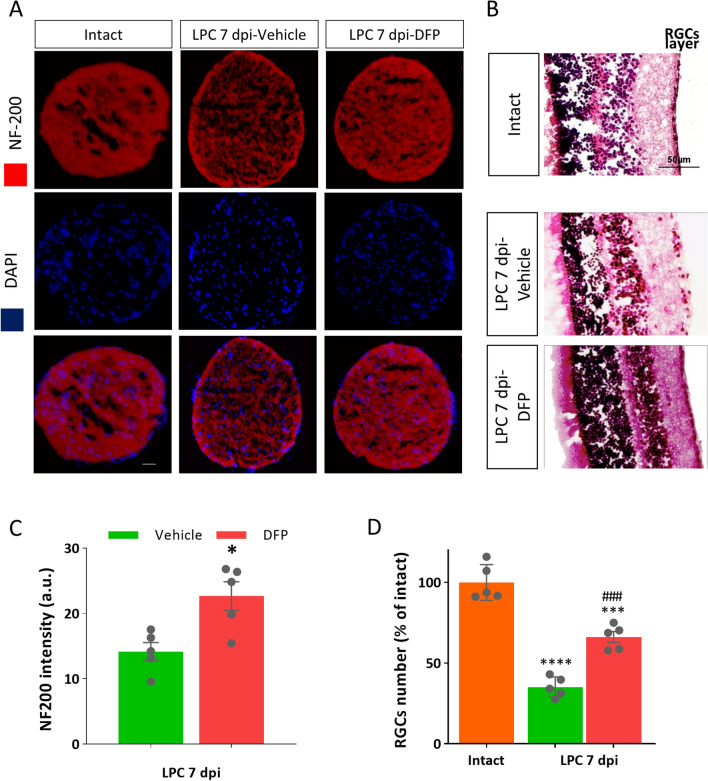
Multiple sclerosis (MS) is a chronic inflammatory disease, which leads to focal demyelination in the brain and spinal cord. Studies showed that iron released during the course of myelin breakdown exacerbates tissue damage, which is in agreement with the features of iron-dependent cell death, ferroptosis. Here, we aimed to investigate the possible contribution of ferroptosis in the demyelinated optic nerve, and to explore the effectiveness of ferroptosis inhibitor, deferiprone (DFP), on the extent of demyelination, inflammation and axonal damage. For this purpose, focal demyelination was induced by injection of lysolecithin (LPC), into the optic nerve of male C57BL/6J mice. Afterward, optic nerves were harvested at different time points from as early as 6 h up to 7 days post-LPC injection. Next, to evaluate the effectiveness of DFP two groups of animals received daily intraperitoneal injection of DFP for 3 or 7 continuous days. Vehicle groups received saline. Iron deposition was observed at different time points post-LPC injection from 6 h to 7 days post injection. Examining ferroptosis markers showed a significant reduction in glutathione content along with increased level of malondialdehyde and upregulated ferroptosis marker genes at early time points after injection. Besides, DFP treatment during the inflammatory phase of the model resulted in decreased microgliosis and inflammation. Reduced demyelination, microgliosis and astrogliosis was shown in mice that received DFP for 7 days. Moreover, DFP protected against axonal damage and retinal ganglion cells loss. Our results suggest the possible contribution of ferroptosis pathway in the process of demyelination. The therapeutic strategies targeting iron deposition, e.g. DFP treatment might thus represent a promising therapeutic target for patients with MS.


Conflict of interest statement

The authors declare no competing interests.

Figures

Figure 1
Figure 1
Iron accumulation in optic nerve following LPC injection. (A) Schematic illustration shows the time line of LPC injection and Perl’s staining in the experimental setting used. (B) DAB-enhanced Perl’s staining shows iron deposition in optic nerve sections at different time points starting at 6 h after LPC injection. (C) Quantification of iron positive fraction of area. Data represent mean ± SEM, one-way ANOVA with Tukey's multiple comparison post-test, n = 3–5 mice per group. **p < 0.01, ***p < 0.001 versus control, ###p < 0.001 versus 3 dpi. Scale bar = 50 µm. hpi: hour post-injection.
Figure 2
Figure 2
Evaluation of ferroptosis markers following LPC injection. (A) Schematic illustration shows the time line of LPC injection and tissue sampling for biochemical and molecular assessments. (B) Spectrophotometric detection of malondialdehyde (MDA) and (C) glutathione (GSH) at different time points after LPC injection. Data represent mean ± SEM, one-way ANOVA with Tukey's multiple comparison test, n = 4–5 mice per group. *p < 0.05, **p < 0.01, ***p < 0.001 versus control, ###p < 0.001 versus 2 dpi. (DM) Ferroptosis related genes expression at different time points following LPC injection. One-way ANOVA with Dunnett's multiple comparisons test, Data represent mean ± SEM, n = 4–5 mice per group (3 animals pooled for each sample; total 12–15 animals used for each time point), *p < 0.05, **p < 0.01 versus control.
Figure 3
Figure 3
Deferiprone (DFP) decreased iron deposition in demyelinated optic nerve. (A) Experimental timeline of LPC injection and DFP treatment. DFP (10 mg/kg twice per day) or vehicle were injected intraperitoneally for 3 continues days. (B) Increased iron deposition was observed on 3 and 7 dpi as revealed by DAB enhanced Perl’s staining, while continuous DFP administration reduced iron deposition. (C) Quantification of iron positive fraction of area (D) Immunostaining against Tfr1, counterstained with DAPI in vehicle and DFP treated mice 3 days post LPC injection. (E) Quantification of Tfr1 immunoreactivity showed significant reduction in Tfr1 intensity in the optic nerve of DFP treated mice at 3 dpi. Data represent mean ± SEM, Student's t-test, n = 3–5 mice per group. *p < 0.05, **p < 0.01 versus vehicle at the same group, ##p < 0.01 versus 3dpi-vehicle, ^p < 0.05, ^^^p < 0.001 vs control. Scale bar = 50 µm.
Figure 4
Figure 4
Deferiprone (DFP) attenuated inflammation and gliosis in demyelinated optic nerve. (A) Representative images of transverse sections from the optic nerve at 3 dpi that stained with hematoxylin and eosin. (B) Immunostaining against Iba1, the microglia marker, counterstained with DAPI in vehicle and DFP treated mice on 3 and 7 dpi. (C) Immunostaining against GFAP, as the astrocyte marker, counterstained with DAPI in vehicle and DFP treated mice 3 or 7 days post LPC injection. (D) Quantification of the number of Iba1+ cells showed that DFP ameliorated microglia activation in the optic nerve following LPC insult at 3 or 7 dpi. (E) Quantification of GFAP immunoreactivity showed non-significant reduction in astrogliosis in the optic nerve following LPC insult at 3 dpi along with significant decrease at 7 dpi. Data represent mean ± SEM. Student's t-test. *p < 0.05, ***p < 0.001 versus vehicle at the same group, ##p < 0.01 versus LPC 3dpi-vehicle, n = 5 mice per group, Scale bar = 50 µm.
Figure 5
Figure 5
Treatment with Deferiprone (DFP) decreased an extensive loss of myelin in the optic nerve. (A) Representative micrographs of optic nerve prepared from intact and LPC-injected mice treated with DFP or vehicle for 3 or 7 days, stained with LFB and crysel violet. (B) Representative images of FluoroMyelin stained sections of intact and LPC-injected mice treated with DFP or vehicle for 3 or 7 continues days. (C) Immunostaining against MBP counterstained with DAPI in intact and LPC-injected mice treated with DFP or vehicle 3 or 7 days. (D) Quantitative analysis of the LFB-stained sections indicated reduced demyelination in mice treated with DFP. The bar graph shows the percentage of demyelination area versus the total area of the transverse section of optic nerve. (E) Quantitative analysis of FluoroMyelin intensity, calculated as the percent of the FM reactivity in intact group. (F) Quantitative analysis of MBP intensity showed significant differences between vehicle and DFP treated groups on 3 and 7 dpi. Data represent mean ± SEM. Student's t-test. *p < 0.05, **p < 0.01, ***p < 0.001 versus vehicle at the same group, n = 5 mice per group, Scale bar = 50 µm.
Figure 6
Figure 6
Deferiprone decreased retinal ganglion cell (RGC) loss in demyelinated optic nerve. (A) Immunostaining against NF-200, as the axonal marker, counterstained with DAPI in vehicle and deferiprone (DFP) treated mice on 7 dpi. (B) H&E staining demonstrated that DFP protects against the loss of RGCs. (C) Quantification of the NF-200 immunoreactivity showed that DFP protected against axonal damage at 7dpi. *p<0.05 versus vehicle group, Student's t-test. (D) Quantification of the number of RGCs. Data represent mean ± SEM. One-way ANOVA with Tukey's multiple comparisions test, ***p< 0.001 versus Intact group, ###p<0.001 versus LPC 7dpi-vehicle, n = 5 mice per group, Scale bar = 50 µm.

Similar articles

Cited by

References

    1. Reich DS, Lucchinetti CF, Calabresi PA. Multiple sclerosis. N. Engl. J. Med. 2018;378:169–180. - PMC - PubMed
    1. Lassmann H, Van Horssen J, Mahad D. Progressive multiple sclerosis: Pathology and pathogenesis. Nat. Rev. Neurol. 2012;8:647–656. - PubMed
    1. Hametner S, et al. Iron and neurodegeneration in the multiple sclerosis brain. Ann. Neurol. 2013;74:848–861. - PMC - PubMed
    1. Möller HE, et al. Iron, myelin, and the brain: Neuroimaging meets neurobiology. Trends Neurosci. 2019;42:384–401. - PubMed
    1. Todorich B, Pasquini JM, Garcia CI, Paez PM, Connor JR. Oligodendrocytes and myelination: The role of iron. Glia. 2009;57:467–478. - PubMed

Publication types