. 2023 May;72(5):1233-1246.
doi: 10.1007/s00262-022-03322-1. Epub 2022 Nov 16.

Expanded natural killer cells potentiate the antimyeloma activity of daratumumab, lenalidomide, and dexamethasone in a myeloma xenograft model

Affiliations

Expanded natural killer cells potentiate the antimyeloma activity of daratumumab, lenalidomide, and dexamethasone in a myeloma xenograft model

Jaya Lakshmi Thangaraj et al. Cancer Immunol Immunother. 2023 May.

Abstract

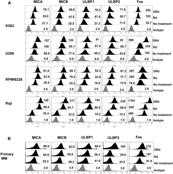
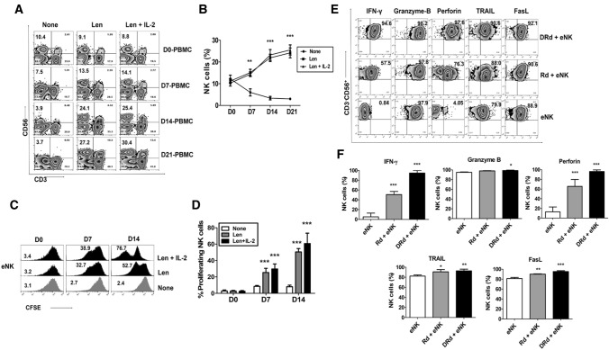
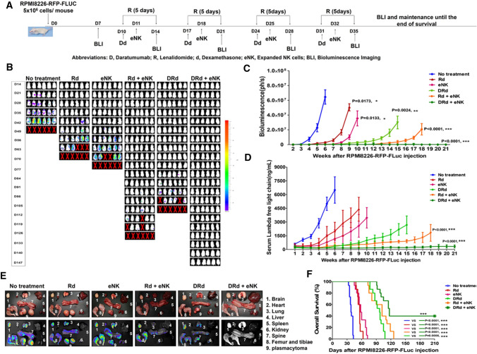
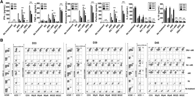
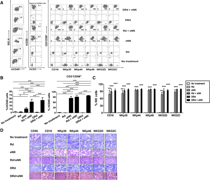
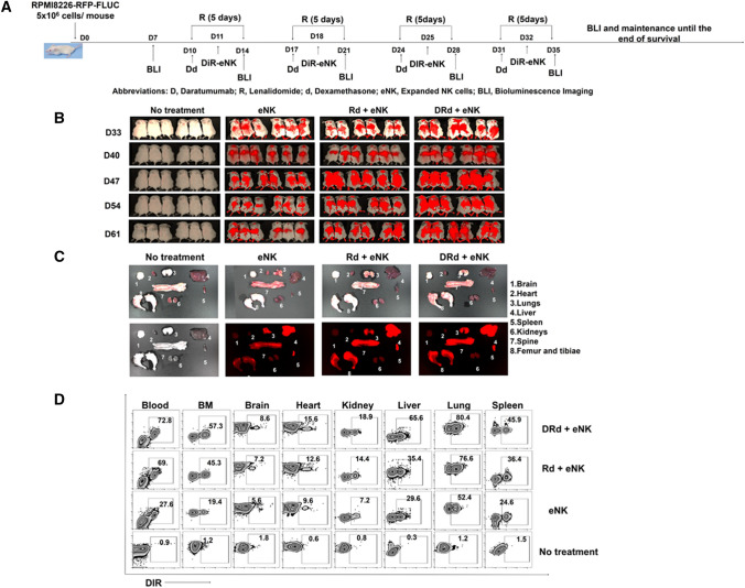
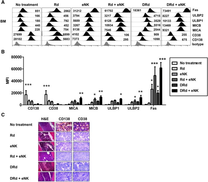
The development of new treatment agents in recent decades has significantly improved the survival of patients with multiple myeloma (MM). Nonetheless, MM remains an incurable disease; therefore, novel combination therapies are required. Natural killer (NK) cells are one of the safest immunotherapeutic options. In this study, we found that the anti-myeloma activity of expanded NK cells (eNKs) was improved by daratumumab, lenalidomide, and dexamethasone (DRd) in an MM xenograft mouse model. NK cells expanded from peripheral blood mononuclear cells collected from MM patients were highly cytotoxic against DRd pretreated tumor cells in vitro. To mimic the clinical protocol, a human MM xenograft model was developed using human RPMI8226-RFP-FLuc cells in NOD/SCID IL-2Rγnull (NSG) mice. MM bearing mice were randomly divided into six groups: no treatment, eNK, Rd, Rd + eNKs, DRd, and DRd + eNKs. DRd significantly enhanced the cytotoxicity of eNKs by upregulating NK cell activation ligands and effector function. DRd in combination with eNKs significantly reduced the serum M-protein level and prolonged mouse survival. In addition, DRd significantly increased the persistence of eNK and homing to MM sites. These results show that the anti-myeloma activity of ex vivo-expanded and activated NK cells is augmented by the immunomodulatory effect of DRd in MM-bearing mice, suggesting the therapeutic potential of this combination for MM patients.

Keywords: Daratumumab; Immunotherapy; Multiple myeloma; Natural killer cells.


Conflict of interest statement

The authors declare no conflicts of interest.

Figures

Fig. 1
Fig. 1
DRd increases NK-activating ligand expression on tumor cells. A and B Flow cytometry histograms showing surface expression (MFI value) patterns of MICA, MICB, ULBP1, ULBP2, and Fas on K562, U266, RPMI8226, Raji, and primary MM cells after treatment with Rd or DRd. DRd significantly increased the expression levels of MICA, MICB, ULBP1, ULBP2, and Fas receptor in both cell lines and primary MM cells. (See also Supplementary Fig. 2)
Fig. 2
Fig. 2
Effect of DRd on effector molecules expression in eNKs. A Representative flow cytometry plots of the expression of NK cell markers in peripheral blood mononuclear cells from patients with MM that had been treated with lenalidomide. B Mean ± standard deviation (SD) NK cell proportion in peripheral blood mononuclear cells from patients with MM (n = 5) after lenalidomide treatment. Lenalidomide significantly increased the NK cell proportion without cytokine stimulation. C Representative histogram showing the proliferation of CFSE-labeled eNKs treated with lenalidomide (1 µm) with or without IL-2. D Mean ± SD CFSE labeled eNK (n = 5) proliferation after lenalidomide treatment. Lenalidomide significantly induced eNK proliferation for 2 weeks with or without IL-2. E Representative FACS plot showing the expression of IFN-γ, granzyme-B, perforin, TRAIL, and FasL in eNKs. eNKs were harvested on day 14 and treated with DRd or Rd for 12 h. F Mean ± SD eNK (n = 5) expression of effector molecules after DRd treatment. DRd significantly increased the expression levels of IFN-γ and perforin in eNKs (mean ± standard deviation; n = 5). *p < 0.05, **p < 0.001, ***p < 0.0001
Fig. 3
Fig. 3
eNKs exert a strong cytotoxic effect on DRd-pretreated tumor cells and primary MM cells. (A and B) Proportion of CD107a-expressing eNKs (CD3CD56.+) after co-culture with DRd-pretreated K562, U266, RPMI8226, Raji cells, or primary MM cells (E:T ratio of 1:1) for 4 h. Flow cytometry revealed a higher level of CD107a in eNKs that had been co-cultured with DRd-treated tumor cells. (C and D) eNK-mediated cytotoxicity in DRd-pretreated tumor cells (K562, U266, RPMI8226, and Raji) and primary MM cells measured using a standard 4-h flow cytometry-based cytotoxicity assay. eNKs showed potent antitumor activity against DRd-pretreated tumor cells at all E:T ratios. *p < 0.05, **p < 0.001, ***p < 0.0001
Fig. 4
Fig. 4
eNKs + DRd inhibited MM progression in RPMI8226-RFP-FLuc xenograft mice. A Treatment of RPMI8226-RFP-FLuc-bearing mice with eNKs + DRd. NSG mice were intravenously injected with 5 × 10.6 RPMI8226-RFP-FLuc cells, and tumor growth was monitored weekly by BLI. B Representative BLI of six mice (n = 15) from each group (dorsal view). C BLI showed that DRd + eNKs treatment provided the strongest antitumor effect at all time points. D Serum M-protein level determined by quantifying the level of human lambda free light chain in mouse peripheral blood. Mice treated with DRd + eNKs had the lowest serum M-protein levels. E Representative ex vivo BLI of mouse tissues. DRd + eNKs treatment did not result in a detectable bioluminescence signal. F Kaplan–Meier survival curves (n = 15 mice per group). Statistical significance was determined by log-rank test. Mice treated with DRd + eNKs exhibited the longest survival. *p < 0.05, **p < 0.01, ***p < 0.001
Fig. 5
Fig. 5
DRd enhanced the in vivo effector function and persistence of eNKs in RPMI8226-RFP-FLuc-bearing mice. A In vivo effector function of circulating eNKs, determined based on the levels of various immune effector and immunosuppressive cytokines at three time points from 1 day after final cell infusion (i.e., D33, D39, and D45). Mice treated with DRd + eNKs had the highest levels of IFN-γ, granzyme-B, perforin, and TNF-α, which decreased over time; they also had the lowest levels of IL-6 and IL-10. B Representative flow cytometry plots showing the in vivo persistence of circulating eNKs expressing NK activation receptors (CD16, NKp30, NKp44, NKp46, NKG2D, and NKG2C) among the hCD45 population with hCD3CD56.+ expression at D33, D39, and D45. *p < 0.01, **p < 0.001, ***p < 0.0001
Fig. 6
Fig. 6
DRd enhanced eNK homing in vivo in RPMI8226-RFP-FLuc-bearing mice. A In vivo homing of eNKs in RPMI8226-RFP-FLuc-bearing mice (n = 10 per group) that were evaluated by flow cytometry. Mice were euthanized and BM samples were analyzed by flow cytometry for human NK cells (CD3CD56+) and activation receptors (CD16, NKp30, NKp44, NKp46, NKG2D, and NKG2C) among the hCD45 population. (B, C) Quantification (mean ± SD) of in vivo eNK homing in the BM based on flow cytometry data. eNKs from mice treated with DRd + eNKs showed the greatest in vivo homing ability. *p < 0.01, **p < 0.001, ***p < 0.0001
Fig. 7
Fig. 7
Trafficking of eNKs in DRd-treated RPMI8226-RFP-FLuc-bearing mice. (A) A separate set of RPMI8226-RFP-FLuc-bearing mice (n = 6 per group) were infused with DIR-labeled eNKs (2 × 107 cells per mouse). (B) Treatment group mice were subjected to fluorescence imaging once weekly after the final eNK infusion for 5 weeks. C and D Mice were euthanized, and BM and other tissues were collected and subjected to fluorescence imaging and flow cytometry. eNK infiltration was greatest in the BM, liver, lung, and spleen
Fig. 8
Fig. 8
DRd + eNKs treatment enhances MM clearance and NK-activating ligand expression levels in RPMI8226-RFP-FLuc xenograft mice. A Residual RPMI8226-RFP-FLuc cells in the BM evaluated by flow cytometry. Representative histograms showing the expression levels (MFI value) of CD138, CD38, MICA, MICB, ULBP1, ULBP 2, and Fas. B Quantification (mean ± SD) of residual myeloma cells in the BM. Mice treated with Rd + eNKs and DRd + eNKs exhibited lower CD138 and CD38 expression levels in the BM, compared with the other groups; they also exhibited higher MICA, MICB, ULBP1, ULBP2, and Fas expression levels. *p < 0.01, **p < 0.001, ***p < 0.0001

References

    1. Bahlis NJ, et al. Daratumumab plus lenalidomide and dexamethasone in relapsed/refractory multiple myeloma: extended follow-up of POLLUX, a randomized, open-label, phase 3 study. Leukemia. 2020;34:1875–1884. doi: 10.1038/s41375-020-0711-6. - DOI - PMC - PubMed
    1. Facon T, et al. Daratumumab plus lenalidomide and dexamethasone for untreated myeloma. N Engl J Med. 2019;380:2104–2115. doi: 10.1056/NEJMoa1817249. - DOI - PMC - PubMed
    1. Mateos MV, et al. Daratumumab, bortezomib, and dexamethasone versus bortezomib and dexamethasone in patients with previously treated multiple myeloma: three-year follow-up of CASTOR. Clin Lymphoma Myeloma Leuk. 2020;20:509–518. doi: 10.1016/j.clml.2019.09.623. - DOI - PubMed
    1. Moreau P, et al. Treatment of relapsed and refractory multiple myeloma: recommendations from the International myeloma working group. Lancet Oncol. 2021;22:e105–e118. doi: 10.1016/S1470-2045(20)30756-7. - DOI - PubMed
    1. Korde N, et al. Treatment with carfilzomib-lenalidomide-dexamethasone with lenalidomide extension in patients with smoldering or newly diagnosed multiple myeloma. JAMA Oncol. 2015;1:746–754. doi: 10.1001/jamaoncol.2015.2010. - DOI - PMC - PubMed