

Allyl ether of mansonone G as a potential anticancer agent for colorectal cancer
- PMID: 36385303
- PMCID: PMC9668903
- DOI: 10.1038/s41598-022-23997-x
Allyl ether of mansonone G as a potential anticancer agent for colorectal cancer
Abstract
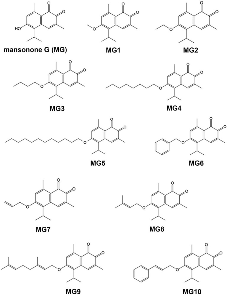
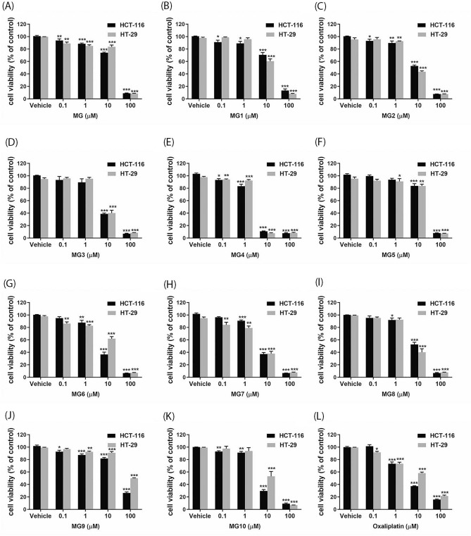
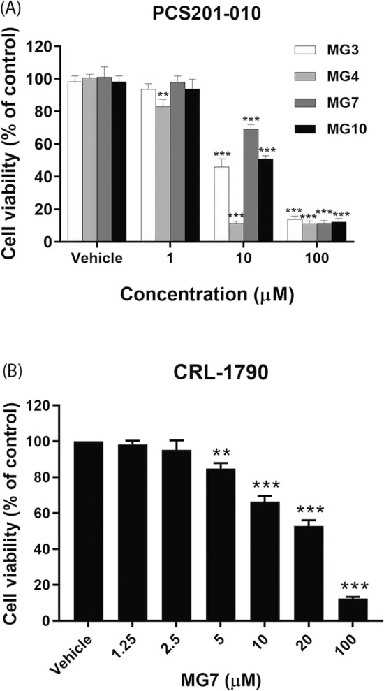
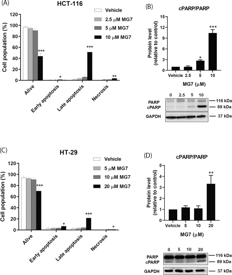
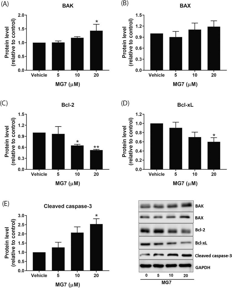
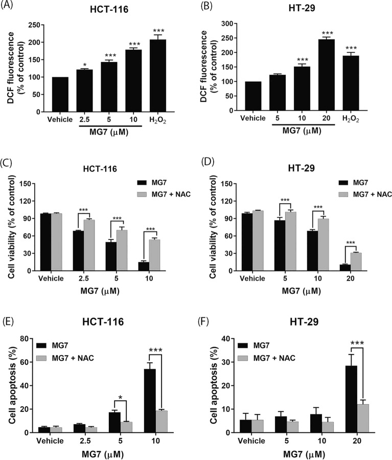
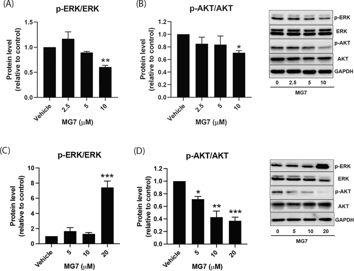
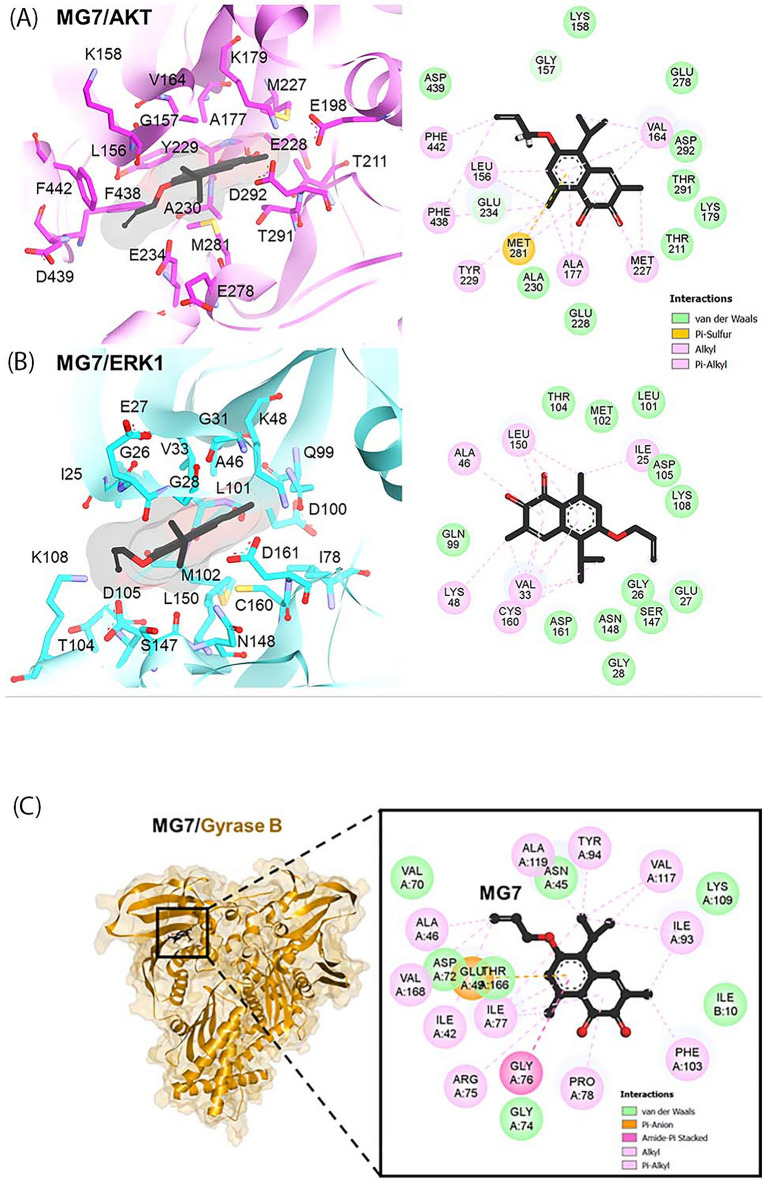
Mansonone G (MG), a 1,2-naphthoquinone isolated from the heartwood of Mansonia gagei Drumm, exhibited several pharmacological activities such as anti-bacterial, anti-estrogenic and anti-adipogenic effect. This study evaluated the cytotoxicity of MG and its derivatives as well as determined the mechanism(s) underlying the cytotoxic activity of the most potent MG derivative on two CRC cell lines, HCT-116 cells carrying p53 wild-type and HT-29 cells carrying p53 mutant. We found that MG and its derivatives could inhibit viability of HCT-116 and HT-29 cells in a concentration-dependent manner. Of all semi-synthetic derivatives of MG, allyl ether mansonone G (MG7) was the most potent cytotoxic agent toward cancer cells and less toxic to normal cells. MG7 could induce ROS generation which was associated with cytotoxicity and apoptosis in both HCT-116 and HT-29 cells. Western blot analysis revealed that MG7 downregulated the expression of Bcl-2 and Bcl-xL proteins in both CRC cell lines and upregulated the expression of BAK protein in HT-29 cells. Moreover, MG7 inhibited AKT signaling pathway in both CRC cell lines and modulated ERK1/2 signaling pathway by inhibiting ERK1/2 phosphorylation in HCT-116 cells and activating ERK1/2 phosphorylation in HT-29 cells. Molecular docking revealed that MG7 could bind to the ATP-binding pocket of AKT and ERK1 via hydrophobic interactions.
© 2022. The Author(s).
Conflict of interest statement
The authors declare no competing interests.
Figures

Similar articles
-
Therapeutic potential of ethoxy mansonone G: A comprehensive exploration of its anticancer actions in breast cancer, colorectal cancer, and non-small cell lung carcinoma.Cell Biol Int. 2024 Sep;48(9):1229-1239. doi: 10.1002/cbin.12207. Epub 2024 Jun 23. Cell Biol Int. 2024. PMID: 38924324 Review.
-
Anti-estrogenic activity of mansonone G and mansorin A derivatives.Pharm Biol. 2013 Aug;51(8):948-54. doi: 10.3109/13880209.2013.771684. Epub 2013 Apr 22. Pharm Biol. 2013. PMID: 23607906
-
Activation of death receptor, DR5 and mitochondria-mediated apoptosis by a 3,4,5-trimethoxybenzyloxy derivative in wild-type and p53 mutant colorectal cancer cell lines.Naunyn Schmiedebergs Arch Pharmacol. 2020 Mar;393(3):405-417. doi: 10.1007/s00210-019-01730-2. Epub 2019 Oct 23. Naunyn Schmiedebergs Arch Pharmacol. 2020. PMID: 31641820
-
Synthetic antiprotozoal thiazolide drug induced apoptosis in colorectal cancer cells: implications of IL-6/JAK2/STAT3 and p53/caspases-dependent signaling pathways based on molecular docking and in vitro study.Mol Cell Biochem. 2020 Jun;469(1-2):143-157. doi: 10.1007/s11010-020-03736-4. Epub 2020 Apr 30. Mol Cell Biochem. 2020. PMID: 32356241
-
Anticancer Profiling for Coumarins and Related O-Naphthoquinones from Mansonia gagei against Solid Tumor Cells In Vitro.Molecules. 2018 Apr 26;23(5):1020. doi: 10.3390/molecules23051020. Molecules. 2018. PMID: 29701706 Free PMC article.
Cited by
-
ATP-competitive inhibitors for cancer treatment - kinases and the world beyond.RSC Med Chem. 2025 Jun 25. doi: 10.1039/d5md00235d. Online ahead of print. RSC Med Chem. 2025. PMID: 40612269 Free PMC article. Review.
References
Publication types
MeSH terms
Substances
Grants and funding
LinkOut - more resources
Full Text Sources
Medical
Research Materials
Miscellaneous
