. 2022 Dec;612(7939):338-346.
doi: 10.1038/s41586-022-05443-0. Epub 2022 Nov 16.

Ferroptosis of tumour neutrophils causes immune suppression in cancer

Affiliations

Ferroptosis of tumour neutrophils causes immune suppression in cancer

Rina Kim et al. Nature. 2022 Dec.

Abstract

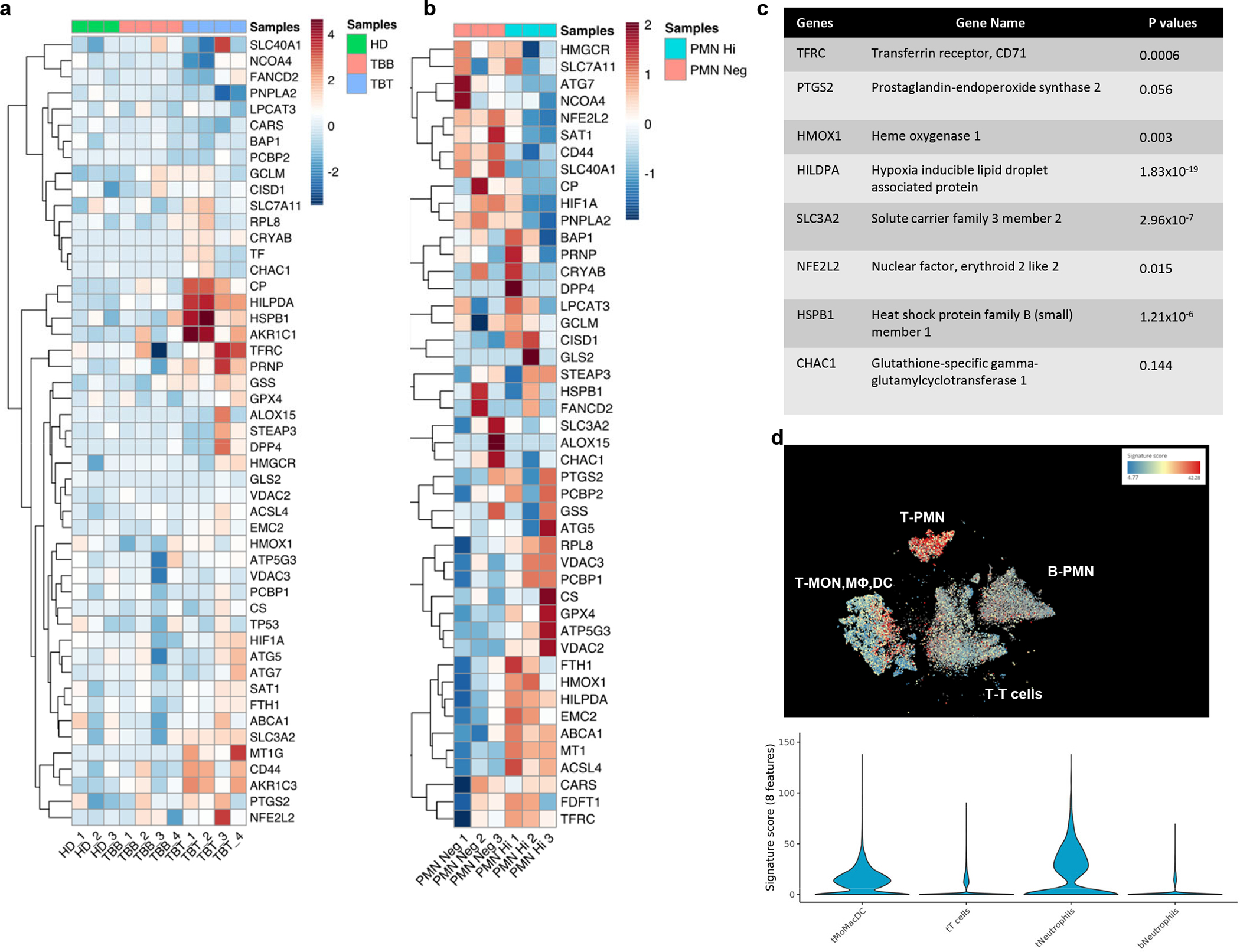
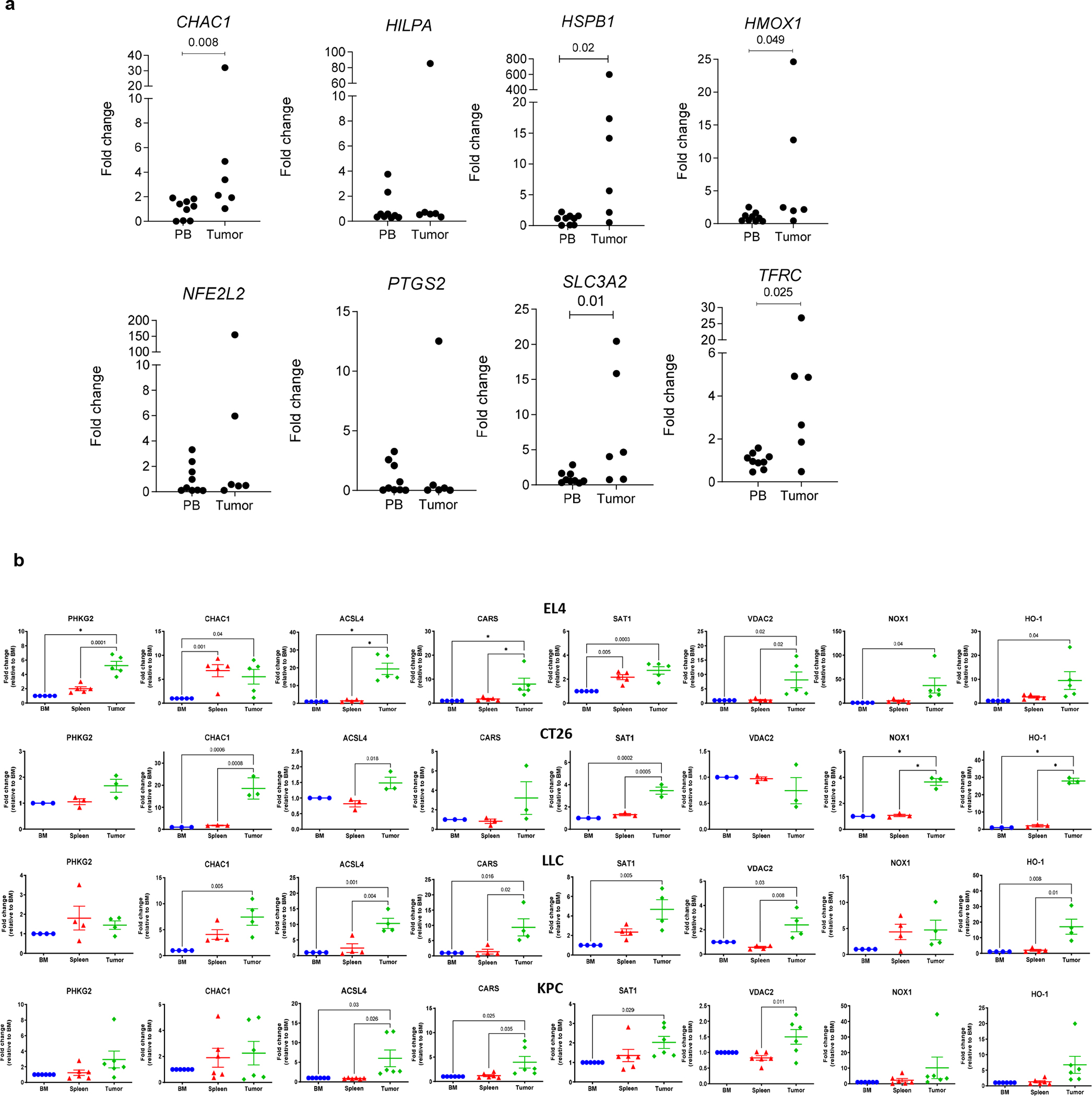
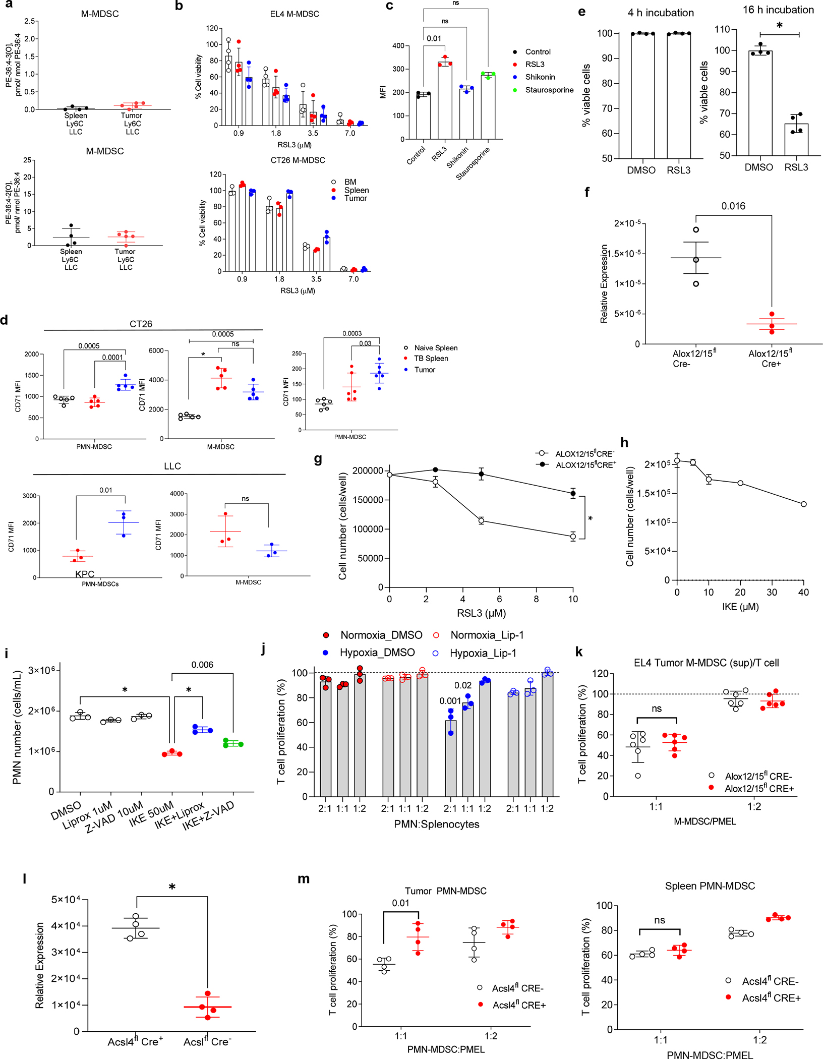
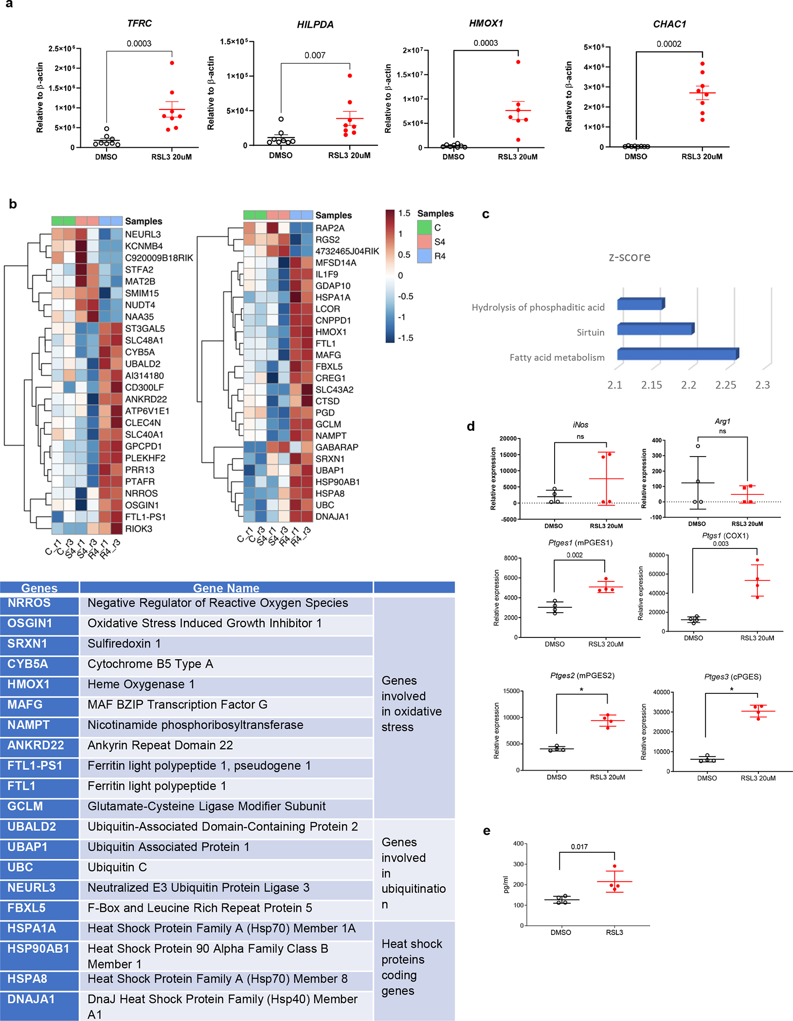
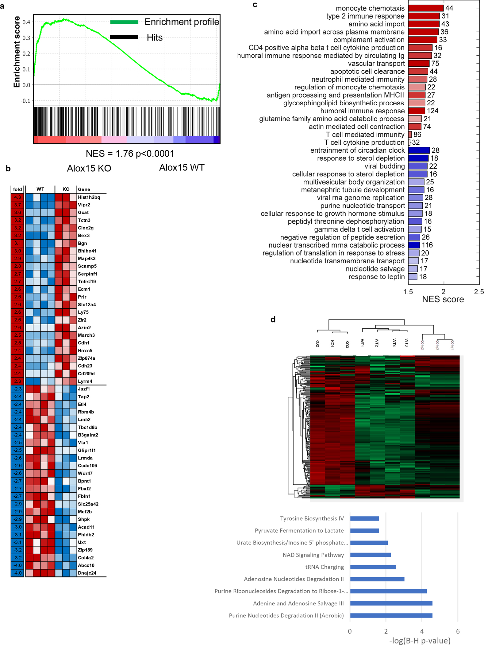
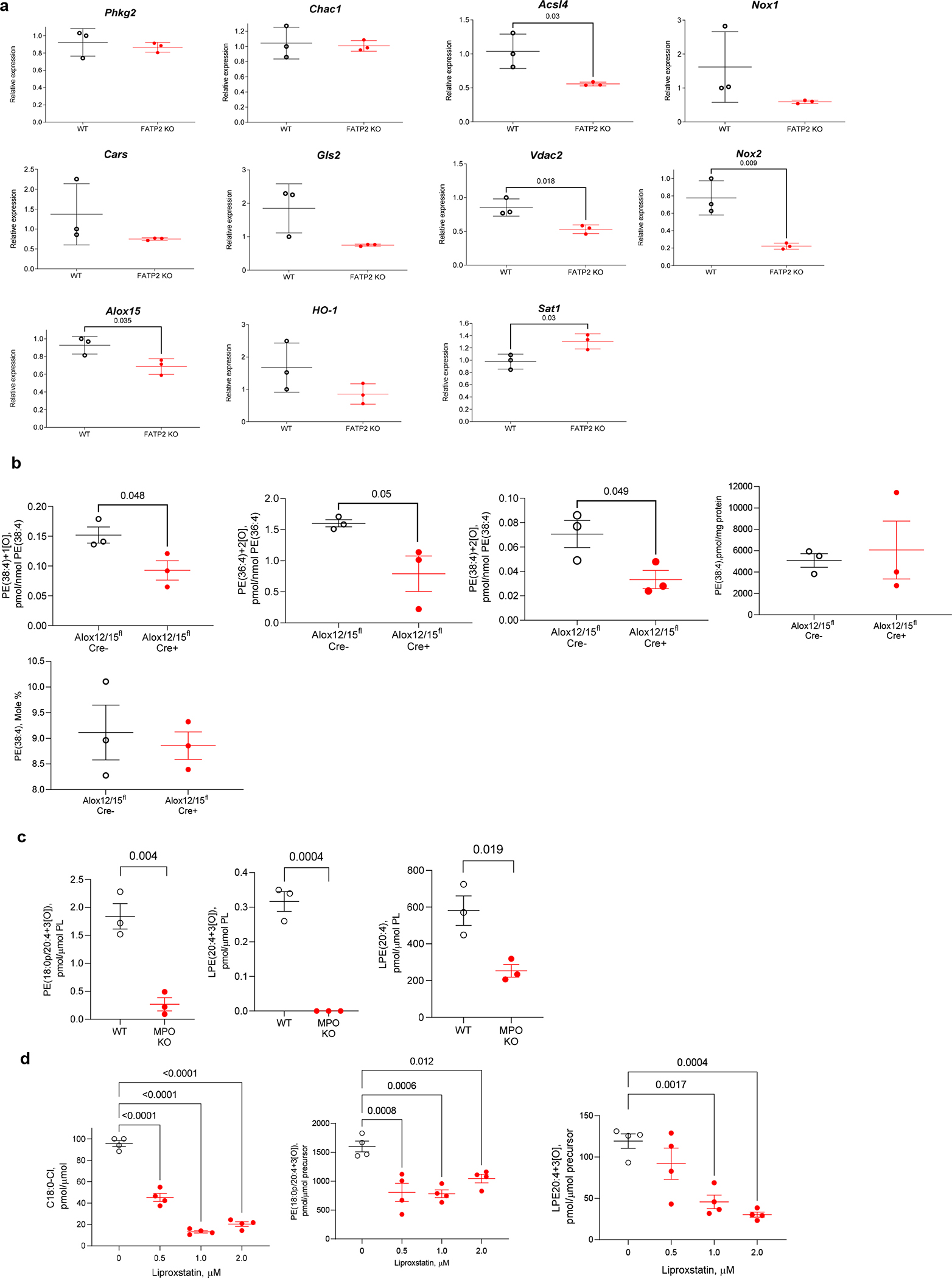
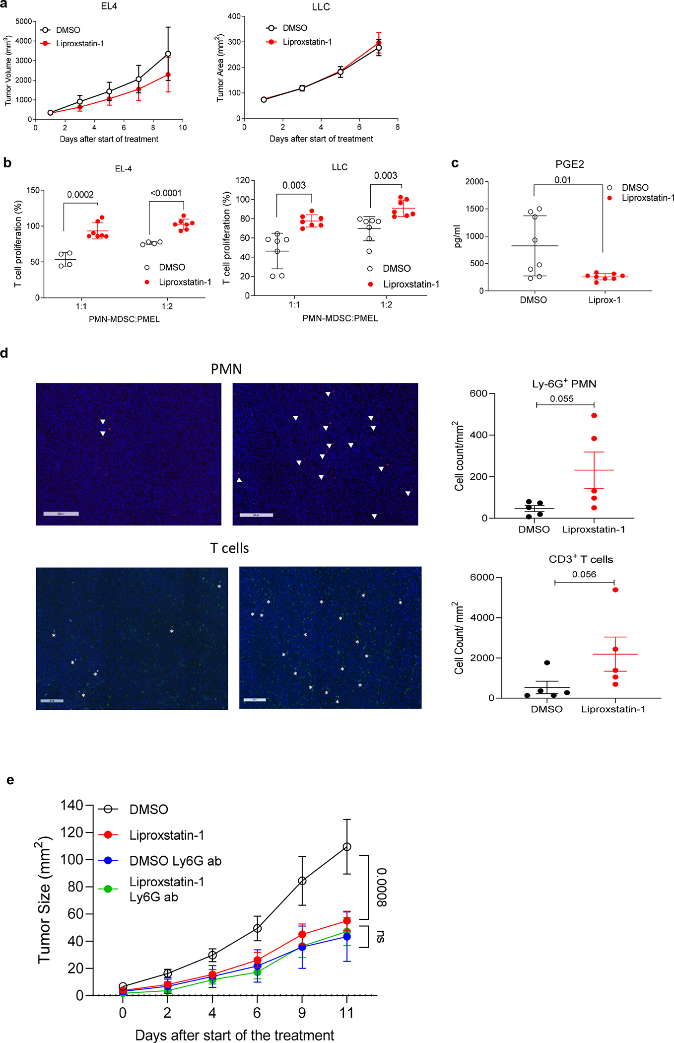
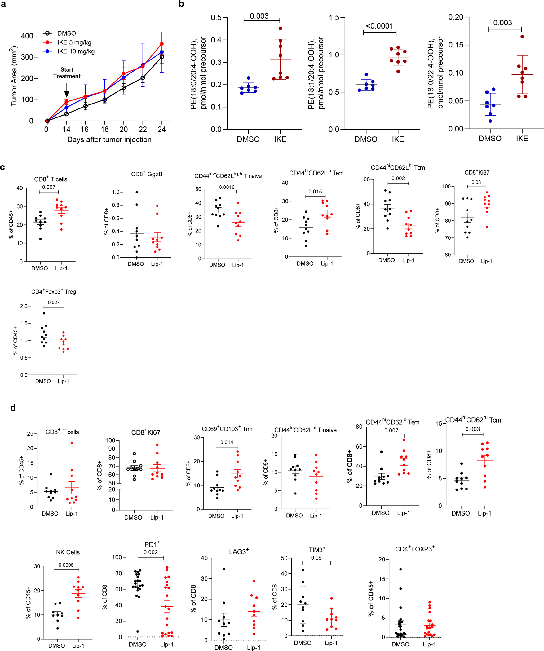
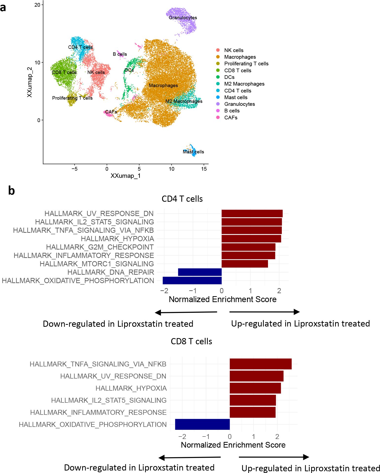
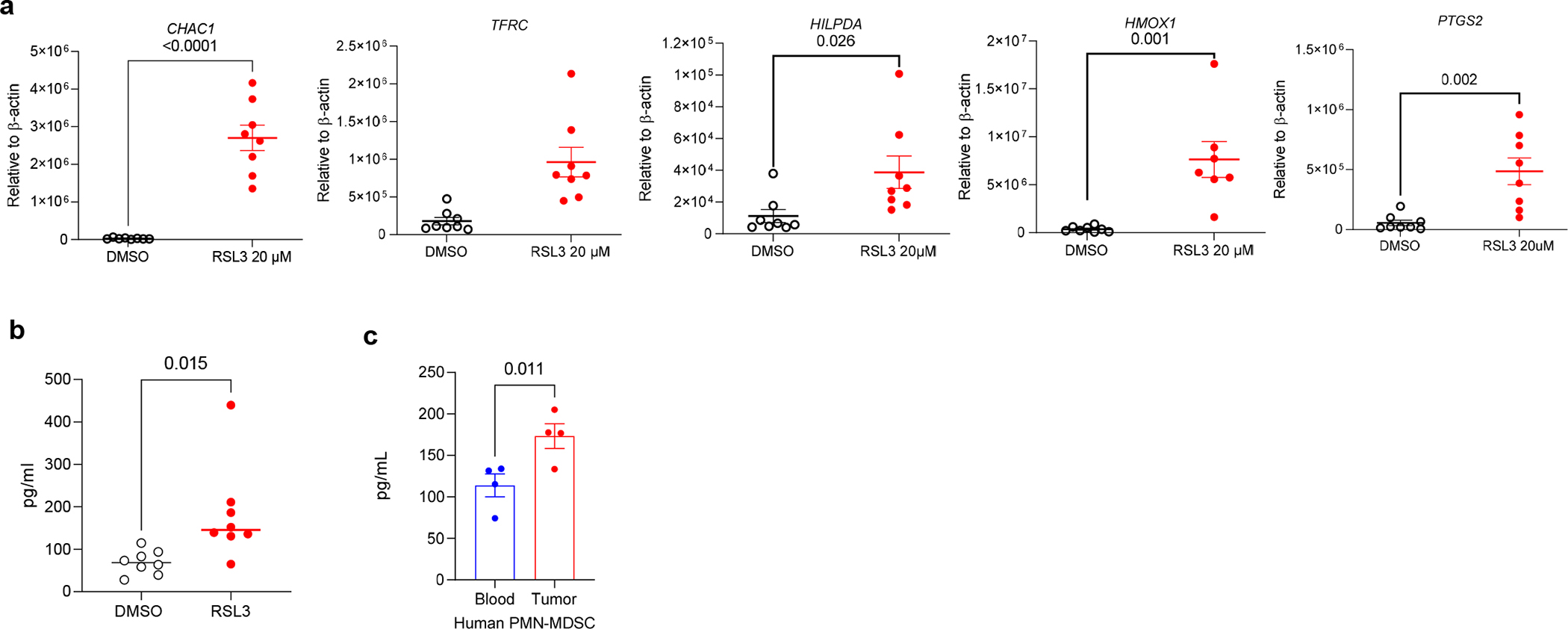
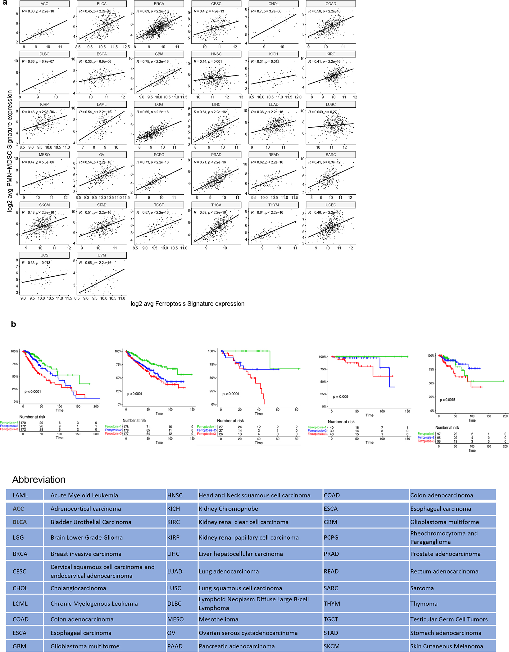
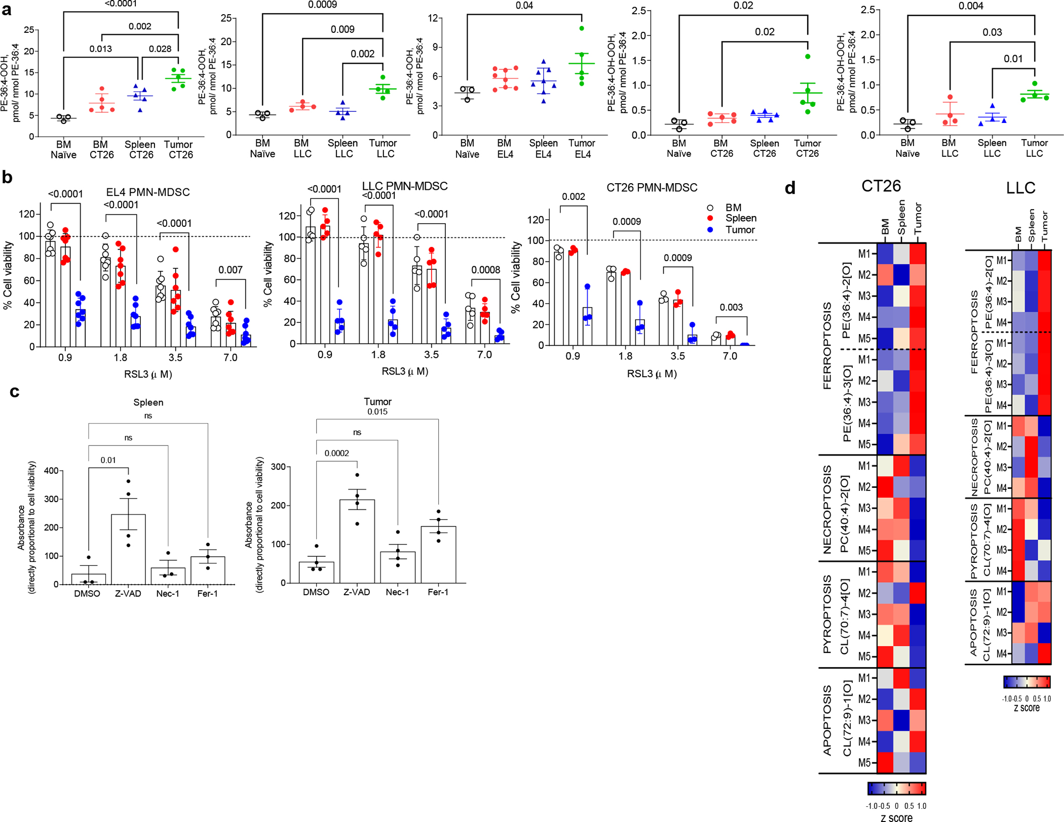
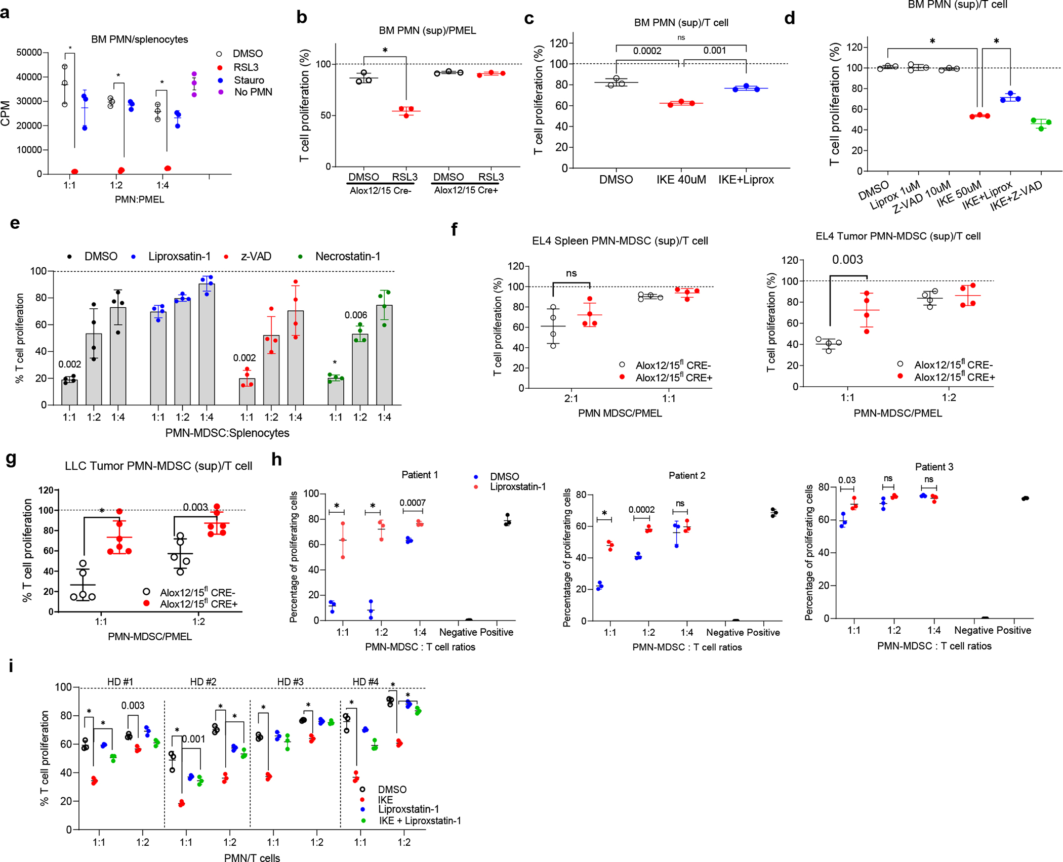
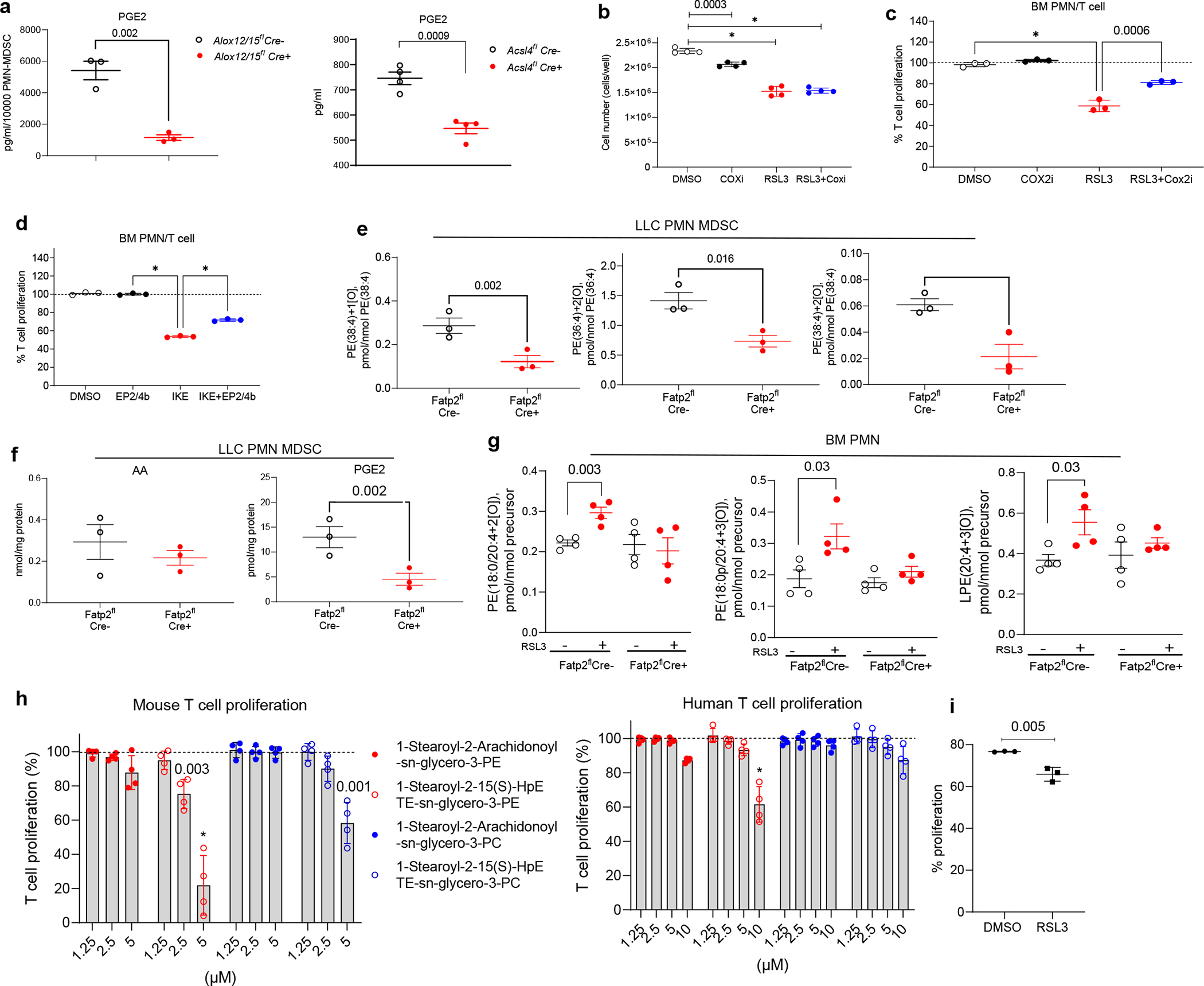
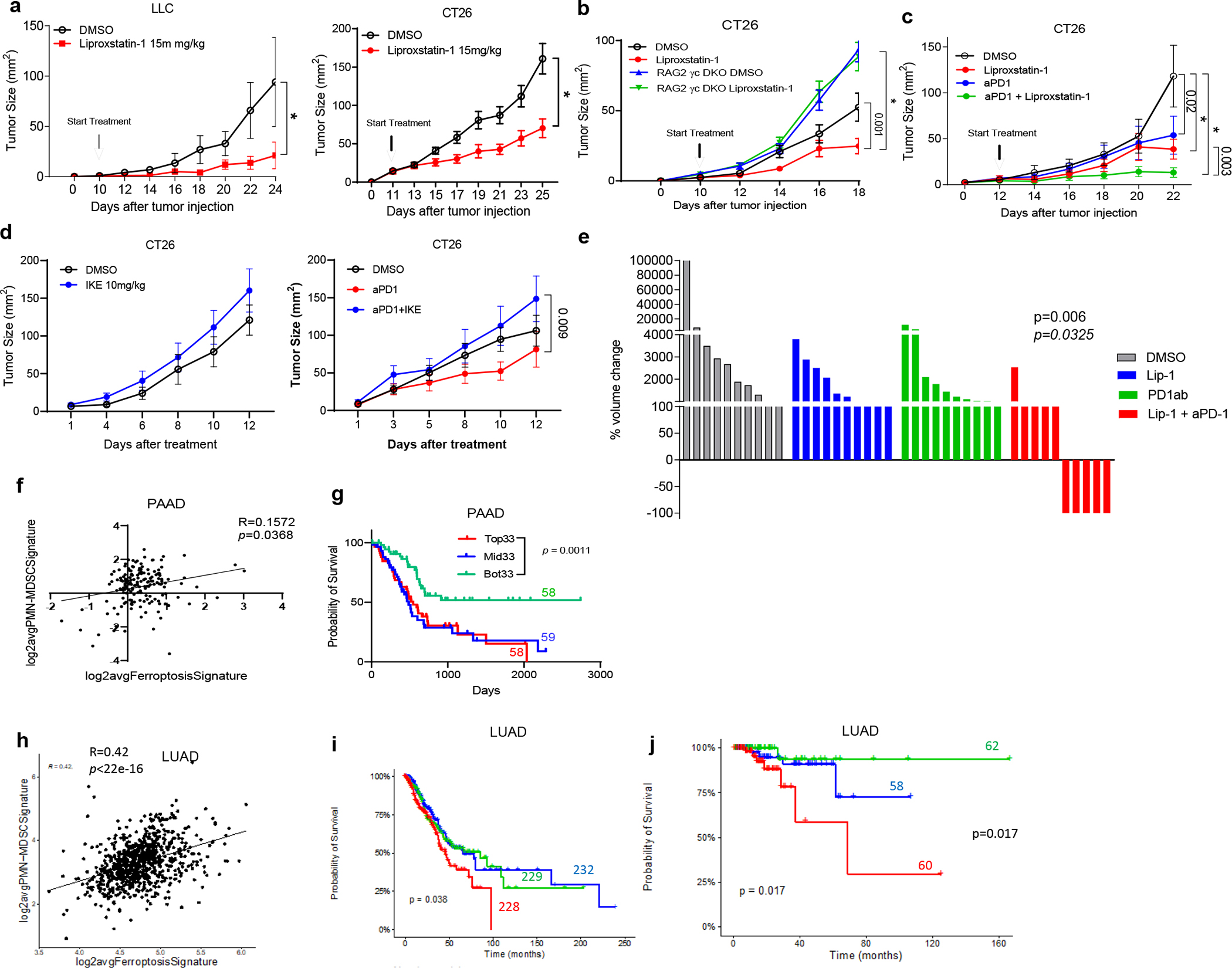
Ferroptosis is a non-apoptotic form of regulated cell death that is triggered by the discoordination of regulatory redox mechanisms culminating in massive peroxidation of polyunsaturated phospholipids. Ferroptosis inducers have shown considerable effectiveness in killing tumour cells in vitro, yet there has been no obvious success in experimental animal models, with the notable exception of immunodeficient mice1,2. This suggests that the effect of ferroptosis on immune cells remains poorly understood. Pathologically activated neutrophils (PMNs), termed myeloid-derived suppressor cells (PMN-MDSCs), are major negative regulators of anti-tumour immunity3-5. Here we found that PMN-MDSCs in the tumour microenvironment spontaneously die by ferroptosis. Although decreasing the presence of PMN-MDSCs, ferroptosis induces the release of oxygenated lipids and limits the activity of human and mouse T cells. In immunocompetent mice, genetic and pharmacological inhibition of ferroptosis abrogates suppressive activity of PMN-MDSCs, reduces tumour progression and synergizes with immune checkpoint blockade to suppress the tumour growth. By contrast, induction of ferroptosis in immunocompetent mice promotes tumour growth. Thus, ferroptosis is a unique and targetable immunosuppressive mechanism of PMN-MDSCs in the tumour microenvironment that can be pharmacologically modulated to limit tumour progression.


Conflict of interest statement

Competing interests:

AH, GK, KH, RAH, NC, and DIG are employee and stakeholders of AstraZeneca.

Figures

Extended data Fig. 1.
Extended data Fig. 1.. Gene expression profile of mouse and human PMN-MDSC.
a. RNA seq of PMN-MDSC from matched blood (TBB) and tumor tissue (TBT) of lung cancer patients and peripheral blood of healthy donors (HD). Color key represents the normalized Z score. b. RNA seq of sorted CD14high and CD14 PMN-MDSC from EL-4 TB mice. Color key represents the normalized Z score. c. Ferroptosis gene signature used in further studies. P values between tumor and blood PMN samples were calculated in two-sided unpaired Student’s t-test. d. Signature score of 8 ferroptosis genes in indicated populations of cells by single cell RNAseq. Data were captured from (22). Analysis was performed using BBrowser (https://bioturing.com/).T– tumor, B – blood.
Extended data Fig. 2.
Extended data Fig. 2.. Expression of ferroptosis related genes.
a. Expression of ferroptosis related genes in PMN from blood and tumor of cancer patients. qRT-PCR performed in CD11b+CD14CD66b+ PMN isolated from blood (PB, n=9) and tumors (Tumor, n=6) of cancer patients. Ct value of housekeeping gene was subtracted from Ct value of gene of interest for each samples (dCt). The mean of dCt for PB group was calculated. Each PB and tumor sample was normalized to this value. P-values are shown in the graph (Two-sided unpaired Mann-Whitney test), non-significant p values are not shown. b. Expression of ferroptosis related genes in PMN-MDSC from indicated tumor models. qRT-PCR performed in CD11b+L6ClowLy6G+ PMN-MDSC isolated from indicated tissues from implanted (EL4, CT26, and LLC) and autochthonous (KPC) tumor bearing mice. Mean ± SEM are shown. P values were calculated in one way ANOVA with Tukey’s test for correction for multiple comparisons.* p<0.0001. Other p values are shown on graphs. N=5 for EL4 model; n=3 for CT26 model; n= 4 for LLC model; n=6 for KPC model.
Extended data Figure 3.
Extended data Figure 3.. Ferroptosis in PMN-MDSCS and M-MDSC.
a. Content of oxygenated PE species PE(36:4-OOH) and PE(36:4-OH-OOH) in M-MDSC isolated from spleen and tumor of LLC TB mice. b. Viability of M-MDSC isolated from BM, spleen, and tumor of implanted EL4 and CT26 TB mice and treated with RSL3. Viability of the cells assessed by cell counts and expressed as a percent of untreated cell counts. c. CD71 expression in BM PMN after treatment with DMSO or various inducers of cell death for 18 hr: 20μM RSL3 for ferroptosis, 1μM Shikonin for necroptosis, and 0.25μM Staurosporine for apoptosis. Mean ± SD are shown. *P < 0.05, in one-way ANOVA. d. CD71 expression in tumor and spleen PMN-MDSC and M-MDSC. e. BM PMN were treated with DMSO or 20 μM RSL3 for 4 hr, washed extensively and proportion of live cells was counted by trypan blue exclusion method.. The washed cells were then incubated in fresh media at for additional 16 hr, and the proportion of viable cells was counted. Proportion of viable cells was calculated based on the number of the DMSO-treated cells. N=4. P values calculated in two-sided Student’s t-test (a, d middle panels), or One-way ANOVA with Tukey’s HSD post-hoc test (b, c, d) and are shown on graphs. * p< 0.0001. In all panels, mean ± SD are shown. f. Expression of Alox12/15 by qRT-PCR in PMN from Alox12/15flCre− and Alox12/15flCre+ mice. N=3. g. BM PMN-MDSC isolated from the LLC tumor bearing Alox12/15flCre− and Alox12/15flCre+ mice (n=6 per group) were treated with 0 – 10 μM RSL3 for 4 h, and then washed 3 times and followed by further 16 h incubation. Cell number was determined by trypan blue exclusion method. h. BM PMN-MDSC from the LLC TB mice were treated with IKE for 6 hr, and then washed 3 times and followed by further 16 h incubation; cell numbers determined by trypan blue exclusion (n=4). i. PMN cell counts by supernatant generated from BM PMN isolated from WT mice and treated with indicated inhibitors or IKE after pre-treatment with inhibitors (n=3/group). Representative experiment of four shown. j. BM PMN of EL-4 TB mice were cultured with 10 ng/mL GM-CSF and tumor explant supernatant for 24 hr in normoxia or hypoxia (0.3% O2) with or without 1 μM liproxstatin-1 (Lip-1) and then cocultured with cell trace-labeled PMEL splenocytes in indicated ratios, in the presence ofcognate peptide (n=3/group). T cell proliferation was evaluated as above. Two experiments with the same results were performed. One-way ANOVA with Tukey’s HSD post-test performed. P values are shown on graphs. k. Suppression of T cell proliferation by supernatants of M-MDSC isolated from EL4 tumors of Alox12/15flCre− and Alox12/15flCre+ mice (n=6/group). T cell proliferation was determined by flow cytometry as CellTrace dye dilution in CD3+CD8+ cells and expressed as a percent of CD8+ T cells stimulated in the absence of supernatants, l, m. Effect of ACSL4 deletion on suppressive activity of PMN-MDSC. l. Expression of Acsl4 in PMN from Acsl4flCre+ and Acsl4flCre− mice. N=4. m. Suppression of T cell proliferation by supernatants of PMN-MDSC isolated from tumors and spleens of Acsl4flCre− and Acsl4flCre+ EL4-TB mice. T cell proliferation was determined by flow cytometry as CellTrace dye dilution in CD3+CD8+ cells and expressed as a percent of CD8+ T cells stimulated in the absence of supernatant. N=4. Mean ± SD are shown. P values were calculated in unpaired two-sided Student’s t-test and shown on graphs. *p < 0.0001.
Extended data Figure 4.
Extended data Figure 4.. Gene changes induced by ferroptosis induction.
a. Expression of ferroptosis related genes measured by qRT-PCR in human BM PMN treated with RSL3. Mean ± SEM shown. P values on the graphs were calculated in unpaired two-sided Mann-Whitney test. N=8, except HMOX1 where n=7. b. RNA seq data of mice BM PMN treated for 4 hr with DMSO (C), 100nM staurosporine (S4) or 20uM RSL3 (R4). Color key represents the normalized Z score. Table shows functional attributes of the genes. c. Pathways/function changed in RSL3 treated BM PMN vs. control (DMSO) and staurosporine treated PMN; Z-score was calculated by Ingenuity Pathway Analysis where the z-score statistic is based on binomial disturution. http://pages.ingenuity.com/rs/ingenuity/images/0812%20downstream_effects_analysis_whitepaper.pdf. Only pathways that were different between groups with p<0.01 adjusted for multiple comparisons are shown. d. Expression of indicated genes in DMSO or 20uM RSL3 treated PMN measured by qRT-PCR. N=4. e. Concentration of released PGE2 levels by ELISA, in the supernatants of DMSO or 20uM RSL3 treated PMN after 8 hours. N=4. Mean ± SD shown in d,e. P values shown on graphs were calculated in unpaired two-sided Student’s t-test. *p < 0.0001
Extended data Figure 5.
Extended data Figure 5.. Effect of ALOX12/15 deletion on gene expression and metabolome of tumor PMN-MDSC.
a. GSEA enrichment analysis of ALOX12/15 KO vs control PMN-MDSC. NES, normalized enrichment score. P-value was calculated by GSEA based on permutation analysis. b, c. Transcription changes in Alox12/15 deficient PMN-MDSC. b. Top 25 up and downregulated genes. c. Gene set enrichment analysis using GSEA. NES score is shown. d. Metabolome of control and ALOX12/15 deficient tumor PMN-MDSC was evaluated by LC-MS. Ingenuity Pathway Analysis (IPA) was performed on differentially expressed metabolites - metabolites names were mapped to KEGG identifiers and given as input to IPA with default settings for metabolomics analysis. Pathways significantly up-regulated in ALOX12/15 deficient PMN-MDSC are shown. Changes considered significant if fold change >2 converted from Log2 ratio and Benjamini-Hochberg q-value corrected for multiple testing < 0.05.
Extended data Figure 6.
Extended data Figure 6.. Ferroptosis in genetically engeneered mice.
a. Ferroptosis related genes in PMN-MDSC from Fatp2flCre+ TB mice. qPCR of tumor PMN-MDSCs from Fatp2flCre− (WT) and Fatp2flCre+ (FATP2 KO) mice. N=3. Mean ± SD are shown. P values shown on graphs were calculated in unpaired two-sided Student’s t-test. b. Oxidized PE in ALOX12/15 deficient mice PMN-MDSC. Content of oxygenated (PE(38:4+1[O]) PE(38:4+2[O]) PE(36:4+2[O]) and non-oxygenated (PE(38:4) PE species in PMN MDSC isolated from LLC TB Alox12/15flCre− and Alox12/15flCre− mice tumors. N=3. Mean ± SD are shown. P values shown on graphs were calculated in unpaired two-sided Student’s t-test. c. Content of PE species containing oxygenated AA in PMN MDSC isolated from LLC TB WT and MPO KO mice. N=3. d. Liproxstatin-1 inhibits MPO (0.05U)/H2O2(50uM) induced formation of C18:0-Cl (left panel), PE-18:0p/20:4+3[O] (middle panel) and LPE(20:4+3[O]) (right panel) from PE(18:0p/20:4) in 20mM phosphate buffer containing 100 mM NaCl and 100uM DTPA, pH=7.4, after 30 min incubation at 37°C. The structure of PGE2 containing PE(18:0p/20+3[O]) was verified by three criteria (retention time (34 min), exact mass (m/z 798.5306 ± 3.3 ppm) and MS/MS fragments (m/z 798→351, 271, 189, 113). LPE(20:4+3[O]) was identified as PGE2 containing species by exact mass (m/z 548.2636 ± 3.4 ppm) and retention time (6 min). N=4. Mean ± SD are shown. P values shown on graphs were calculated in unpaired two-sided Student’s t-test (a) or one-way ANOVA with correction for multiple comparsons.
Extended data Figure 7.
Extended data Figure 7.. Ferroptosis induced PMN suppression is abrogated by inhibition of MPO.
a. BM PMNs were treated with 5 μM of MPO inhibitor (iMPO, 4-Aminobenzoic Acid Hydrazide, Cayman Chemical) for an hour followed by 6 h treatment with 40 μM IKE. The cells were washed 3 times then incubated with fresh media for 16 h. Supernatant was used for T cell proliferation assay. PMEL splenocytes were labeled with CellTrace dye and stimulated with cognate peptide in the presence of 50% of supernatant for 48 hr. Left panel - T cell proliferation measured by flow cytometry N=3. Four separate experiments are shown. Right panel – number of cells recovered after the incubation with IKE. N=4. Mean ± SD are shown. P values shown on graphs were calculated by one-way ANOVA with correction for multiple comparisons. * p<0.0001. b, c. GPX4 expression in tumor PMN-MDSC. b. GPX4 protein expression by Western blotting, in PMN-MDSC from BM, spleen and tumor of CT26 and EL4 TB mice. Results from individual mice are shown. c. GPX4 protein expression in BM PMN treated with increasing concentrations of supernatants obtained from tumor explants (TES) maintained under normoxic or hypoxic conditions. Three experients witt the same results were performed. For gel source data, see Supplementary Figure 1. d–f. Regulation of ferroptosis and suppressive activity in TAM by PMN-MDSC. TAM (CD11b+Ly6G Ly6Clow F4/80+) were sorted from EL4 tumor. d. Contents of oxygenated phospholipid ferroptotic cell death signals, PE(38:4+2[O]) and PE (38:5+2[O]), in TAM isolated from WT, ALOX12/15 KO and MPO KO mice. n=4, p values were calculated in unpaired two-sided Student’s t-test, * - p<0.05, ** - p<0.01, *** - p<0.001. e. PGE2 contents in TAM from WT and ALOX KO mice (left panel) and WT and MPO KO mice (right panel). n=4, Mean ± SD are shown. P values were calculated in unpaired two-sided Student’s t-test. f. TAM isolated from S100A8-cre x ALOX12/15fl mice and their littermate controls were co-cultured with cell trace-labeled OT-1 splenocytes in the presence of 0.025 ng/mL SIINFEKL peptide. T cell proliferation was analyzed by flow cytometry after 2 days incubation. Proliferation of T cells in the absence of TAM in each experiment was set as 100%. Mean ± SD are shown. P values were calculated in two-sided unpaired Student’s t-test. (n=4)
Extended data Figure 8.
Extended data Figure 8.. Effect of liproxstatin-1 treatment on PMN-MDSC in vivo.
a. Tumor growth curve of DMSO and 15mg/kg liproxstatin-1 treated EL-4 and LLC TB mice (n=4/group). b. Suppression of T cell proliferation by PMN-MDSC isolated from tumors of EL-4 or LLC TB mice treated with liproxstatin-1. T cell proliferation was determined by flow cytometry as CellTrace dye dilution in CD3+CD8+ cells and expressed as a percent of CD8+ T cells stimulated in the absence of PMN-MDSC. N=4 DMSO group, n=7 Liproxstatin-1 group. c. PGE2 amount in supernatants after 3 hr incubation of PMN-MDSC isolated from CT-26 TB mice treated with DMSO or liproxstatin-1 mice for 8 days. N=8 d. Numbers of PMN (red staining, some stained cells indicated by white aroowheads) and T cells (green staining, some stained cells indicated by white astersks) by IF in tumors from DMSO and Liproxstatin-1 treated TB mice. N=5. Mean ± SEM shown in a and d, and mean ± SD in b-c. Unpaired, two-sided Stident’s t test was used in b and c, and two-sided Mann-Whitney test in d. e. Effect of PMN depletion on antitumor activity of liproxstatin-1. CT26 tumor cells were implanted subcutaneously into Balb/c mice. Mice were treated with DMSO or 15 mg/kg Liproxstatin-1 with or without Ly6G depletion starting from day 10 post inoculation (DMSO and Lirpoxstatin-1 groups n=10; DMSO Ly6G and Lyproxstatin-1 Ly6G groups n=8). Ly6G depletion was initiated 7 days post inoculation with 200μg/mouse anti-Ly6G (1A8) and 50μg/mouse anti-rat kappa light chain (MAR 18.5) every 3 days. Mean ± SD are shown. P values were calculated in two-way ANOVA.
Extended data Figure 9.
Extended data Figure 9.. Effect of IKE treatment on tumor growth and phenotype of T cells in liproxstatin-1 treated mice.
a. EG7 tumor growth in C57BL/6 mice treated with IKE (n=5/group). b. LC/MS quantitative assessment of ferroptotic cell death signals (PE(18:0/20:4-OOH), PE(18:1–20:4-OOH) and PE(18:0/22:4-OOH) in tumors from IKE treated CT26 TB mice. N=7 in DMSO group; N=8 in IKE group. Mean ± SEM shown in a and mean ± SD in b. P were determined by unpaired two-sided Student’s t-test. c,d. Phenotype of T cells in liproxstatin-1 treated mice. Flow cytometric analysis of percentages of subsets of T cells in CT26 TB mice c. lymph nodes and d. tumors 8 days after treatment with DMSO or liproxstatin-1. (n=10–20/group). Mean ± SD are shown. P values calculated in two-sided unpaired Student’s t test. Tem – effector memory T cells, Tcm – central memory T cell, and Trm-tissue resident memory T cells.
Extended data Figure 10.
Extended data Figure 10.. Single cell RNAseq of tumor tissues.
CT26 TB mice were treated with liproxstatin-1 for 8 days. Tumors were collected and analyzed by scRNAseq. a. UMAP visualization of cell populations; b. Gene set enrichment analysis of differentially expressed genes at adjusted p value <0.01 for liproxstatin-1 treated vs DMSO treated CD4+ and CD8+ T cells. Enrichment p-values were calculated as described in fgsea R package and p-values were adjusted using Benjamini-Hochberg method.
Extended data Figure 11.
Extended data Figure 11.. Induction of ferroptosis in human PMN.
a. qRT-PCR measurements of ferroptosis related genes in DMSO or 20uM RSL3 treated PMN isolated from peripheral blood of healthy individuals. N=8. b. PGE2 levels measured by ELISA in supernatants of DMSO or 20uM RSL3 treated PMN isolated from peripheral blood of healthy individuals. N=8 c. PGE2 levels measured by ELISA in supernatants of PMN-MDSC isolated from peripheral blood and tumors of patients with NSCLC. N=4. Mean ± SD are shown. P values were determined by unpaired two-sided Student’s t-test.
Extended data Figure 12.
Extended data Figure 12.. Correlation of ferroptosis signature with PMN-MDSC signature and clinical outcome in cancer patients.
The data were obtained from TCGA database. a. Correlation between ferroptosis and PMN-MDSC signatures in patients with various tumors. Spearman’s correlation coefficient and associated probability (p value) was calculated. b. Association of ferroptosis signature and clinical outcome. Patient survival in top, mid, and bottom thirds of ferroptosis genes expression levels based on TCGA dataset. The number of patients in each group are shown on the graph. P values were calculated between high and low or intermediate third of gene expression using log-rank (Mantel-Cox) test.
Figure 1.
Figure 1.. Ferroptosis in tumor-associated PMN-MDSC.
a. PE containing oxidized AA in PMN from naïve, and PMN-MDSC from bone marrow (BM) and spleen of indicated TB mice (for PE-36:4-OOH: n=3 BM naïve; n=5 CT26; n=4 LLC; n=8 EL-4 BM and spleen; n=5 EL4 tumor; for PE-36:4-OH-OOH n= 3 BM naïve; n=5 CT26; n=4 LLC). In all figures, each data point represents a biological replicate. b. PMN-MDSC collected from BM, spleenss, or tumors were incubated with RSL3 for 16 hr. Cell viability was assessed with alamarBlue Cell Viability Assay and expressed as a percent of untreated cells viability shown as a dotted line. N=7 for EL4; n=5 for LLC; n=3 for CT26. c. PMN-MDSC from the spleens and tumors of EL4 TB mice were cultured with indicated inhibitors for 24 hr: Ferrostatin-1 (Fer-1, 1 μM), Necrostatin-1 (Nec-1, 1 μM), and Z-VAD-FMK (zVad, 10 μM). Cell viability was measured using alamarBlue Cell Viability Assay. N=3 spleen, n=4 tumor. Mean ± SEM (a,c) and mean ± SD (b) are shown. P values were calculated in one-way ANOVA with Tukey’s HSD post-test. ns – not significant. d. Changes in the contents of oxygenated phospholipids associated with ferroptosis - PE(36:4)-2[O], PE(36:4)-3[O], necroptosis - PC(40:4)-2[O], pyroptosis - CL(70:7)-4[O] and apoptosis - CL(72:9)-1[O] in PMN-MDSC isolated from BM, spleens and tumors of CT26 and LLC bearing mice. Heat maps auto-scaled to z scores and coded blue (low values) to red (high values) are shown. N=5 for CT26; n=4 for LLC. The data obtained from individual mice are shown.
Figure 2.
Figure 2.. Ferroptosis confers immunosuppressive activity to PMN.
a. Suppression of Pmel splenocyte proliferation stimulated with cognate peptide by BM PMN treated with DMSO, RSL3 (20 μM), or staurosporine (100 nM) (n=3). Incorporation of [3H]thymidine: CPM - counts per min. Representative experiment of two is shown. b. Suppression of T cell proliferation by supernatant (sup) generated from BM PMN isolated from ALox12/15flCre− and ALox12/15flCre+ mice and treated with DMSO or 20 μM RSL3 for 4 hr (n=3). Dotted line - T cell proliferation stimulated with cognate peptide in the absence of supernatant. Representative experiment of three is shown. c. Suppression of T cell proliferation by supernatants generated from BM PMN from WT mice treated as shown on the graph (n=3). Representative experiment of three shown. d. Suppression of T cell proliferation by supernatant generated from BM PMN from WT mice and treated with indicated inhibitors or IKE (n=3). Representative experiment of four is shown. e. PMN-MDSC from EL4 TB mice (n=4), pre-incubated with inhibitors, and cocultured with Pmel splenocytes in the presence of cognate peptide. Dotted line - T cell proliferation in the absence of PMN-MDSC. f. Suppression of T cell proliferation by supernatants of PMN-MDSC from Alox12/15flCre− or Alox12/15flCre+ EL4-TB mice (n=4). g. Suppression of T cell proliferation by supernatants of PMN-MDSC from LLC Alox12/15flCre− and Alox12/15flCre+ TB mice (n=6). Dotted line - T cells proliferation in the absence of supernatants. h. PMN-MDSC isolated from tumors of cancer patients were cocultured with T cells in triplicates in the plates with immobilized CD3/CD28 antibodies in the presence of DMSO or Liproxstatin-1 (1 μM). Negative – no CD3/CD28 antibody. Positive – CD3/CD28 antibodies but no PMN-MDSC. i. Suppression of T cell proliferation by blood PMN isolated from healthy donors and treated with indicated compounds (n=3). The dotted line indicates T cell proliferation in the absence of PMN. In all panels, Mean ± SD are shown. One-way ANOVA with Tukey’s HSD post-test (c, d, e, i) or unpaired two-sided Student’s t-test (a, b, f, g, h) were performed. * p<0.0001.
Figure 3.
Figure 3.. Mechanisms of ferroptosis induction in tumor PMN-MDSC.
a. PGE2 in the supernatants of LLC tumor PMN-MDSC of Alox12/15flCre− and Alox12/15flCre+ mice and EL4 tumors of Acsl4flCre− and Acsl4flCre+ mice, after 48 hr incubation (n=3). b. BM PMN were treated with 20 nM of ketorolac and rofecoxib (COXi) for 1hr and then with 20 μM RSL3 for 4 hr (n=4), washed and viable cells were counted after 16 hr. c. Suppression of T cell proliferation by supernatants generated from BM PMN and treated as indicated on the graph for 4 hr (n=3). Representative experiment of three is shown. d. Suppression of T cell proliferation by supernatants generated from DMSO or 40 μM IKE treated BM PMN (for 6 hr). Cognate peptide stimulated Pmel splenocytes were incubated with supernatants for 48 hr with or without EP2 and EP4 blockers (100 nM ONO-AE3–208 and 100 nM PF-04418948, EP2/4b). T cell proliferation in the absence of PMN (100%) indicated by the dotted line. n=3. e,f. Contents of PEox molecular species (e) and free AA and PGE2 (f) assessed by LC/MS in PMN-MDSC isolated from LLC tumors of Fatp2flCre− and Fatp2flCre+ mice (n=3). g. Contents of indicated oxygenated PE assessed by LC/MS in PMN obtained from BM of Fatp2flCre− and Fatp2flCre+ mice with and without 20 μM RSL3 treatment (n=4). h. Left – Proliferation of Pmel splenocytes in the presence of cognate peptide and indicated lipids (n=4). Right – proliferation of human T cells with CD3/28 Dynabeads in the presence of indicated lipids (n=4). Dotted lines indicate T cell proliferation in the absence of lipids. i. BM PMNs were treated with DMSO or 20 μM RSL3 for 6 hr, lipids were extracted, resuspended in ethanol at 10 μM, and used for the treatment of Pmel splenocytes stimulated with cognate peptide (n=3). Mean ± SD are shown. Unpaired two-sided Student’s t-test performed in panels a, e, f, g, i, and one-way ANOVA with Tukey’s HSD post-test performed on panels b, c, d, h. * p<0.0001.
Figure 4.
Figure 4.. Antitumor effect of ferroptosis inhibition.
a. LLC (n=6) and CT26 (n=10) tumor growth with DMSO and 15 mg/kg liproxstatin-1 treatment. b. CT26 tumor growth in WT and RAG DKO mice with DMSO and liproxstatin-1 treatment (n=10/group). c. CT26 tumor growth in mice with indicated treatments (n=7/group). d. CT26 tumor growth in mice treated with DMSO, 10 mg/kg IKE, or anti-PD1+IKE (n=9/group). e. Subcutaneous KPC tumors growth in mice treated with DMSO, 15mg/kg liproxstatin-1, 200 μg/dose anti-PD1, or anti-PD1+liproxstatin-1 (n=10/group). Treatment started 7 days post implantation. DMSO or liproxstatin-1 group received treatment daily while PD1 group received treatment every 3 days (6 doses total). P value of proportion of mice rejected tumors calculated in Fisher exact test. P value proportion of mice with more than 200% increase in tumor volume between combination and control groups are shown. f. Correlation between ferroptosis and PMN MDSC gene signatures in pancreatic adenocarcinoma (PAAD, TCGA datasets, n=177). Spearman’s correlation coefficient was calculated. g. PAAD patient survival in thirds of ferroptosis genes expression levels based on TCGA dataset. The number of patients in each group are shown on the graph. h. Correlation between ferroptosis and PMN MDSC gene signatures in lung adenocarcinomas (LUAD, Tempus dataset, n=689). Spearman’s correlation coefficient was calculated. i. Overall survival in lung cancer patients (Tempus dataset) in top (red), mid (blue), and bottom (green) thirds of the expression level of the ferroptosis signature genes. The number of patients in each group are shown on the graph. j. Overall survival in lung cancer patients treated with immunotherapy (Tempus dataset). In panels a-d, error bars indicate SEM. Two-way ANOVA with multiple comparisons performed on panels a-d. In g, i, and j, p values were calculated between high and low thirds of gene expression using log-rank (Mantel-Cox) test. * p<0.0001.

Comment in

References

    1. Dierge E et al. Peroxidation of n-3 and n-6 polyunsaturated fatty acids in the acidic tumor environment leads to ferroptosis-mediated anticancer effects. Cell metabolism 33, 1701–1715 e1705, doi:10.1016/j.cmet.2021.05.016 (2021). - DOI - PubMed
    1. Zou Y et al. Plasticity of ether lipids promotes ferroptosis susceptibility and evasion. Nature 585, 603–608, doi:10.1038/s41586-020-2732-8 (2020). - DOI - PMC - PubMed
    1. Condamine T, Mastio J & Gabrilovich DI Transcriptional regulation of myeloid-derived suppressor cells. J Leukoc Biol, doi:10.1189/jlb.4RI0515-204R (2015). - DOI - PMC - PubMed
    1. Wang W, Xia X, Mao L & Wang S The CCAAT/Enhancer-Binding Protein Family: Its Roles in MDSC Expansion and Function. Frontiers in immunology 10, 1804, doi:10.3389/fimmu.2019.01804 (2019). - DOI - PMC - PubMed
    1. Ostrand-Rosenberg S, Beury DW, Parker KH & Horn LA Survival of the fittest: how myeloid-derived suppressor cells survive in the inhospitable tumor microenvironment. Cancer Immunol Immunother 69, 215–221, doi:10.1007/s00262-019-02388-8 (2020). - DOI - PMC - PubMed

References in Methods

    1. Killion EA et al. A role for long-chain acyl-CoA synthetase-4 (ACSL4) in diet-induced phospholipid remodeling and obesity-associated adipocyte dysfunction. Mol Metab 9, 43–56, doi:10.1016/j.molmet.2018.01.012 (2018). - DOI - PMC - PubMed
    1. Langmead B & Salzberg SL Fast gapped-read alignment with Bowtie 2. Nat Methods 9, 357–359, doi:10.1038/nmeth.1923 (2012). - DOI - PMC - PubMed
    1. Li B & Dewey CN RSEM: accurate transcript quantification from RNA-Seq data with or without a reference genome. BMC bioinformatics 12, 323, doi:10.1186/1471-2105-12-323 (2011). - DOI - PMC - PubMed
    1. Love MI, Huber W & Anders S Moderated estimation of fold change and dispersion for RNA-seq data with DESeq2. Genome biology 15, 550, doi:10.1186/s13059-014-0550-8 (2014). - DOI - PMC - PubMed
    1. Wolf FA, Angerer P & Theis FJ SCANPY: large-scale single-cell gene expression data analysis. Genome biology 19, 15, doi:10.1186/s13059-017-1382-0 (2018). - DOI - PMC - PubMed

Publication types