. 2022 Dec;612(7938):132-140.
doi: 10.1038/s41586-022-05444-z. Epub 2022 Nov 16.

Direct activation of a bacterial innate immune system by a viral capsid protein

Affiliations

Direct activation of a bacterial innate immune system by a viral capsid protein

Tong Zhang et al. Nature. 2022 Dec.

Abstract

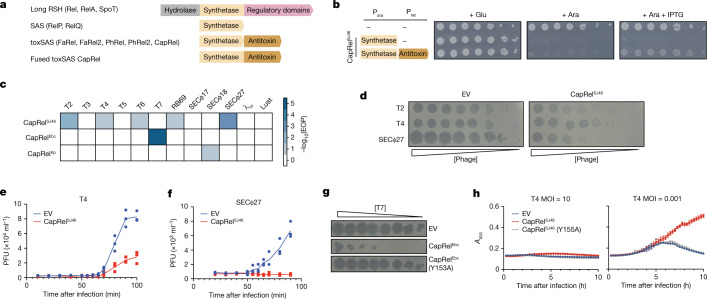
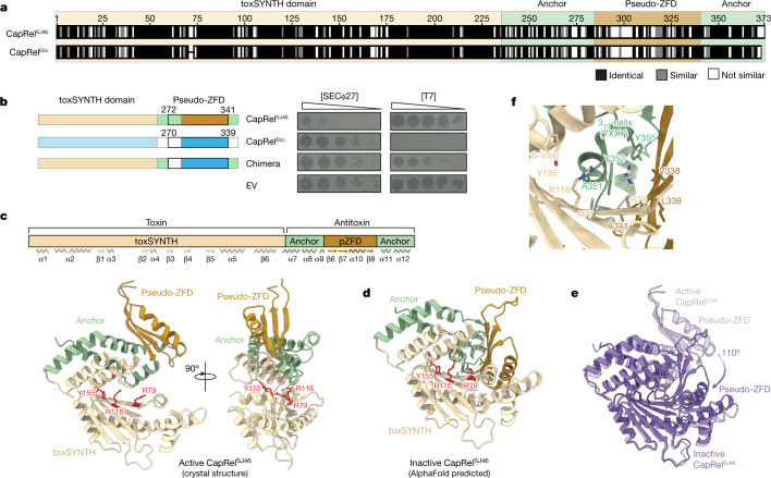
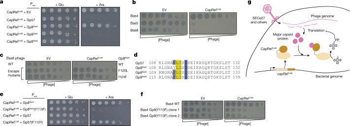
Bacteria have evolved diverse immunity mechanisms to protect themselves against the constant onslaught of bacteriophages1-3. Similar to how eukaryotic innate immune systems sense foreign invaders through pathogen-associated molecular patterns4 (PAMPs), many bacterial immune systems that respond to bacteriophage infection require phage-specific triggers to be activated. However, the identities of such triggers and the sensing mechanisms remain largely unknown. Here we identify and investigate the anti-phage function of CapRelSJ46, a fused toxin-antitoxin system that protects Escherichia coli against diverse phages. Using genetic, biochemical and structural analyses, we demonstrate that the C-terminal domain of CapRelSJ46 regulates the toxic N-terminal region, serving as both antitoxin and phage infection sensor. Following infection by certain phages, newly synthesized major capsid protein binds directly to the C-terminal domain of CapRelSJ46 to relieve autoinhibition, enabling the toxin domain to pyrophosphorylate tRNAs, which blocks translation to restrict viral infection. Collectively, our results reveal the molecular mechanism by which a bacterial immune system directly senses a conserved, essential component of phages, suggesting a PAMP-like sensing model for toxin-antitoxin-mediated innate immunity in bacteria. We provide evidence that CapRels and their phage-encoded triggers are engaged in a 'Red Queen conflict'5, revealing a new front in the intense coevolutionary battle between phages and bacteria. Given that capsid proteins of some eukaryotic viruses are known to stimulate innate immune signalling in mammalian hosts6-10, our results reveal a deeply conserved facet of immunity.


Conflict of interest statement

A.G.-P. is co-founder and stockholder of Santero Therapeutics. the other authors declare no competing interests.

Figures

Fig. 1
Fig. 1. Fused CapRel homologues are toxin–antitoxin systems that can provide E. coli with robust defence against phages.
a, Domain organization of long RelA-SpoT homologues (RSH), SAS, toxSAS and the fused subclass of toxSAS toxin–antitoxin systems including CapRelSJ46. b, Cell viability assessed by serial dilutions for strains expressing the N-terminal toxin domain of CapRelSJ46 alone or with the C-terminal antitoxin domain. Ara, arabinose; glu, glucose; Para, arabinose-inducible, glucose-repressible promoter; Plac, IPTG-inducible promoter. c, EOP data for the indicated phages when infecting cells producing CapRelSJ46, CapRelEbc or CapRelKp. d, Serial, tenfold dilutions of the indicated phages spotted on lawns of cells harbouring plasmid expressing CapRelSJ46 or an empty vector (EV). Relative phage concentration is indicated by the height of the wedge. e,f, One-step growth curves measuring plaque-forming units (PFU) during the first round of infection by T4 (e) or SECΦ27 (f) in cells harbouring plasmid expressing CapRelSJ46 or an empty vector. g, Serial dilutions of T7 phage spotted on lawns of cells harbouring plasmids expressing CapRelEbc, CapRelEbc(Y153A) or an empty vector. h, Growth of cells producing CapRelSJ46 or CapRelSJ46(Y155A) or harbouring an empty vector, following infection with T4 at a MOI of 10 or 0.001. Data are mean ± s.d. of eight plate replicates and representative of three independent experiments. Source data
Fig. 2
Fig. 2. The pseudo-ZFD of CapRel confers phage specificity.
a, Sequence alignment of CapRelSJ46 and CapRelEbc, with the more variable pseudo-ZFD labelled. b, Serial dilutions of the indicated phages spotted on lawns of cells harbouring the indicated CapRel constructs. Left, schematic of the CapRel constructs. c, Cartoon representation of the crystal structure of CapRelSJ46 with active site G-loop Y155 and the ATP-coordination residues R79 and R116 highlighted in red. Structural elements (toxSYNTH, pseudo-ZFD and the anchors) are coloured as in a. Wavy lines and arrows indicate alpha helices and beta strands, respectively. d, The closed conformation of CapRelSJ46 predicted by AlphaFold and coloured as in c. e, Superposition of the active (open, light purple) and inactive (closed, dark purple) states of CapRelSJ46 as observed in the crystal structure and predicted by AlphaFold. f, Details of the autoinhibited active site of CapRelSJ46 in the closed state. In this conformation, the YXXY neutralization motif blocks the adenine coordination site, preventing catalysis. Source data
Fig. 3
Fig. 3. CapRelSJ46 is activated by the major capsid protein of SECΦ27 to pyrophosphorylate tRNAs and block translation.
a, Immunoblot of His6–CapRelSJ46 following infection with SECΦ27 compared with an uninfected control. Representative of two biological replicates. b, Schematic of the experimental evolution approach to identify SECΦ27 escape mutants that can infect CapRelSJ46-containing cells. White wells indicate clearing by phages and brown wells indicate bacterial growth. c, Serial dilutions of five independently evolved populations of SECΦ27 and a control population spotted on cells harbouring an empty vector or a CapRelSJ46 expression vector. d, Summary of identified escape mutants, all of which map to a hypothetical protein encoded by gene 57 of SECΦ27. The fraction of each escape mutant is indicated. e, AlphaFold-predicted structure of Gp57 compared with the major capsid protein Gp5 from phage HK97. f, Mass spectrometry analysis of SECΦ27 phage lysates (wild type or a mutant producing Gp57(L114P)). Spectrum count normalized to molecular mass is shown for individual phage proteins. g, Serial dilutions of cells expressing CapRelSJ46 and the indicated variant of Gp57 from an arabinose-inducible promoter on media containing glucose or arabinose. h, Cells expressing CapRelSJ46 and Gp57 (wild type (WT) or L114P) from an arabinose-inducible promoter or harbouring empty vector were pulse-labelled with 35S-cysteine and 35S-methionine after addition of arabinose. i, As in h, but for cells harbouring a CapRelSJ46 expression vector or empty vector and after infection with SECΦ27 (top) or the escape mutant expressing Gp57(L114P) (bottom). Asterisks indicate P = 0.022 (45 min) or P = 0.004 (60 min) (unpaired two-tailed t-test). j, In vitro transcription–translation assays using DHFR production from a DNA template as the readout of expression activity. Purified CapRelSJ46 was added along with a template for producing Gp57. Representative of two biological replicates. k, Autoradiography of reactions in which purified CapRelSJ46 was incubated with [γ-32P]ATP, bulk E. coli tRNAs and Gp57. SYBR Gold staining of bulk tRNAs serves as a loading control. Representative of two biological replicates. Source data
Fig. 4
Fig. 4. The SECΦ27 major capsid protein Gp57 binds directly to the pseudo-ZFD of CapRelSJ46.
a, CapRelSJ46–Flag was immunoprecipitated from cells expressing CapRelSJ46–Flag and Gp57–HA (wild-type or indicated mutant) and probed for the presence of the indicated Gp57 variant via the HA tag. Lysates used as input for the immunoprecipitation (IP) were probed as controls for expression levels. Images are representatives of three biological replicates. b, Binding of CapRelSJ46 to Gp57 monitored by ITC. Kd, binding affinity; N, stoichiometry. c, Structural model of the CapRelSJ46–Gp57 complex predicted by AlphaFold. According to the model, the P-domain of Gp57 (pink) recognizes the pseudo-ZFD (orange) and anchor regions (green) of CapRelSJ46. This interaction prevents the recoil of pseudo-ZFD to the active site and activates the enzyme. d, Differential HDX (ΔHDX) between CapRelSJ46 and CapRelSJ46–Gp57 displayed as a difference heat map. Red indicates increased deuteration of CapRelSJ46 in the presence of Gp57; blue indicates lower deuteration. Grey bars indicate peptides identified in mass spectrometry analysis. e, Topological representation of CapRelSJ46, coloured according to the ΔHDX. The active site of the enzyme is marked by a black dashed outline and the catalytic toxSYNTH domain and the phage-recognition pseudo-ZFD are shadowed in light yellow and light orange, respectively. f, Serial dilutions on media containing glucose or arabinose of cells expressing the indicated mutant of CapRelSJ46 from its native promoter and the wild-type Gp57 from an arabinose-inducible promoter. g, Serial dilutions of SECΦ27 phage spotted on cells expressing the indicated mutant of CapRelSJ46 or an empty vector. h, As in a, but with the indicated mutants of CapRelSJ46–Flag. Images shown are representatives of three independent biological replicates. Source data
Fig. 5
Fig. 5. Evidence for the coevolution of CapRelSJ46 and the major capsid protein of SECΦ27 and related phages.
a, Serial dilutions on media containing glucose or arabinose of cells expressing CapRelSJ46 from its native promoter and the major capsid protein homologue from the phage indicated from an arabinose-inducible promoter. b, Serial dilutions of the phages indicated spotted on lawns of cells expressing CapRelSJ46 or an empty vector. c, Serial dilutions of wild-type Bas8 phage or the escape mutants bearing the major capsid mutations indicated spotted on lawns of cells harbouring CapRelSJ46 or an empty vector. d, Alignment of the region of the major capsid protein in SECΦ27, Bas5 and Bas8 that triggers CapRelSJ46, along with Bas4, which has a tyrosine at position 113 instead of phenylalanine. e, Serial dilutions on media containing glucose or arabinose of cells expressing CapRelSJ46 from its native promoter and the Bas4 or SECΦ27 major capsid protein variant indicated from an arabinose-inducible promoter. f, Serial dilutions of wild-type Bas4 or two mutant clones containing Y113F in the major capsid protein Gp8 spotted on lawns of cells expressing CapRelSJ46 or an empty vector. g, Model for the direct activation of CapRelSJ46 by the major capsid protein of SECΦ27 and related phages. After genome injection, the production of the major capsid protein triggers relief of autoinhibition by the C-terminal antitoxin of CapRelSJ46, leading to pyrophosphorylation (PPi) of tRNAs by activated CapRelSJ46, which inhibits translation and restricts viral infection. Source data
Extended Data Fig. 1
Extended Data Fig. 1. CapRel is broadly distributed in different bacteria, and is usually a fused TA system.
FlaGs output for CapRel and PhRel representatives (black arrows). Those proteins studied in this paper are in bold and underlined. Most CapRel systems are fused, with the exception of Actinobacteria where the TA pair is usually unfused. The unfused state is the most common form of the system in the closely related PhRel subfamily of toxSASs, and fusion and/or fission events appear to have occurred independently multiple times. Conserved flanking open reading frames are colored and numbered by homologous clusters: 1 (red): antitoxin homologous to the CapRel C-terminus, and 2 (purple): Panacea/SocA (DUF4065) domain-containing proteins. For other coloured open reading frames see the legend on the lower left. Arrows with no fill colour and with blue and green outlines are, respectively, pseudogenes and non-coding RNA genes. Red numbers on branches show Maximum Likelihood bootstrap support on a scale of 0-1, where 1 is 100% support.
Extended Data Fig. 2
Extended Data Fig. 2. Analysis of CapRel homologs.
(a) Sequence alignment comparing fused CapRel systems with related, unfused systems. Alignment of toxSAS PhRel and ATphRel from the Mycobacterium phage Phrann, non-fused CapRel and ATcapRel from Mycobacterium terramassilience, and the three fused systems CapRelSJ46, CapRelEbc and CapRelKp. The N-terminal region of fused CapRel systems is a toxSAS toxin domain, while the C-terminal region is homologous to the antitoxins of the PhRel and unfused CapRel TA systems. Substituted sites that rendered CapRelSJ46 toxic  (see Extended Data Fig. 3j) are indicated with black arrowheads. The inset diagram summarises the homologous regions of the bicistronic toxin-antitoxin and fused toxin-antitoxin systems considered here. (b) Genome maps of native locations of CapRelSJ46, CapRelEbc and CapRelKp (+/− 10 kb) with predicted flanking prophage and phage genes. (c) Summary of 3 independent replicates of cell viability assay in Fig. 1b. Asterisks indicate p = 0.007 (unpaired two-tailed t-test). (d) Serial dilutions of the phages indicated spotted on lawns of cells producing CapRelSJ46, CapRelEbc, or CapRelKp or harboring an empty vector (EV). (e) Summary of 3 independent replicates of phage spotting assay in Fig. 1d. Asterisks indicate p = 10−10 (T2, SECΦ27), 10−6 (T4) (unpaired two-tailed t-test). (f) Serial dilutions of the phages indicated spotted on lawns of cells containing genomic CapRelSJ46, CapRelSJ46 (Y155A) or His6-CapRelSJ46. (g) Summary of 3 independent replicates of phage spotting assay in Fig. 1g. Asterisk indicates p = 10−22 (unpaired two-tailed t-test).
Extended Data Fig. 3
Extended Data Fig. 3. Structural analysis of CapRelSJ46.
(a) Alignment of CapRelSJ46 and diverse fused CapRel homologs, with labels indicating the pseudo-ZFD and location of substitutions that render CapRelSJ46 constitutively active or unable to be activated by Gp57, the SECΦ27 major capsid protein. (b) Summary of 3 independent replicates of phage spotting assay in Fig. 2b. (c) Immunoblot of untagged CapRel chimera or FLAG-tagged CapRelSJ46, CapRelEbc or CapRel chimera. GyrA is included as a loading control. Image shown is a representative of 2 biological replicates. (d) Topology of CapRelSJ46. The toxSYNTH domain is colored in light yellow, the pseudo-ZFD in dark gold and the regions that anchor pseudo-ZFD to toxSYNTH are in green. The adenine coordinating R79 and R116 are shown as red dots and the G-loop is colored in red. (e) Superposition of the toxSYNTH domain of CapRelSJ46 (colored in light yellow) onto RelQ (PDB ID: 5DEC, colored in light orange) from Bacillus subtilis. (f) Superposition of the pseudo-ZFD of CapRelSJ46 (colored in dark gold) onto the ZFD transcription factor of Acidianus hospitalis (2LVH, colored in purple). (g) Analysis of asymmetric unit and symmetry-related partners of CapRelSJ46 crystal packing. Black arrow indicates steric clash that would arise if CapRelSJ46 were in a closed conformation. (h) Superposition of the crystal structure of CapRelSJ46 (colored in light yellow) onto the structure of the open state predicted by AlphaFold (colored in green). (i) Structures of the open (left; from crystal structure) or closed (right; AlphaFold prediction) conformations of CapRelSJ46 color coded by the conservation score of each amino acid calculated by ConSurf. Substitutions that render CapRelSJ46 constitutively active or unable to be activated by Gp57 are labeled as spheres. (j) Serial dilutions of cells expressing the indicated variant of CapRelSJ46 from an arabinose-inducible promoter on media containing glucose (left) or arabinose (right). (k) Summary of 3 independent replicates of cell viability assay in Extended Data Fig. 3j under arabinose induction. Asterisks indicate p = 0.007 (unpaired two-tailed t-test).
Extended Data Fig. 4
Extended Data Fig. 4. Gp57 from SECΦ27 triggers CapRelSJ46 to inhibit translation, not transcription.
(a) Serial dilutions of phage SECΦ27 spotted on lawns of cells producing CapRelSJ46, His6-CapRelSJ46, or CapRelSJ46-FLAG, or harboring an empty vector (EV). (b) Loading control for Fig. 3a shown as total protein levels stained by Coomassie stain. Image shown is a representative of 2 biological replicates. (c) Immunoblot of His6-CapRelSJ46 expressed from the bacterial genome or a low-copy number plasmid following infection with SECΦ27 (MOI = 100) compared to an uninfected control. Total protein levels stained by Coomassie stain is included as a loading control. Images shown are representatives of 2 biological replicates. (d) Summary of 3 independent replicates of phage spotting assay in Fig. 3c. Asterisk indicates p = 10−10 (unpaired two-tailed t-test). (e) Serial dilutions on media containing glucose (left) or arabinose (right) of cells expressing CapRelSJ46 from its native promoter in the bacterial genome and expressing Gp57 from an arabinose-inducible promoter or an empty vector. (f) Summary of 3 independent replicates of cell viability assay in Fig. 3g under arabinose induction. Asterisk indicates p = 10−30 (unpaired two-tailed t-test). (g) Serial dilutions on media containing glucose (left) or arabinose (right) of cells expressing CapRelSJ46(Y155A) from its native promoter or an empty vector and expressing the indicated variant of Gp57 from an arabinose-inducible promoter. (h) Cells harboring CapRelSJ46 and producing the wild-type or L114P variant of Gp57 (expressed from an arabinose-inducible promoter) or harboring an empty vector were pulse-labeled with 3H-uridine at the times indicated post-addition of arabinose. (i) Cells producing the CapRelSJ46 N-terminal toxin domain (expressed from an arabinose-inducible promoter) or harboring an empty vector were pulse-labeled with 35S-Cys/Met (left) or 3H-uridine (right) at the times indicated post-addition of arabinose. (j) Same as (h) but for cells carrying CapRelSJ46 or an empty vector and at times post-infection with SECΦ27 at MOI = 100. (k) Adsorption of T4 and SECΦ27 with cells containing CapRelSJ46 or an empty vector. Data reported are the mean +/− SD from 3 biological replicates. (l) Immunoblot of Gp57-HA expressed from its native promoter in the bacterial genome following infection with SECΦ27. Total protein level stained by Coomassie stain is included as a loading control (left). Quantification of the relative intensities of both Gp57-HA bands normalized to the loading control from 3 independent replicates (right). Asterisks indicate p = 0.004 (30 min), 0.0003 (40 min), 0.0002 (50 min) (unpaired two-tailed t-test). Source data
Extended Data Fig. 5
Extended Data Fig. 5. Characterization of the CapRelSJ46-Gp57 interaction.
(a) Immunoprecipitation of CapRelSJ46-FLAG from cells infected with wild-type SECΦ27 or mutant phage that produces Gp57(L114P), followed by mass spectrometry. Spectrum counts (SC) of Gp57 that had co-precipitated with CapRelSJ46 were normalized to the spectrum counts of CapRelSJ46. Data reported are 2 biological replicates. (b) Same as in (a) but showing spectrum counts of CapRelSJ46 and Gp57 in two independent replicates. (c) Serial dilutions on media containing glucose (left) or arabinose (right) of cells producing CapRelSJ46 or CapRelSJ46-FLAG, each expressed from its native promoter, and the indicated variant of untagged or HA-tagged version of Gp57, expressed from an arabinose-inducible promoter. (d) SEC-MALS analysis of CapRelSJ46-Gp57 complex, revealing a molecular weight of 74 kDa. The monomers of CapRelSJ46 and Gp57 are predicted to be 42 and 36 kDa, respectively. (e) Binding of CapRelSJ46 to Gp57 (L114P I115F) (left) or CapRelSJ46(L280P) to Gp57 (right) monitored by isothermal titration calorimetry (ITC). (f) Topology and cartoon representation of SECΦ27 Gp57. The P-domain is colored in pink and the A-domain in violet. (g) Heat maps representing the HDX of CapRelSJ46 (top) and CapRelSJ46-Gp57 complex (center) and the ΔHDX (bottom). Regions involved in strong uptake such as residues 115-145 and 225-235 (which includes the active site β-strand β2 and the G-loop) are shaded in red and regions involved in strong protection 240-268 and 288-366 (which include both anchors and the pseudo-ZFD) are shaded in blue. (h) Heat map representing the HDX of Gp57 in the complex with CapRelSJ46. Shaded regions highlight areas of variable HDX signal that indicate these regions are involved in the CapRelSJ46-Gp57 interface. (i) Summary of 3 independent replicates of cell viability assay in Fig. 4f under arabinose induction. (j) Summary of 3 independent replicates of phage spotting assay in Fig. 4g. (k) Serial dilutions of T2 and T4 phage spotted on cells producing the indicated mutant of CapRelSJ46 or harboring an empty vector.
Extended Data Fig. 6
Extended Data Fig. 6. The major capsid proteins from multiple, related phages activate CapRelSJ46.
(a) Multiple sequence alignment of the major capsid proteins from phages SECΦ27, Bas4, Bas5 and Bas8. (b) Summary of 3 independent replicates of cell viability assay in Fig. 5a under arabinose induction. Asterisks indicate p = 10−34 (unpaired two-tailed t-test). (c) Summary of 3 independent replicates of phage spotting assay in Fig. 5b. (d) Summary of 3 independent replicates of phage spotting assay in Fig. 5c. (e) Serial dilutions on media containing glucose (left) or arabinose (right) of cells expressing CapRelSJ46 from its native promoter or harboring an empty vector and producing the indicated variant of the Bas8 major capsid protein (Gp8Bas8) from an arabinose-inducible promoter. (f) Summary of 3 independent replicates of cell viability assay in Extended Data Fig. 6e under arabinose induction. (g) Serial dilutions on media containing glucose (left) or arabinose (right) of cells containing an empty vector and producing the indicated variant of the Bas8 major capsid protein from an arabinose-inducible promoter. (h) Serial dilutions of the phages indicated spotted on lawns of cells harboring CapRelEcHT or an empty vector. (i) Summary of 3 independent replicates of cell viability assay in Fig. 5e under arabinose induction. (j) Summary of 3 independent replicates of phage spotting assay in Fig. 5f. Asterisks indicate p = 10−6 (unpaired two-tailed t-test).

References

    1. Bernheim A, Sorek R. The pan-immune system of bacteria: antiviral defence as a community resource. Nat. Rev. Microbiol. 2020;18:113–119. doi: 10.1038/s41579-019-0278-2. - DOI - PubMed
    1. Hampton HG, Watson BNJ, Fineran PC. The arms race between bacteria and their phage foes. Nature. 2020;577:327–336. doi: 10.1038/s41586-019-1894-8. - DOI - PubMed
    1. Rostøl JT, Marraffini L. (Ph)ighting phages: how bacteria resist their parasites. Cell Host Microbe. 2019;25:184–194. doi: 10.1016/j.chom.2019.01.009. - DOI - PMC - PubMed
    1. Fitzgerald KA, Kagan JC. Toll-like receptors and the control of immunity. Cell. 2020;180:1044–1066. doi: 10.1016/j.cell.2020.02.041. - DOI - PMC - PubMed
    1. Daugherty MD, Malik HS. Rules of engagement: molecular insights from host–virus arms races. Annu. Rev. Genet. 2012;46:677–700. doi: 10.1146/annurev-genet-110711-155522. - DOI - PubMed

Publication types

MeSH terms

Substances