

The effects of the recurrent social isolation stress on fear extinction and dopamine D2 receptors in the amygdala and the hippocampus
- PMID: 36385611
- PMCID: PMC9889440
- DOI: 10.1007/s43440-022-00430-8
The effects of the recurrent social isolation stress on fear extinction and dopamine D2 receptors in the amygdala and the hippocampus
Abstract
Background: The present study assessed the influence of recurrent social isolation stress on the aversive memory extinction and dopamine D2 receptors (D2R) expression in the amygdala and the hippocampus subnuclei. We also analyzed the expression of epigenetic factors potentially associated with fear extinction: miRNA-128 and miRNA-142 in the amygdala.
Methods: Male adult fear-conditioned rats had three episodes of 48 h social isolation stress before each fear extinction session in weeks intervals. Ninety minutes after the last extinction session, the D2R expression in the nuclei of the amygdala and the hippocampus (immunocytochemical technique), and mRNA levels for D2R in the amygdala were assessed (PCR). Moreover, we evaluated the levels of miRNA-128 and miRNA-142 in the amygdala.
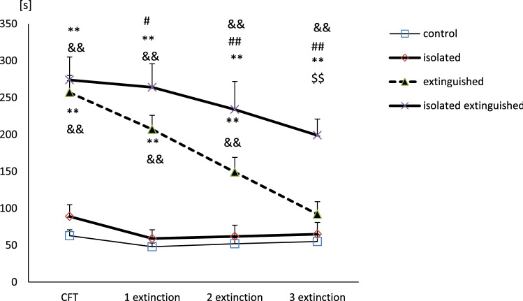
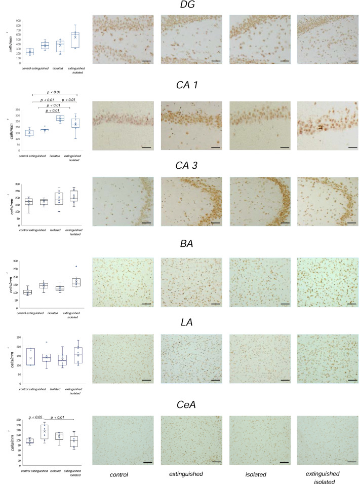
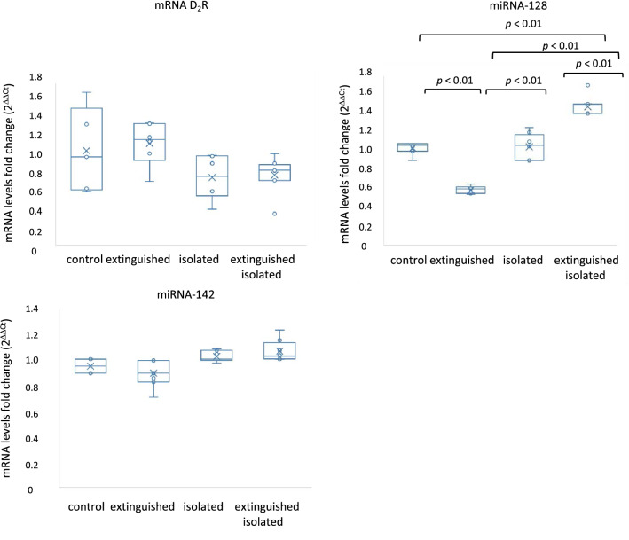
Results: It was found that recurrent social isolation stress decreased the fear extinction rate. The extinguished isolated rats were characterized by higher expression of D2R in the CA1 area of the hippocampus compared to the extinguished and the control rats. In turn, the isolated group presented higher D2R immunoreactivity in the CA1 area compared to the extinguished, the control, and the extinguished isolated animals. Moreover, the extinguished animals had higher expression of D2R in the central amygdala than the control and the extinguished isolated rats. These changes were accompanied by the increase in miRNA-128 level in the amygdala in the extinguished isolated rats compared to the control, the extinguished, and the isolated rats. Moreover, the extinguished rats had lower expression of miRNA-128 compared to the control and the isolated animals.
Conclusions: Our results suggest that social isolation stress impairs aversive memory extinction and coexists with changes in the D2R expression in the amygdala and hippocampus and increased expression of miRNA-128 in the amygdala.
Keywords: Amygdala; Dopamine D2 receptors; Fear extinction; Hippocampus; Social isolation.
© 2022. The Author(s).
Conflict of interest statement
The authors declare that they have no conflict of interest.
Figures

Similar articles
-
Reciprocal patterns of c-Fos expression in the medial prefrontal cortex and amygdala after extinction and renewal of conditioned fear.Learn Mem. 2009 Jul 24;16(8):486-93. doi: 10.1101/lm.1463909. Print 2009 Aug. Learn Mem. 2009. PMID: 19633138 Free PMC article.
-
Early life social experience affects adulthood fear extinction deficit and associated dopamine profile abnormalities in a rat model of PTSD.Prog Neuropsychopharmacol Biol Psychiatry. 2020 Jul 13;101:109914. doi: 10.1016/j.pnpbp.2020.109914. Epub 2020 Mar 9. Prog Neuropsychopharmacol Biol Psychiatry. 2020. PMID: 32165120
-
Investigating the role of dopamine receptor- and parvalbumin-expressing cells in extinction of conditioned fear.Neurobiol Learn Mem. 2017 Nov;145:7-17. doi: 10.1016/j.nlm.2017.08.009. Epub 2017 Aug 31. Neurobiol Learn Mem. 2017. PMID: 28842281
-
Neural circuits involved in the renewal of extinguished fear.IUBMB Life. 2017 Jul;69(7):470-478. doi: 10.1002/iub.1636. Epub 2017 May 2. IUBMB Life. 2017. PMID: 28464461 Review.
-
Brain sites involved in fear memory reconsolidation and extinction of rodents.Neurosci Biobehav Rev. 2015 Jun;53:160-90. doi: 10.1016/j.neubiorev.2015.04.003. Epub 2015 Apr 14. Neurosci Biobehav Rev. 2015. PMID: 25887284 Review.
Cited by
-
Increased threat learning after social isolation in human adolescents.R Soc Open Sci. 2024 Nov 13;11(11):240101. doi: 10.1098/rsos.240101. eCollection 2024 Nov. R Soc Open Sci. 2024. PMID: 39539503 Free PMC article.
References
-
- Furini C, Myskiw J, Izquierdo I. The learning of fear extinction. Neurosci Biobehav Rev. 2014;47:670–683. - PubMed
MeSH terms
Substances
Grants and funding
LinkOut - more resources
Full Text Sources
Miscellaneous