

Quantitative muscle MRI captures early muscle degeneration in calpainopathy
- PMID: 36385624
- PMCID: PMC9669006
- DOI: 10.1038/s41598-022-23972-6
Quantitative muscle MRI captures early muscle degeneration in calpainopathy
Erratum in
-
Author Correction: Quantitative muscle MRI captures early muscle degeneration in calpainopathy.Sci Rep. 2024 May 6;14(1):10381. doi: 10.1038/s41598-024-61083-6. Sci Rep. 2024. PMID: 38710928 Free PMC article. No abstract available.
Abstract
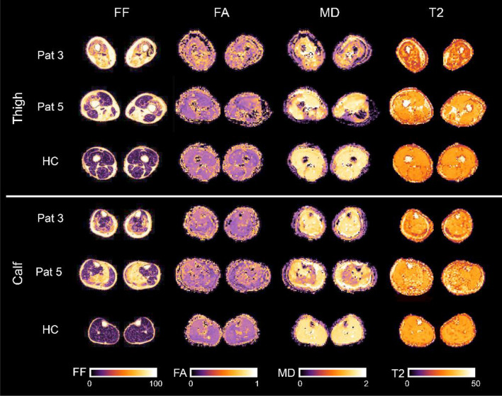
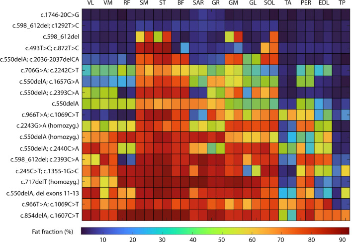
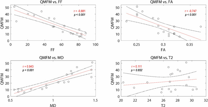
To evaluate differences in qMRI parameters of muscle diffusion tensor imaging (mDTI), fat-fraction (FF) and water T2 time in leg muscles of calpainopathy patients (LGMD R1/D4) compared to healthy controls, to correlate those findings to clinical parameters and to evaluate if qMRI parameters show muscle degeneration in not-yet fatty infiltrated muscles. We evaluated eight thigh and seven calf muscles of 19 calpainopathy patients and 19 healthy matched controls. MRI scans were performed on a 3T MRI including a mDTI, T2 mapping and mDixonquant sequence. Clinical assessment was done with manual muscle testing, patient questionnaires (ACTIVLIM, NSS) as well as gait analysis. Average FF was significantly different in all muscles compared to controls (p < 0.001). In muscles with less than 8% FF a significant increase of FA (p < 0.005) and decrease of RD (p < 0.004) was found in high-risk muscles of calpainopathy patients. Water T2 times were increased within the low- and intermediate-risk muscles (p ≤ 0.045) but not in high-risk muscles (p = 0.062). Clinical assessments correlated significantly with qMRI values: QMFM vs. FF: r = - 0.881, p < 0.001; QMFM versus FA: r = - 0.747, p < 0.001; QMFM versus MD: r = 0.942, p < 0.001. A good correlation of FF and diffusion metrics to clinical assessments was found. Diffusion metrics and T2 values are promising candidates to serve as sensitive early and non-invasive methods to capture early muscle degeneration in non-fat-infiltrated muscles in calpainopathies.
© 2022. The Author(s).
Conflict of interest statement
The authors declare no competing interests.
Figures

References
Publication types
MeSH terms
Substances
Supplementary concepts
Grants and funding
LinkOut - more resources
Full Text Sources
Medical
Miscellaneous
