. 2022 Nov 16;12(1):19658.
doi: 10.1038/s41598-022-23923-1.

Association between SARS-CoV-2 RNAemia and dysregulated immune response in acutely ill hospitalized COVID-19 patients

Affiliations

Association between SARS-CoV-2 RNAemia and dysregulated immune response in acutely ill hospitalized COVID-19 patients

Roberta Rovito et al. Sci Rep. .

Abstract

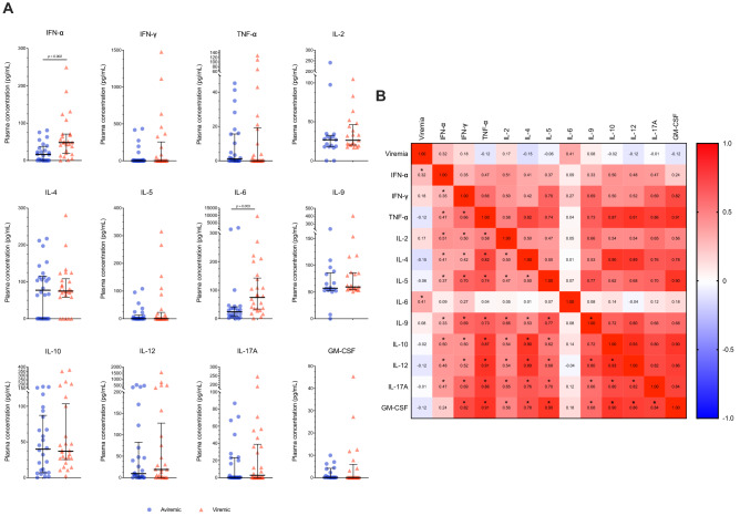
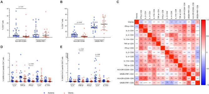
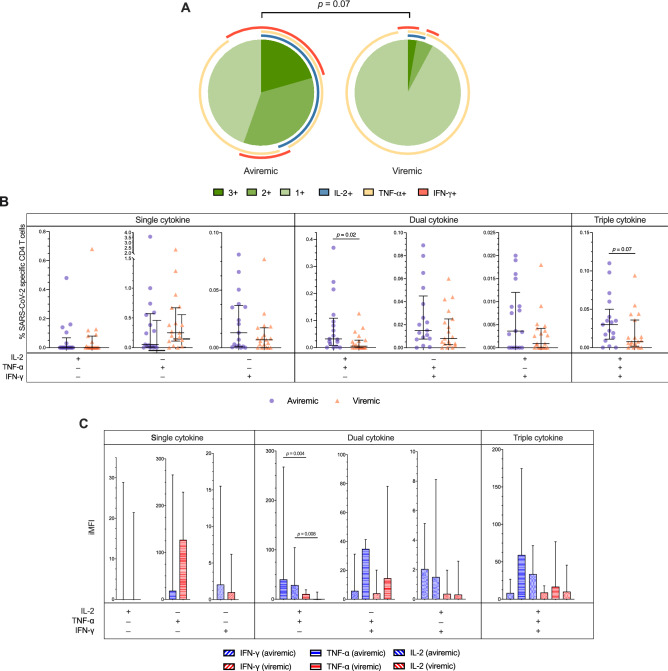
Severe/critical COVID-19 is associated with immune dysregulation and plasmatic SARS-CoV-2 detection (i.e. RNAemia). We detailed the association of SARS-CoV-2 RNAemia with immune responses in COVID-19 patients at the end of the first week of disease. We enrolled patients hospitalized in acute phase of ascertained SARS-CoV-2 pneumonia, and evaluated SARS-CoV-2 RNAemia, plasmatic cytokines, activated/pro-cytolytic T-cells phenotypes, SARS-CoV-2-specific cytokine-producing T-cells (IL-2, IFN-γ, TNF-α, IL-4, IL-17A), simultaneous Th1-cytokines production (polyfunctionality) and amount (iMFI). The humoral responses were assessed with anti-S1/S2 IgG, anti-RBD total-Ig, IgM, IgA, IgG1 and IgG3, neutralization and antibody-dependent cellular cytotoxicity (ADCC). Out of 54 patients, 27 had detectable viremia (viremic). Albeit comparable age and co-morbidities, viremic more frequently required ventilatory support, with a trend to higher death. Viremic displayed higher pro-inflammatory cytokines (IFN-α, IL-6), lower activated T-cells (HLA-DR+CD38+), lower functional SARS-CoV-2-specific T-cells (IFN-γ+CD4+, TNF-α+CD8+, IL-4+CD8+, IL-2+TNF-α+CD4+, and IL-2+TNF-α+CD4+ iMFI) and SARS-CoV-2-specific Abs (anti-S IgG, anti-RBD total-Ig, IgM, IgG1, IgG3; ID50, %ADCC). These data suggest a link between SARS-CoV-2 RNAemia at the end of the first stage of disease and immune dysregulation. Whether high ab initium viral burden and/or intrinsic host factors contribute to immune dysregulation in severe COVID-19 remains to be elucidated, to further inform strategies of targeted therapeutic interventions.


Conflict of interest statement

The authors declare no competing interests.

Figures

Figure 1
Figure 1
Plasmatic cytokines in COVID-19 patients according to SARS-CoV-2 viremia. (A) Plasmatic cytokines in aviremic and viremic COVID-19 patients. IFN-α (n = 24 and n = 26), IFN-γ (n = 26 and n = 25), IL-2 (n = 17 and n = 21), IL-4 (n = 26 and n = 26), IL-5 (n = 26 and n = 26), IL-6 (n = 24 and n = 26), IL-9 (n = 17 and n = 21), IL-10 (n = 26 and n = 26), IL-12p70 (n = 26 and n = 25), IL-17A (n = 25 and n = 26), TNF- α (n = 26 and n = 26), GM-CSF (n = 17 and n = 21). (B) Heatmap of correlations between SARS-CoV-2 viremia and plasmatic cytokines. Median and interquartile range (IQR) are shown for each group of patients. Mann–Whitney U test and Spearman’s correlation test, *statistical significance at p-value < 0.05.
Figure 2
Figure 2
Immunophenotypes and SARS-CoV-2-specific T cells in COVID-19 patients according to SARS-CoV-2 viremia. (A) Frequencies of circulating HLA-DR + CD38 + (activated) and GRZB + PRF + (pro-cytolitic) CD4 T cells in aviremic and viremic COVID-19 patients (n = 25 and n = 21). (B) Frequencies of circulating HLA-DR + CD38 + (activated) and GRZB + PRF + (pro-cytolitic) CD8 T cells in aviremic and viremic COVID-19 patients (n = 25 and n = 21). (C) Heatmap of correlations between SARS-CoV-2 viremia, immunophenotypes, and SARS-CoV-2-specific T cells. (D) Frequencies of CD4 T cells producing cytokines upon PBMCs challenge for 5 h with 1 μg/ml of a pool of 15-mer peptides of SARS-CoV-2 S, N- and M-proteins in aviremic and viremic COVID-19 patients (n = 19 and n = 17). (E) Frequencies of CD8 T cells producing cytokines upon PBMCs challenge for 5 h with 1 μg/ml of a pool of 15-mer peptides of SARS-CoV-2 S-, N- and M-proteins in aviremic and viremic COVID-19 patients (n = 19 and n = 17). Median and interquartile range (IQR) are shown for each group of patients. Mann–Whitney U test and Spearman’s correlation test, *statistical significance at p-value < 0.05.
Figure 3
Figure 3
SARS-CoV-2 specific CD4 Th1 cells functionality in COVID-19 patients according to SARS-CoV-2 viremia. (A) Pie charts showing the median distribution of polyfunctionality profiles in SARS-CoV-2–specific cytokine-producing Th1 cells of aviremic and viremic individuals. The pie slices represent median percentages of tri- (3 +), bi- (2 +), and mono- (1 +) functional T-cells. The arches around the circumference indicate the cytokine (IFN-g, TNF-a, or IL-2) produced by the portion of T-cells that lie under the arc; parts of the pie surrounded by multiple arches represent polyfunctional cells. Statistics: permutation test performed by SPICE 6.0. (B) Th1 polyfunctionality: frequencies of CD4 T cells producing one Th1 cytokine (Single cytokine), two Th1 cytokines (Dual cytokine), or three Th1 cytokines (Triple cytokine) upon PBMCs challenge for 5 h with 1 μg/ml of a pool of 15-mer peptides of SARS-CoV-2 S-, N- and M-proteins in aviremic and viremic COVID-19 patients (n = 19 and n = 17). (C) Magnitude of Th1 cytokines produced: the integrated median intensity fluorescence (iMFI) was calculated by multiplying the frequency of Single cytokine, Dual cytokine or Triple cytokine SARS-CoV-2-specific Th1 cells with the MFI for a given cytokine in aviremic and viremic COVID-19 patients (n = 19 and n = 17). Statistics: Mann–Whitney U test performed by SPICE 6.0.
Figure 4
Figure 4
Elicitation and functionality of S1/S2- and RBD-specific antibodies in COVID-19 patients according to SARS-CoV-2 viremia. (A) Anti-S1/S2 IgG (AU/ml) were assessed by means of a commercially available ELISA (LIAISON SARS-CoV-2 S1/S2 IgG, DiaSorin) in aviremic and viremic COVID-19 patients (n = 18 and n = 22). (B) Anti-RBD Ig (AUC) were assessed in aviremic and viremic COVID-19 patients by means of an in-house ELISA (n = 18 and n = 22 for total Ig; n = 17 and n = 21 for IgM, IgA, IgG1 and IgG3). (C) Neutralization activity was measured by incubating pseudoviruses bearing the D614G SARS-CoV-2 Spike with serial dilutions of plasma for 1 h at 37 °C before infecting 293 T-ACE2 target cells. Neutralization half maximal inhibitory serum dilution (ID50) values were determined using a normalized non-linear regression. (D) %ADCC in the presence of plasma at a 1/500 dilution. CEM.NKr parental cells were mixed at a 1:1 ratio with CEM.NKr-Spike cells and were used as target cells. PBMCs from uninfected donors were used as effector cells in a FACS-based ADCC assay. (E) Heatmap of correlations between SARS-CoV-2 viremia and humoral response. Median and interquartile range (IQR) are shown for each group of patients. S1/S2-specific total IgG, RBD-specific total Abs, IgM, IgA, IgG1 and IgG3 were negatively correlated with SARS-CoV-2 viremia (Spearman r = − 0.67, − 0.58, − 0.54, − 0.35, − 0.51 and − 0.34; p-value = 0.000003, 0.0001, 0.0005, 0.032, 0.001, 0.04, respectively). Mann–Whitney U test and Spearman’s correlation test, *statistical significance at p-value < 0.05.

References

    1. Guan W-J, et al. Clinical characteristics of coronavirus disease 2019 in China. N. Engl. J. Med. 2020;382:1708–1720. doi: 10.1056/nejmoa2002032. - DOI - PMC - PubMed
    1. Wu Z, Mcgoogan JM. Characteristics of and important lessons from the coronavirus disease 2019 (COVID-19) outbreak in China. JAMA. 2020;323:1239. doi: 10.1001/jama.2020.2648. - DOI - PubMed
    1. De Biasi S, et al. Marked T cell activation, senescence, exhaustion and skewing towards TH17 in patients with COVID-19 pneumonia. Nat. Commun. 2020 doi: 10.1038/s41467-020-17292-4. - DOI - PMC - PubMed
    1. Song J-W, et al. Immunological and inflammatory profiles in mild and severe cases of COVID-19. Nat. Commun. 2020 doi: 10.1038/s41467-020-17240-2. - DOI - PMC - PubMed
    1. Tincati C, et al. Heightened circulating interferon-inducible chemokines, and activated Pro-Cytolytic Th1-Cell phenotype features Covid-19 aggravation in the second week of illness. Front. Immunol. 2020;11:580987. doi: 10.3389/fimmu.2020.580987. - DOI - PMC - PubMed