. 2022 Nov 29;80(1):ftac044.
doi: 10.1093/femspd/ftac044.

Impact of nutrients on the function of the chlamydial Rsb partner switching mechanism

Affiliations

Impact of nutrients on the function of the chlamydial Rsb partner switching mechanism

Shiomi Kuwabara et al. Pathog Dis. .

Abstract

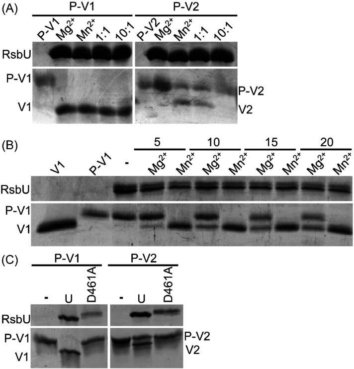
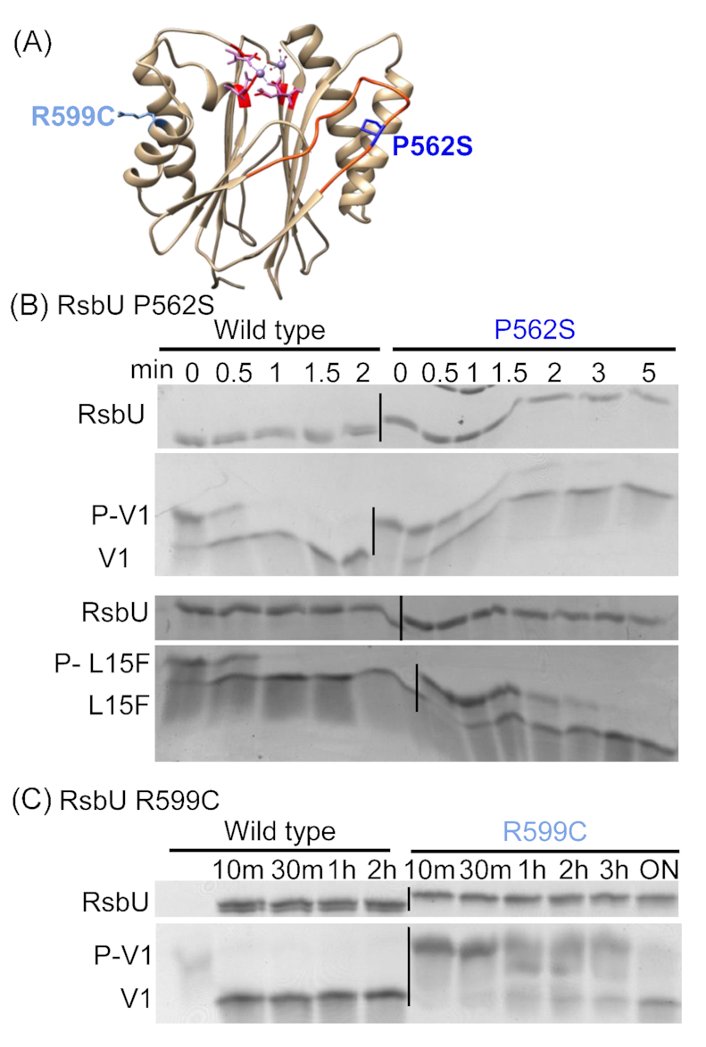
The obligate intracellular bacterial pathogen Chlamydia trachomatis is a leading cause of sexually transmitted infections and infectious blindness. Chlamydia undergo a biphasic developmental cycle alternating between the infectious elementary body (EB) and the replicative reticulate body (RB). The molecular mechanisms governing RB growth and RB-EB differentiation are unclear. We hypothesize that the bacterium senses host cell and bacterial energy levels and metabolites to ensure that development and growth coincide with nutrient availability. We predict that a partner switching mechanism (PSM) plays a key role in the sensing and response process acting as a molecular throttle sensitive to metabolite levels. Using purified wild type and mutant PSM proteins, we discovered that metal type impacts enzyme activity and the substrate specificity of RsbU and that RsbW prefers ATP over GTP as a phosphate donor. Immunoblotting analysis of RsbV1/V2 demonstrated the presence of both proteins beyond 20 hours post infection and we observed that an RsbV1-null strain has a developmental delay and exhibits differential growth attenuation in response to glucose levels. Collectively, our data support that the PSM regulates growth in response to metabolites and further defines biochemical features governing PSM-component interactions which could help in the development of novel PSM-targeted therapeutics.

Keywords: Chlamydia; PP2C; Rsb; glucose; metalation; phosphorylation.


Conflict of interest statement

The authors declare no competing financial interest.

Figures

Figure 1.
Figure 1.
RsbW prefers ATP over GTP as a phosphate donor for RsbV1. Kinase reactions were performed with RsbW using RsbV1 as a substrate and either ATP (A) or GTP (B) as the phosphate donor. Reactions were performed at 30°C with a substrate::enzyme molar ratio of 1 to 0.19. Phosphorylation status of RsbV1 was confirmed by running products on 15% Phos-tag gels followed by staining with Coomassie Brilliant Blue. The relative affinity of RsbW for each phosphate donor was measured through time course analysis (left panels in A and B) and by using different concentrations of the phosphate donor with reactions halted at 3 minutes (right panels in A and B). The expected positions of products are shown to the left of the panels. Representative gels are shown from at least three biological replicates.
Figure 2.
Figure 2.
Amino acid sequence alignment of PP2C-type phosphatases. Group I and Group II phosphatases were selected to highlight divergence in the flap regions associated with substrate binding. Only the predicted PP2C domains were used for GII phosphatases. Sequences were aligned using Multiple Sequence Comparison by Log- Expectation and displayed using BioEdit with an 80% identity cut-off (black shading). Protein sequences: RsbU (CTL0851) from C. trachomatis (CtRsbU), SpoIIE from B. subtilis (BsSpoIIE), RsbU from B. subtilis (BsRsbU), RsbX from B. subtilis (BsRsbX), Rv1364c from Mycobacterium tuberculosis (MtRv1364c), IcfG from Synechocystis sp. PCC6803 (SIcfG), CppA (CLT0511) from C. trachomatis (CtCppA), PrpC from B. subtilis (BsPrpC), MspP from Mycobacterium smegmatis (MsMspP), PstP from M. tuberculosis (MtPstP), Stp1 from Streptococcus agalactiae (SaStp1) and tPphA from Thermosynechococcus elongatus (TetPphA). The eight conserved residues amongst PP2C phosphatases are denoted with asterisks. The predicted secondary structure of RsbU from C. trachomatis is indicated with green (α helices) or purple (β sheets) arrows. The vertical arrows mark residues mutated in this study: D461A (red, essential for metal coordination), P562S (blue, (Kokes et al. 2015)), and R599C (light blue, (Kokes et al. 2015)). The predicted flap region involved in substrate binding and used to differentiate Group I and II PP2Cs (Kerk et al. 2015) is highlighted in orange between the seventh and eighth beta sheets.
Figure 3.
Figure 3.
RsbU phosphatase activity is enhanced by Mn2+. Dephosphorylation of P-RsbV1 and P-RsbV2 by RsbU was assessed in the presence of Mg2+ and/or Mn2+. RsbU was incubated with phosphorylated substrates and dephosphorylation was measured using 15% Phos-tag gels stained with Coomassie Brilliant Blue. Expected product locations are listed to the left of the gels. In (A), we determined that RsbU can dephosphorylate both P-RsbV1 and P-RsbV2 in the presence of Mn2+. The introduction of Mg2+ or use of Mg2+ alone blocks dephosphorylation of P-RsbV2, but not dephosphorylation of P-RsbV1. (B) Metal dependence and metal impact on phosphatase activity was further assessed versus P-RsbV1. Time course analysis showed that Mn2+ enhances phosphatase activity compared to Mg2+. Thirty minutes was used for the no metal time point (-). (C) Mutation of the metal-coordinating D461 to Ala leads to loss of phosphatase activity towards both RsbV1 and RsbV2. Representative gels are shown from at least three biological replicates.
Figure 4.
Figure 4.
RsbW can phosphorylate an RsbV1 L15F mutant. A phosphorylation time course experiment was used to assess the impact of the L15F mutation on RsbV1 as an RsbW substrate compared to wild type RsbV1. (A) A protein structure model of RsbV1 was constructed using Phyre2 and processed with UCSF Chimera. The phosphorylation site (S56) is highlighted in red and the L15F point mutation is shown in blue. (B) RsbV1 and RsbV1 L15F were incubated with RsbW and ATP, samples were processed at the indicated times, and products were separated on Phos-tag gels and visualized with Coomassie Brilliant Blue. Expected product positions are indicated on the sides of the gels. Representative gels are shown from at least three biological replicates.
Figure 5.
Figure 5.
P562S and R599C mutations in RsbU reduces phosphatase activity towards P-RsbV1. Nonconservative RsbU amino acid mutations in the PP2C domain present in growth impaired C. trachomatis mutant strains derived by chemical mutagenesis (Kokes et al.) were modeled using Phyre2 for the PP2C domain of RsbU. The model was further processed using UCSF Chimera to superimpose the catalytic active site of RsbU from M. tuberculosis Rv1364c (3KE6, purple) onto the C. trachomatis RsbU PP2C domain (red). Mn2+ (purple spheres) and water molecules (red spheres) are also depicted in the active site. The substrate binding flap region is modeled in orange and the two blue residues reflect the constructed mutations: blue (P562S) and light blue (R599C). Mutant proteins were purified and incubated with phosphorylated RsbV1 substrates. Reactions were stopped at the indicated times and products were resolved on Phos-tag gels and visualized with Coomassie Brilliant Blue. (B) The RsbU P562S mutant was tested versus both wild type P-RsbV1 and the P-RsbV1 L15F mutant and P562S showed equivalent reduced activity towards both substrates. (C) RsbU R599C showed significantly reduced activity requiring hours instead of minutes to dephosphorylate the substrate. Representative gels are shown from at least three biological replicates. Expected protein positions are listed on the sides of each gel.
Figure 6.
Figure 6.
Glucose levels more strongly impact IFU yields than total bacterial numbers. As we hypothesize that the PSM governs growth and development in response to nutritional and energy cues, EB production (IFU) and total bacteria (RBs and EBs, gDNA) were measured in HeLa cells grown under high or low glucose conditions. Cells were infected with the parental Chlamydia strain (DFCT28) or rsbV1-mutants (RsbV1-null, DFCT34; RsbV1-complemented, DFCT35; and RsbV1-overexpression, DFCT36, Fig. S4). Replicate samples were collected at the indicated time points and analyzed for IFU production and gDNA levels. IFU production was reduced for the RsbV1-null strain under both glucose conditions compared to the wild type strain (panels A and B) and all strains grown under low glucose (panel B) had reduced IFUs compared to high glucose conditions (panel A). Fold yield of IFUs for DFCT28 and DFCT34 comparing input versus output at various times between the high glucose and low glucose conditions are shown in (C). In (D), samples paired with (C) were assessed for gDNA levels with data plotted for the fold yield of gDNA versus input. Growth assays were performed twice with replicates for each individual experiment. Error bars report standard deviation. Cross bars report significant differences between groups calculated using two-way ANOVA with Tukey's multiple comparison test (P-value < 0.05).

Similar articles

Cited by

References

    1. Abdelrahman YM, Belland RJ. The chlamydial developmental cycle. FEMS Microbiol Rev. 2005;29:949–59. - PubMed
    1. Beaufay F, Coppine J, Hallez R. When the metabolism meets the cell cycle in bacteria. Curr Opin Microbiol. 2021;60:104–13. - PubMed
    1. Belland RJ, Zhong G, Crane DD, et al. Genomic transcriptional profiling of the developmental cycle of Chlamydia trachomatis. Proc Natl Acad Sci USA. 2003;100:8478–83. - PMC - PubMed
    1. Benson AK, Haldenwang WG. Regulation of sigma b levels and activity in Bacillus subtilis. J Bacteriol. 1993;175:2347–56. - PMC - PubMed
    1. Borel N, Leonard C, Slade J, et al. Chlamydial antibiotic resistance and treatment failure in veterinary and human medicine. Curr Clin Microbiol Rep. 2016;3:10–8. - PMC - PubMed