

ChimeraNet: U-Net for Hair Detection in Dermoscopic Skin Lesion Images
- PMID: 36385676
- PMCID: PMC10039207
- DOI: 10.1007/s10278-022-00740-6
ChimeraNet: U-Net for Hair Detection in Dermoscopic Skin Lesion Images
Abstract
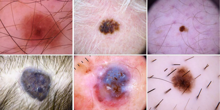
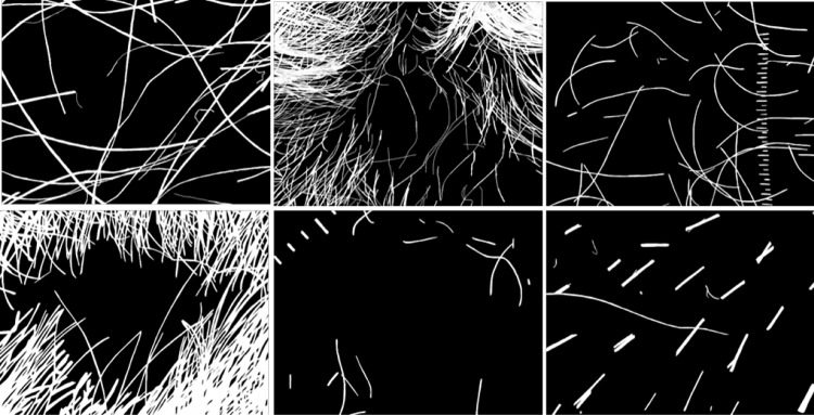
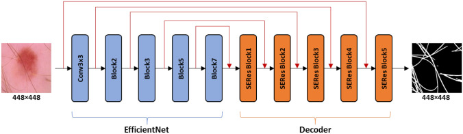
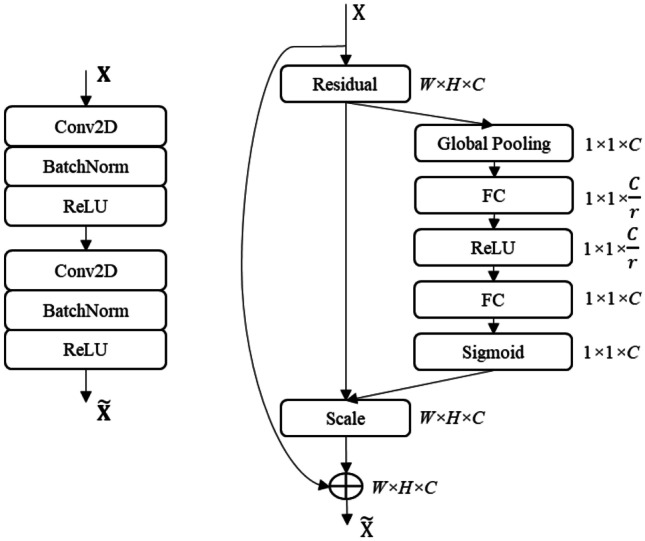
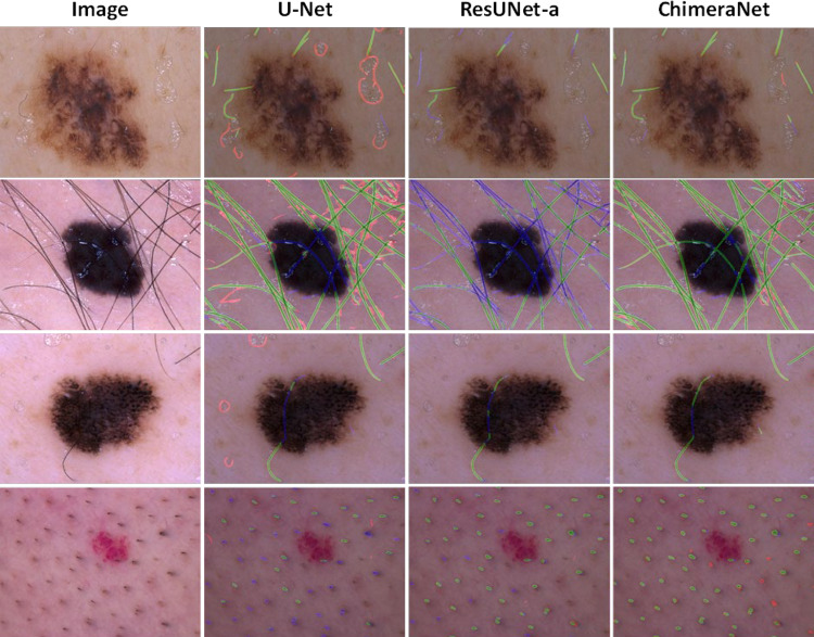
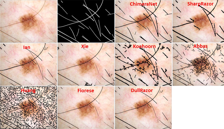
Hair and ruler mark structures in dermoscopic images are an obstacle preventing accurate image segmentation and detection of critical network features. Recognition and removal of hairs from images can be challenging, especially for hairs that are thin, overlapping, faded, or of similar color as skin or overlaid on a textured lesion. This paper proposes a novel deep learning (DL) technique to detect hair and ruler marks in skin lesion images. Our proposed ChimeraNet is an encoder-decoder architecture that employs pretrained EfficientNet in the encoder and squeeze-and-excitation residual (SERes) structures in the decoder. We applied this approach at multiple image sizes and evaluated it using the publicly available HAM10000 (ISIC2018 Task 3) skin lesion dataset. Our test results show that the largest image size (448 × 448) gave the highest accuracy of 98.23 and Jaccard index of 0.65 on the HAM10000 (ISIC 2018 Task 3) skin lesion dataset, exhibiting better performance than for two well-known deep learning approaches, U-Net and ResUNet-a. We found the Dice loss function to give the best results for all measures. Further evaluated on 25 additional test images, the technique yields state-of-the-art accuracy compared to 8 previously reported classical techniques. We conclude that the proposed ChimeraNet architecture may enable improved detection of fine image structures. Further application of DL techniques to detect dermoscopy structures is warranted.
Keywords: Deep learning; Dermoscopy; Hair removal; Image segmentation; Melanoma; Transfer learning.
© 2022. The Author(s) under exclusive licence to Society for Imaging Informatics in Medicine.
Conflict of interest statement
The authors declare no competing interests.
Figures
Similar articles
-
Skin Lesion Segmentation in Dermoscopic Images with Noisy Data.J Digit Imaging. 2023 Aug;36(4):1712-1722. doi: 10.1007/s10278-023-00819-8. Epub 2023 Apr 5. J Digit Imaging. 2023. PMID: 37020149 Free PMC article.
-
Melanoma segmentation using deep learning with test-time augmentations and conditional random fields.Sci Rep. 2022 Mar 10;12(1):3948. doi: 10.1038/s41598-022-07885-y. Sci Rep. 2022. PMID: 35273282 Free PMC article.
-
Machine learning based skin lesion segmentation method with novel borders and hair removal techniques.PLoS One. 2022 Nov 10;17(11):e0275781. doi: 10.1371/journal.pone.0275781. eCollection 2022. PLoS One. 2022. PMID: 36355845 Free PMC article.
-
Deep Learning Approaches Towards Skin Lesion Segmentation and Classification from Dermoscopic Images - A Review.Curr Med Imaging. 2020;16(5):513-533. doi: 10.2174/1573405615666190129120449. Curr Med Imaging. 2020. PMID: 32484086 Review.
-
ACCPG-Net: A skin lesion segmentation network with Adaptive Channel-Context-Aware Pyramid Attention and Global Feature Fusion.Comput Biol Med. 2023 Mar;154:106580. doi: 10.1016/j.compbiomed.2023.106580. Epub 2023 Jan 25. Comput Biol Med. 2023. PMID: 36716686 Review.
Cited by
-
Hybrid Topological Data Analysis and Deep Learning for Basal Cell Carcinoma Diagnosis.J Imaging Inform Med. 2024 Feb;37(1):92-106. doi: 10.1007/s10278-023-00924-8. Epub 2024 Jan 12. J Imaging Inform Med. 2024. PMID: 38343238 Free PMC article.
-
Basal Cell Carcinoma Diagnosis with Fusion of Deep Learning and Telangiectasia Features.J Imaging Inform Med. 2024 Jun;37(3):1137-1150. doi: 10.1007/s10278-024-00969-3. Epub 2024 Feb 8. J Imaging Inform Med. 2024. PMID: 38332404 Free PMC article.
-
Improving Automatic Melanoma Diagnosis Using Deep Learning-Based Segmentation of Irregular Networks.Cancers (Basel). 2023 Feb 16;15(4):1259. doi: 10.3390/cancers15041259. Cancers (Basel). 2023. PMID: 36831599 Free PMC article.
-
Skin Lesion Segmentation in Dermoscopic Images with Noisy Data.J Digit Imaging. 2023 Aug;36(4):1712-1722. doi: 10.1007/s10278-023-00819-8. Epub 2023 Apr 5. J Digit Imaging. 2023. PMID: 37020149 Free PMC article.
-
Weakly supervised deep learning for diagnosis of multiple vertebral compression fractures in CT.Eur Radiol. 2024 Jun;34(6):3750-3760. doi: 10.1007/s00330-023-10394-9. Epub 2023 Nov 16. Eur Radiol. 2024. PMID: 37973631
References
Publication types
MeSH terms
Grants and funding
LinkOut - more resources
Full Text Sources
Medical
