

Ligand-gated ion channel P2X7 regulates hypoxia-induced factor-1α mediated pain induced by dental pulpitis in the medullary dorsal horn
- PMID: 36385758
- PMCID: PMC9644926
- DOI: 10.3389/fnmol.2022.1015751
Ligand-gated ion channel P2X7 regulates hypoxia-induced factor-1α mediated pain induced by dental pulpitis in the medullary dorsal horn
Abstract
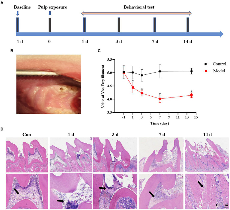
Dental pulpitis often induces severe pain, and the molecular immune response is remarkable in both peripheral and central nervous system. Accumulating evidence indicates that activated microglia in the medullary dorsal horn (MDH) contribute to dental pulpitis induced pain. The P2X7 receptor plays an important role in driving pain and inflammatory processes, and its downstream target hypoxia-induced factor-1α (HIF-1α) has a crucial role in maintaining inflammation. However, the relationship between P2X7 and HIF-1α in dental inflammatory pain remains unclear. This study demonstrated that the degree of inflammation in the dental pulp tissue became more severe in a time-dependent manner by establishing a rat dental pulpitis model via pulp exposure. Meanwhile, the expression of P2X7, HIF-1α, IL-1β, and IL-18 in the MDH increased most on the seventh day when the pain threshold was the lowest in the dental pulpitis model. Furthermore, lipopolysaccharides (LPS) increased P2X7-mediated HIF-1α expression in microglia. Notably, the suppression of P2X7 caused less IL-1β and IL-18 release and lower HIF-1α expression, and P2X7 antagonist Brilliant Blue G (BBG) could alleviate pain behaviors of the dental pulpitis rats. In conclusion, our results provide further evidence that P2X7 is a key molecule, which regulates HIF-1α expression and inflammation in dental pulpitis-induced pain.
Keywords: HIF-1α; P2X7; dental pulpitis; medullary dorsal horn; microglia; pain.
Copyright © 2022 Zhang, Si, Liang, Lu, Shang, Li, Sun and Wu.
Conflict of interest statement
The authors declare that the research was conducted in the absence of any commercial or financial relationships that could be construed as a potential conflict of interest.
Figures


Similar articles
-
Enhanced exosome secretion regulated by microglial P2X7R in the medullary dorsal horn contributes to pulpitis-induced pain.Cell Biosci. 2025 Feb 22;15(1):28. doi: 10.1186/s13578-025-01363-4. Cell Biosci. 2025. PMID: 39987146 Free PMC article.
-
Ligand-gated ion channel P2X7 regulates NLRP3/Caspase-1-mediated inflammatory pain caused by pulpitis in the trigeminal ganglion and medullary dorsal horn.Brain Res Bull. 2023 Jan;192:1-10. doi: 10.1016/j.brainresbull.2022.10.020. Epub 2022 Oct 31. Brain Res Bull. 2023. PMID: 36328143
-
Central sensitization of nociceptive neurons in rat medullary dorsal horn involves purinergic P2X7 receptors.Neuroscience. 2011 Sep 29;192:721-31. doi: 10.1016/j.neuroscience.2011.06.083. Epub 2011 Jul 14. Neuroscience. 2011. PMID: 21763757 Free PMC article.
-
Autophagy induced by hypoxia in pulpitis is mediated by HIF-1α/BNIP3.Arch Oral Biol. 2024 Mar;159:105881. doi: 10.1016/j.archoralbio.2024.105881. Epub 2024 Jan 5. Arch Oral Biol. 2024. PMID: 38199116
-
Hypoxia-inducible factor 1 alpha (HIF-1α) stimulated and P2X7 receptor activated by COVID-19, as a potential therapeutic target and risk factor for epilepsy.Hum Cell. 2022 Sep;35(5):1338-1345. doi: 10.1007/s13577-022-00747-9. Epub 2022 Jul 13. Hum Cell. 2022. PMID: 35831562 Free PMC article. Review.
Cited by
-
Activation of the microglial P2X7R/NLRP3 inflammasome mediates central sensitization in a mouse model of medication overuse headache.Front Mol Neurosci. 2023 Jun 12;16:1177171. doi: 10.3389/fnmol.2023.1177171. eCollection 2023. Front Mol Neurosci. 2023. PMID: 37377770 Free PMC article.
-
Combined Transcriptomic and Proteomic Profiling of the Mouse Anterior Cingulate Cortex Identifies Potential Therapeutic Targets for Pulpitis-Induced Pain.ACS Omega. 2024 Jan 25;9(5):5972-5984. doi: 10.1021/acsomega.3c09759. eCollection 2024 Feb 6. ACS Omega. 2024. PMID: 38343959 Free PMC article.
-
Enhanced exosome secretion regulated by microglial P2X7R in the medullary dorsal horn contributes to pulpitis-induced pain.Cell Biosci. 2025 Feb 22;15(1):28. doi: 10.1186/s13578-025-01363-4. Cell Biosci. 2025. PMID: 39987146 Free PMC article.
-
Transcriptome Analysis of Trigeminal Ganglion and Medullary Dorsal Horn in Mice to Identify Potential Targets for Pulpitis-Induced Pain.ACS Omega. 2025 Mar 14;10(11):11439-11453. doi: 10.1021/acsomega.4c11486. eCollection 2025 Mar 25. ACS Omega. 2025. PMID: 40160770 Free PMC article.
-
Involvement of microglial P2X7 receptor in pain modulation.CNS Neurosci Ther. 2024 Jan;30(1):e14496. doi: 10.1111/cns.14496. Epub 2023 Nov 10. CNS Neurosci Ther. 2024. PMID: 37950524 Free PMC article. Review.
References
-
- Chakfe Y., Seguin R., Antel J. P., Morissette C., Malo D., Henderson D., et al. (2002). ADP and AMP induce Interleukin-1 release from microglial cells through activation of ATP-primed P2X7 receptor channels. J. Neurosci. 22, 3061–3069. doi: 10.1523/JNEUROSCI.22-08-03061.2002, PMID: - DOI - PMC - PubMed
LinkOut - more resources
Full Text Sources
Miscellaneous