. 2022 Oct 28:15:962974.
doi: 10.3389/fnmol.2022.962974. eCollection 2022.

The transforming growth factor beta ligand TIG-2 modulates the function of neuromuscular junction and muscle energy metabolism in Caenorhabditis elegans

Affiliations

The transforming growth factor beta ligand TIG-2 modulates the function of neuromuscular junction and muscle energy metabolism in Caenorhabditis elegans

Xinran Cheng et al. Front Mol Neurosci. .

Abstract

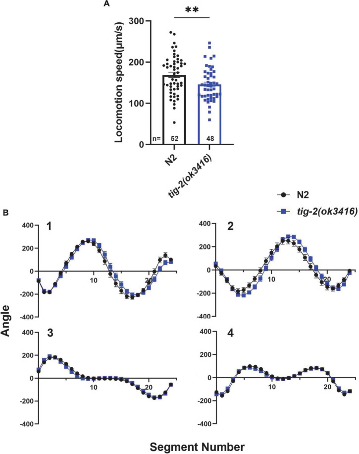
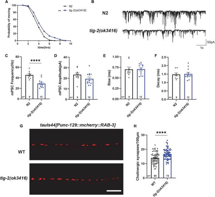
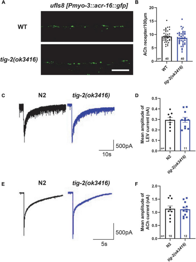
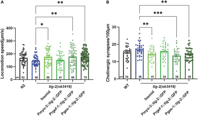
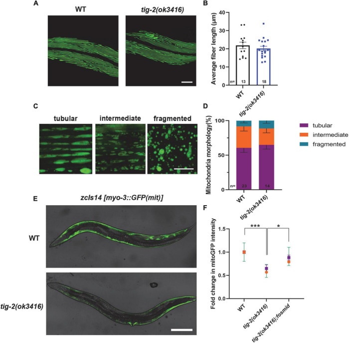
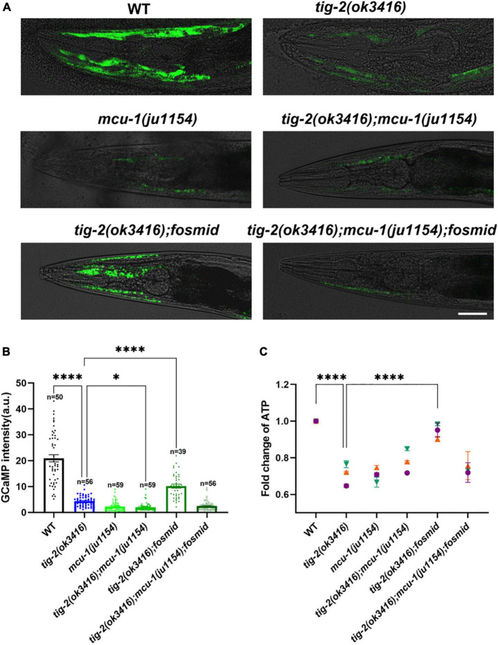
Deciphering the physiological function of TGF-β (the transforming growth factor beta) family ligands is import for understanding the role of TGF-β in animals' development and aging. Here, we investigate the function of TIG-2, one of the ligands in Caenorhabditis elegans TGF-β family, in animals' behavioral modulation. Our results show that a loss-of-function mutation in tig-2 gene result in slower locomotion speed in the early adulthood and an increased density of cholinergic synapses, but a decreased neurotransmitter release at neuromuscular junctions (NMJs). Further tissue-specific rescue results reveal that neuronal and intestinal TIG-2 are essential for the formation of cholinergic synapses at NMJs. Interestingly, tig-2(ok3416) mutant is characterized with reduced muscle mitochondria content and adenosine triphosphate (ATP) production, although the function of muscle acetylcholine receptors and the morphology muscle fibers in the mutant are comparable to that in wild-type animals. Our result suggests that TIG-2 from different neuron and intestine regulates worm locomotion by modulating synaptogenesis and neurotransmission at NMJs, as well as energy metabolism in postsynaptic muscle cells.

Keywords: Caenorhabditis elegans; TIG-2; acetylcholine; locomotion; mitochondria; neuromuscular junction.


Conflict of interest statement

The authors declare that the research was conducted in the absence of any commercial or financial relationships that could be construed as a potential conflict of interest.

Figures

FIGURE 1
FIGURE 1
tig-2 mutant displays lower locomotion speed than wild-type worms. (A) tig-2(ok3416) mutant has a lower locomotion speed is lower in on day 3 compared to wild-type animals (p = 0.0099). (B) The four eigenworms derived from wild-type worms and tig-2(ok3416) mutants are similar. A student’s t-test was performed to determine the significance of differences in locomotion speed. Error bars represent SEM. **p < 0.01.
FIGURE 2
FIGURE 2
tig-2 mutant has decreased acetylcholine (ACh) release but increased density of cholinergic synapses. (A) Comparison of aldicarb induced paralysis of tig-2(ok3416) mutant and wild-type worms. In early adulthood, tig-2(ok3416) mutant is more aldicarb-resistant than wild-type worms (p = 0.0055), indicating that tig-2 mutant releases less ACh on day 3. n > 60. (B) Representative traces of miniature post-synaptic currents (mPSCs) on day 3. (C,D) The frequency of mPSC in tig-2(ok3416) mutant is lower than that in wild-type worms (p < 0.0001), but the amplitude of mPSC is similar between tig-2 mutant and wild-type worms (p = 0.1384). (E,F) Quantifications of the 25–75% rise time and decay time of mPSCs. The data shows that no significant difference between wild-type and tig-2(ok3416) mutant. (G) Images of the cholinergic synapses on the nerve code. (H) Images of tauIs44 strain show that the density of cholinergic synapses on the nerve cord is increased in tig-2(ok3416) mutants (p < 0.0001). A student’s t-test was performed to determine the significance of differences in mean density. Error bars represent SEM. **p < 0.01, ****p < 0.0001.
FIGURE 3
FIGURE 3
tig-2 mutants and wild-type worms display similar density and function of postsynaptic AChRs. (A) Images of the post-synapse on the dorsal nerve cord. Scale bar is 10 μm. (B) Quantification of the density of post-synapses. The density of postsynaptic AChRs is unchanged in tig-2(ok3416) mutant (p = 0.3327). (C,E) Postsynaptic currents at NMJs evoked by levamisole (C) or ACh (E). (D,F) Levamisole-evoked current (p = 0.9811) and ACh-evoked current (p = 0.8671) are similar in tig-2(ok3416) mutants and wild-type worms. A Student’s t-test was performed for each statistical analysis. Error bars represent SEM.
FIGURE 4
FIGURE 4
Neuronal or intestinal expression of tig-2 abolishes locomotion speed defects and decreased density of cholinergic synapse in tig-2(ok3416). (A) Locomotion speed on day 3 in tig-2(ok3416) mutants and tissue-specific tig-2 rescue alleles. Green columns indicate transgenic lines that carry plasmids restoring the expression of tig-2 in muscle cells (p = 0.9768), neurons (p = 0.0060), intestine (p = 0.0089) and globally rescue (p = 0.0139). Neuronal TIG-2 and intestinal TIG-2 abolish the decrease in locomotion speed in the mutant. (B) Qualification of the density of cholinergic synapse in tig-2(ok3416) mutants and tissue-specific tig-2 rescue alleles. Neuronal tig-2 (p = 0.0003) and intestinal tig-2 (p = 0.0062) suppress the increase of presynaptic puncta. p-values were calculated by one-way ANOVA with multiple comparisons. Error bars represent SEM. *p < 0.05, **p < 0.01, ***p < 0.001.
FIGURE 5
FIGURE 5
tig-2 mutant has decreased mitochondrial content and mitochondrial calcium. (A) Images represent myosin fibers in body wall muscles. Scale bar is 20 μm. (B) Quantification of average muscle fiber length of individual worms. tig-2(ok3416) mutants and wild-type worms exhibit similar muscle fiber morphology (p = 0.4244). A student’s t-test was performed. (C) Representative images of mitochondria in body wall muscles showing three classes of cells. Scale bar is 5 μm. (D) Quantification of three classes of muscle cells in wild-type worms and tig-2(ok3416) mutants. Data from day 3 adult worms suggested similar mitochondrial morphology in tig-2 mutants and wild-type worms. p-values were calculated by two-way ANOVA. (E) Representative images of mitochondria targeted green fluorescent protein (GFP) expressed in body wall muscles. Scale bar is 200 μm. (F) Quantification of the intensity of mitoGFP in muscle cells. The intensity in tig-2(ok3416) is lower than in wild-type worms (p = 0.0007), indicating fewer mitochondria content in tig-2 mutants. All data are normalized to wild-type worms. n = 3 technical replicates. p-values were calculated by two-way ANOVA. Error bars represent SEM. *p < 0.05, ***p < 0.001.
FIGURE 6
FIGURE 6
The content of mitochondrial Ca2+ and adenosine triphosphate (ATP) production are lower in tig-2 mutants than in wild-type worms. (A) Representative images of GCaMP3 expression. Scale bar = 10 μm. (B) Quantification of mitochondrial Ca2+. The mitochondrial calcium content in tig-2(ok3416) mutant, mcu-1(ju1154) mutant and tig-2; mcu-1 double mutants is similar, and much lower than that in wild-type worms (p < 0.0001). Restoring the expression of tig-2 eliminates the decreased Ca2+ level in tig-2 signal mutants (p < 0.0001), but not in tig-2 mcu-1 double mutants (p = 0.1268). p-values were calculated by one-way ANOVA with multiple comparisons. (C) Relative ATP levels in worms expressing mito:GCaMP3 in their muscle cells. Data represents results from three independent tests. Each test was done in triplicate wells. n = 3 technical replicates. p-values were calculated by two-way ANOVA with multiple comparisons. Error bars represent SEM. ***p < 0.001, ****p < 0.0001.

Similar articles

Cited by

References

    1. Aberle H., Haghighi A. P., Fetter R. D., McCabe B. D., Magalhães T. R., Goodman C. S. (2002). Wishful thinking encodes a BMP type II receptor that regulates synaptic growth in Drosophila. Neuron 33 545–558. 10.1016/s0896-6273(02)00589-5 - DOI - PubMed
    1. Álvarez-Illera P., García-Casas P., Fonteriz R. I., Montero M., Alvarez J. (2020). Mitochondrial Ca2+ dynamics in MCU knockout C. elegans worms. Int. J. Mol. Sci. 21:8622. - PMC - PubMed
    1. Baltaci O., Pedersen M. E., Sherry T., Handley A., Snieckute G., Cao W., et al. (2022). Atypical TGF-β signaling controls neuronal guidance in Caenorhabditis elegans. Iscience 25:103791. 10.1016/j.isci.2022.103791 - DOI - PMC - PubMed
    1. Baughman J. M., Perocchi F., Girgis H. S., Plovanich M., Belcher-Timme C. A., Sancak Y., et al. (2011). Integrative genomics identifies MCU as an essential component of the mitochondrial calcium uniporter. Nature 476 341–345. 10.1038/nature10234 - DOI - PMC - PubMed
    1. Berke B., Wittnam J., McNeill E., Van Vactor D. L., Keshishian H. (2013). Retrograde BMP signaling at the synapse: A permissive signal for synapse maturation and activity-dependent plasticity. J. Neurosci. 33 17937–17950. 10.1523/JNEUROSCI.6075-11.2013 - DOI - PMC - PubMed

LinkOut - more resources