Review
. 2022 Oct 25:9:793267.
doi: 10.3389/fcvm.2022.793267. eCollection 2022.

The evolving role of multi-modality imaging in transcatheter tricuspid valve interventions

Affiliations
Review

The evolving role of multi-modality imaging in transcatheter tricuspid valve interventions

Livia Luciana Gheorghe et al. Front Cardiovasc Med. .

Abstract

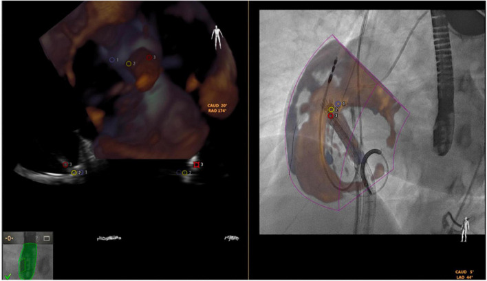
Tricuspid valve pathophysiology is not well-understood. Emergence of novel transcatheter tricuspid therapies has fueled the requirements for improved imaging visualization techniques and interventional imaging physician skillsets in guiding these complex transcatheter procedures. There is growing understanding on the clinical significance of tricuspid regurgitation which expanded the interest for percutaneous tricuspid valve interventions. The present review concentrates on three essential aspects of tricuspid valve pathophysiology: anatomical considerations for tricuspid interventions, optimal timing of tricuspid interventions by imaging guidance, and the role of interventional imaging physicians' skillset and knowledge in this field.

Keywords: MDCT (multidetector cardiac computed tomography); echo; imaging; percutaneous intervention; structural heart intervencions; tricuspid valve.


Conflict of interest statement

Author MS was proctor/lecturer for Abbott Vascular, Boston Scientific, Edwards Lifesciences, Philips Healthcare and Bioventrix Inc. Author DW has acted as a consultant for Abbott, Boston Scientific, Edwards Lifesciences, and has received a research grant support from Boston Scientific assigned to employer Henry Ford Health.

Figures

FIGURE 1
FIGURE 1
Tricuspid regurgitation grading.
FIGURE 2
FIGURE 2
Case of dynamic changes of TR during medical therapy. Patient with previous aortic surgical replacement admitted for right heart failure and torrential tricuspid regurgitation. (A1, A2) Show the transesophageal transgastric view with torrential tricuspid regurgitation and no-coaptation between anterior and septal leaflet. (B1–E1) Images during the admission and (B2–E2) images during the follow-up with intensive diuretic treatment. The transthoracic echocardiography showed a torrential TR with a jet which filled the entire atrium (B1), vena contracta of 12 mm (C1) and a tricuspid annulus of 44 mm (D1). The thoracic radiography at admission with serious right pleural effusion (E1). Three months follow-up transthoracic echocardiography showed a mild TR (B2) with a vena contracta of 4 mm (C2) and a regression of tricuspid annulus size of 28 mm (D2). The thoracic radiography at follow-up showed no pleural effusion.
FIGURE 3
FIGURE 3
Imaging tools for tricuspid valve.
FIGURE 4
FIGURE 4
Tricuspid regurgitation treated with 3 TriClips implantation. Transesophageal evaluation of the tricuspid regurgitation (2D/color). (A) 2D mid-esophageal view at 0° showing the anterior and septal leaflet. (B) Deep-esophageal view 0° showing the jet between anterior and posterior leaflet. (C) Mid-esophageal view at 90° showing the jet between the anterior and septal; (D) transgastric view short axis, in systole, the valve is closed, and the jet is mainly located between the anterior and septal leaflet. Anterior leaflet: green; Septal leaflet: yellow; Posterior leaflet: red.
FIGURE 5
FIGURE 5
Triclip procedure: (A) bicaval view, delivery catheter advancement. (B) Deep esophageal 60° and X-plane view (150°) color showing the first clip in the middle of the main regurgitant jet. (C) 2D mid esophageal view at 60° with the clip localized between anterior and septal leaflet (D) 2D deep esophageal view at 140°, the clip opened at 180°. (E) Mid-esophageal view at 90° showing first clip implanted and residual tricuspid regurgitation. (F) Transgastric view, placing second clip at anterior and septal leaflet. (G) Mid esophageal view at 80° (2D and color) showing the results after second clip implantation with a residual jet between anterior and posterior leaflet. (F) 2D enface view (and X-plane) with the tricuspid valve opened and a third clip located between anterior and posterior leaflet. (I) Color enface view (and X-plane) of the tricuspid valve with trace tricuspid regurgitation. (J) Angiographic view with the presence of 3 implanted clips.
FIGURE 6
FIGURE 6
Tricuspid regurgitation before (A–C) and after procedure (D–F). (A) Mid-esophageal view at 90° showing the main jet between the anterior and septal. (B) Transgastric enface view 3D showing a gap between anterior and septal leaflet and anterior and posterior leaflet. (C) Annular dimension before first clip implantation. (D) Mid-esophageal view at 90° showing the final result with 3 implanted clips and trace tricuspid regurgitation. (E) Transgastric enface view 3D showing the localization of the clips: 2 clips between anterior and septal leaflet (yellow arrow) and 1 between anterior and posterior leaflet (red arrow). (F) Reduction of annular dimensions after clips implantation.
FIGURE 7
FIGURE 7
Tricuspid apparatus analysis of Cardioband procedure. (A) Projection of the virtual Cardioband device at the level of TA. (B) Measurement of the angle between TA and VCI (128°). (C) Enface view of TA showing a simulation of the Cardioband implantation (17 anchors). (D) Measurement of the distance between the closest anchor (A14) to the RCA. (E) Enface view of TA with the Cardioband device and trajectory of the RCA. (F) Measurement of the distance between the last anchor and coronary sinus (13 mm).
FIGURE 8
FIGURE 8
Heart evaluation in patient with significant TR and prior mechanical MV replacement using CMR. (A–C) Cine imaging of several views of the RA/RV. (D) Late gadolinium enhancement imaging show basal lateral infarct (red arrows). (E) Extracellular volume fraction (ECV) mapping showing upper normal value for LV septum and in the corresponding lateral area which matches the replacement fibrosis seen on (D), significantly elevated supportive of prior infarct.
FIGURE 9
FIGURE 9
Quantification of LV, RV, TR volume using CMR. (A–C) Quantification of biventricular volumes, ejection fraction and stroke volume using semi-automated tracing of short-axis cine. (D,E) 2D phase contrast quantifying forward flow through main pulmonary artery. The delta (RVSV-PA flow) is the TR volume (118 ml). TR fraction (63%) is the TRVol/RVSV.
FIGURE 10
FIGURE 10
Fusion of 3-dimensional echocardiography and fluoroscopy (using EchoNavigator) in a transcatheter tricuspid Cardioband direct annuloplasty.

References

    1. Nath J, Foster E, Heidenreich PA. Impact of tricuspid regurgitation on long-term survival. J Am Coll Cardiol. (2004) 43:405–9. 10.1016/j.jacc.2003.09.036 - DOI - PubMed
    1. Ruel M, Rubens FD, Masters RG, Pipe AL, Bédard P, Mesana TG. Late incidence and predictors of persistent or recurrent heart failure in patients with mitral prosthetic valves. J Thorac Cardiovasc Surg. (2004) 128:278–83. 10.1016/j.jtcvs.2003.11.048 - DOI - PubMed
    1. Topilsky Y, Nkomo VT, Vatury O, Michelena HI, Letourneau T, Suri RM, et al. Clinical outcome of isolated tricuspid regurgitation. JACC Cardiovasc Imaging. (2014) 7:1185–94. 10.1016/j.jcmg.2014.07.018 - DOI - PubMed
    1. Zack CJ, Fender EA, Chandrashekar P, Reddy YNV, Bennett CE, Stulak JM, et al. National trends and outcomes in isolated tricuspid valve surgery. J Am Coll Cardiol. (2017) 70:2953–60. 10.1016/j.jacc.2017.10.039 - DOI - PubMed
    1. Alqahtani F, Berzingi CO, Aljohani S, Hijazi M, Al-Hallak A, Alkhouli M. Contemporary trends in the use and outcomes of surgical treatment of tricuspid regurgitation. J Am Heart Assoc. (2017) 6:e007597. 10.1161/jaha.117.007597 - DOI - PMC - PubMed