. 2022 Nov 11:32:101380.
doi: 10.1016/j.bbrep.2022.101380. eCollection 2022 Dec.

Autologous NK cells propagated and activated ex vivo decrease senescence markers in human PBMCs

Affiliations

Autologous NK cells propagated and activated ex vivo decrease senescence markers in human PBMCs

Nickolas Chelyapov et al. Biochem Biophys Rep. .

Abstract

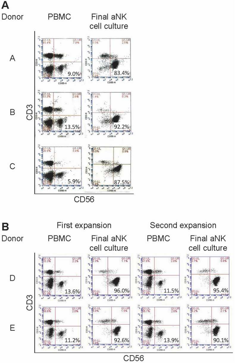
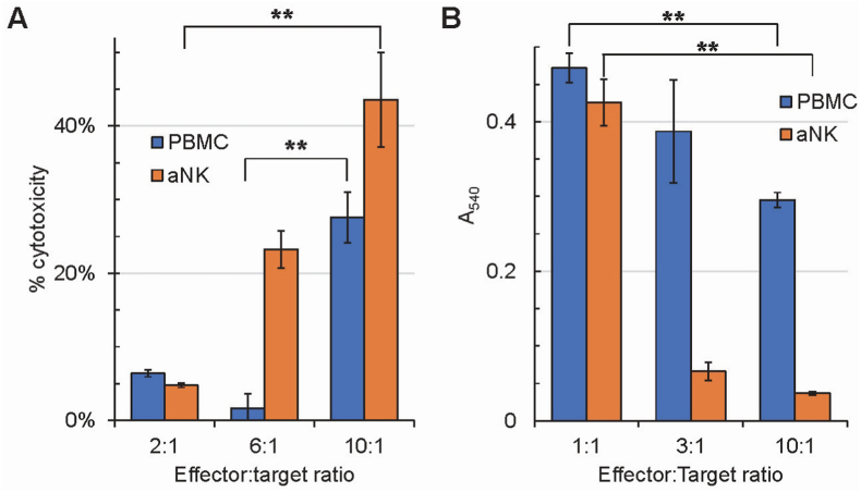
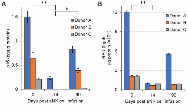
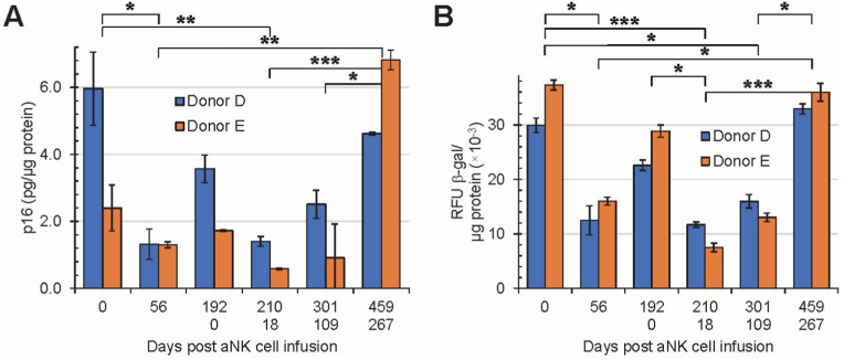
Aging is a multifactorial process involving many steps including senescence. The immune system plays a critical role in aging where chronic inflammation and senescence has been shown to be detrimental. Natural killer (NK) cells are the predominant innate lymphocyte subset that mediate various responses to include surveillance and elimination of senescent cells. Here, we use autologous propagated and activated NK (aNK) cells from 5 patients to demonstrate that aNK cells decrease senescent cells in vitro and immunosenescence in humans based on markers p16 and β-galactosidase. In addition, inflammatory cytokine panel data suggest a role for removal of immunosenescence to reduce the aging-related inflammatory response.

Keywords: Aging; Inflammation; NK cells; Senescence; aNK, Autologous propagated and activated natural killer cells.


Conflict of interest statement

The authors declare the following financial interests/personal relationships which may be considered as potential competing interests: Nickolas Chelyapov, Toai Nguyen, Rafael Gonzalez report a relationship with Thebiobox, LLC that includes: employment. Nickolas Chelyapov, Rafael Gonzalez have patent #16/178,369 METHODS AND COMPOSITIONS OF NATURAL KILLER CELL ADOPTIVE TRANSFER THERAPY issued to RESTEM.

Figures

Fig. 1
Fig. 1
Flow cytometry CD3 vs. CD56 dot plots of PBMCs and aNK cells. A: Results from cohort 1 with donors A, B, and C. B: Results from cohort 2 with donors D and E. The 5 patients had a median frequency of 11.2% CD3(−)CD56(+) NK cells (range 5.9–13.6) in their PBMCs, and following culture expansion, there was an increase of the median frequency to 91.0% CD3(−)CD56(+) aNK cells (range 83.4–96.0).
Fig. 2
Fig. 2
Cytotoxic effects of PBMCs and aNK cells on K562 cells (A) and senescent human skin fibroblasts (B). Based on flow cytometry results, PBMC numbers were adjusted to contain equivalent NK cell numbers in reactions with aNK cells. A: Kruskal-Wallis test indicates that there is significant difference between at least two experimental PBMC groups (p = 0.02651) and aNK groups (p = 0.02732); post-hoc Dunn's test identifies PBMC groups 6:1 vs 10:1 and aNK groups 2:1 vs 10:1 to have significant differences (p = 0.007049 and 0.00729, respectively). B: Kruskal-Wallis test indicates that there is significant difference between at least two experimental PBMC groups (p = 0.02732) and aNK groups (p = 0.02732); post-hoc Dunn's test identifies PBMC groups 1:1 vs 10:1 and aNK groups 1:1 vs 10:1 to have significant differences (p = 0.00729 and 0.00729, respectively). The cytotoxicity effects on K562 cells and senescent human skin fibroblasts are enhanced in aNK cells in comparison to PBMCs, and dose-dependent. Data were obtained in triplicates for each effector to target ratio, for PBMC and aNK cells; error bar = standard deviation. **: 0.001 < p ≤ 0.01 (Kruskal-Wallis test followed by post-hoc Dunn's test). If not indicated, results are not statistically significant.
Fig. 3
Fig. 3
Effects of aNK cell infusion on senescence markers p16 (A) and β-galactosidase (B) in human PBMCs isolated from donors A, B, and C, of ages, 70, 51, and 41 years old, respectively. Donors were infused with 109 aNK cells on day 0. Data was obtained in duplicates for each donor at each time point. Data within each time point were pooled from the three donors and analyzed with Kruskal-Wallis test followed by post-hoc Dunn's test. Error bar = Standard deviation. Expression of senescent markers p16 and β-galactosidase decreases following infusion of aNK cells and trends toward pre-infusion levels by 90 days post-infusion. Significant differences were denoted by *: 0.01 < p ≤ 0.05; **: 0.001 < p ≤ 0.01. If not indicated, results are not statistically significant.
Fig. 4
Fig. 4
Effects of aNK cell infusion on senescence markers p16 (A) and β-galactosidase (B) for human PBMCs in vivo. Donors D and E were infused with 2 × 109 of aNK cells twice, as indicated by the upper row (Days 0 through 192, first infusion) and lower row (Days 0 through 267, second infusion) of the x-axis label. Total study period 459 days. Error bar = Standard deviation. Data was obtained in duplicates for each donor at each time point. Data within each time point were pooled from the two donors and analyzed with Kruskal-Wallis test followed by post-hoc Dunn's test. Expression of senescent markers p16 and β-galactosidase decreases following infusion of aNK cells and trends toward pre-infusion levels by 6 months post-infusion; the pattern is repeated with a second infusion. Significant differences were denoted by *: 0.01 < p ≤ 0.05; **: 0.001 < p ≤ 0.01; ***: p ≤ 0.001. If not indicated, results are not statistically significant.

References

    1. Liochev S.I. Which is the most significant cause of aging? Review, Antioxidants. 2015;4:793–810. doi: 10.3390/antiox4040793. - DOI - PMC - PubMed
    1. Antonangeli F., Zingoni A., Soriani A., Santoni A. Senescent Cells: living or dying is a matter of NK cells. J. Leukoc. Biol. 2019;105(6):1275–1283. doi: 10.1002/JLB.MR0718-299R. - DOI - PubMed
    1. He S., Sharpless N.E. Senescence in health and disease. Cell. 2017;169(6):1000–1011. doi: 10.1016/j.cell.2017.05.015. - DOI - PMC - PubMed
    1. Boccardi V., Mecocci P. The importance of cellular senescence in frailty and cardiovascular diseases. Adv. Exp. Med. Biol. 2020;1216:79–86. doi: 10.1007/978-3-030-33330-0_9. - DOI - PubMed
    1. Baker D., Wijshake T., Tchkonia T., LeBrasseur N., et al. Clearance of p16Ink4a-positive senescent cells delays ageing-associated disorders. Nature. 2011;479(7372):232–236. doi: 10.1038/nature10600. - DOI - PMC - PubMed

LinkOut - more resources