

Circ_0084043-miR-134-5p axis regulates PCDH9 to suppress melanoma
- PMID: 36387162
- PMCID: PMC9641620
- DOI: 10.3389/fonc.2022.891476
Circ_0084043-miR-134-5p axis regulates PCDH9 to suppress melanoma
Abstract
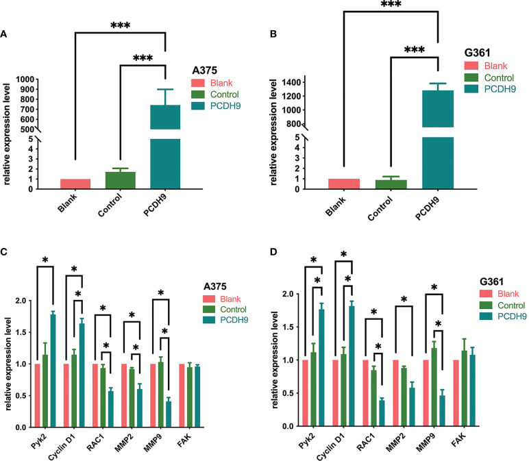
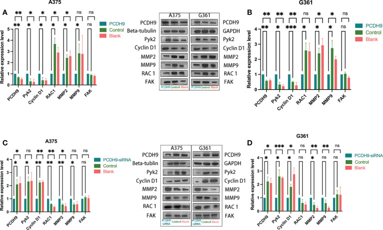
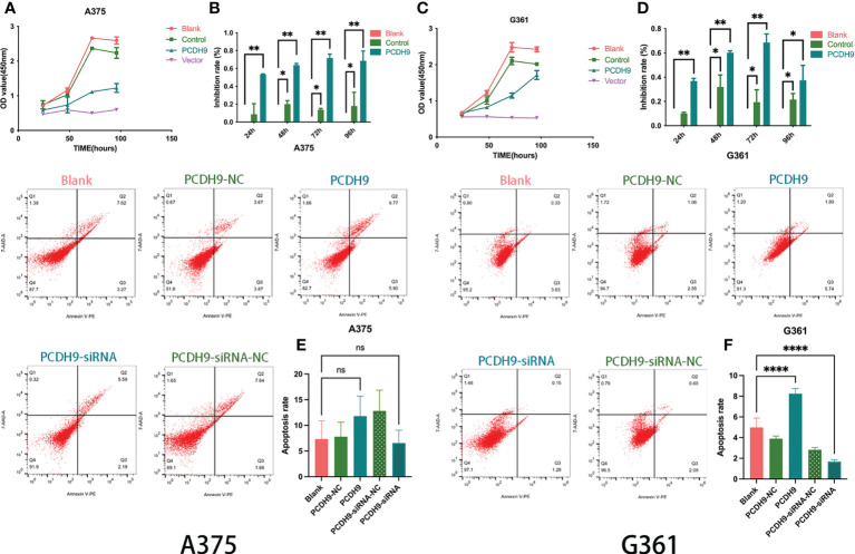
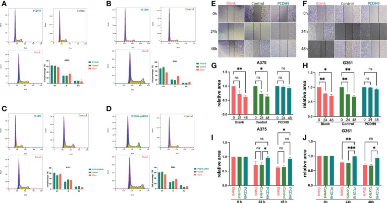
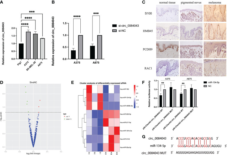
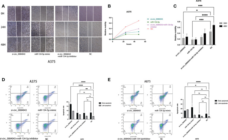
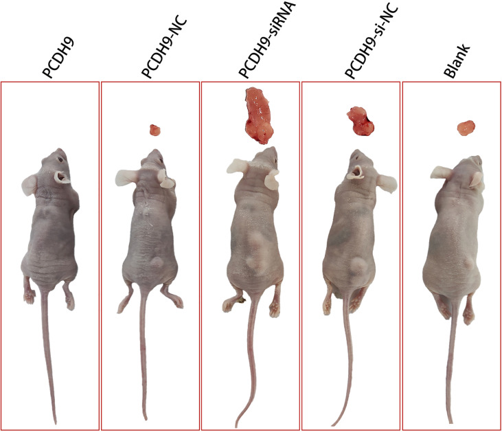
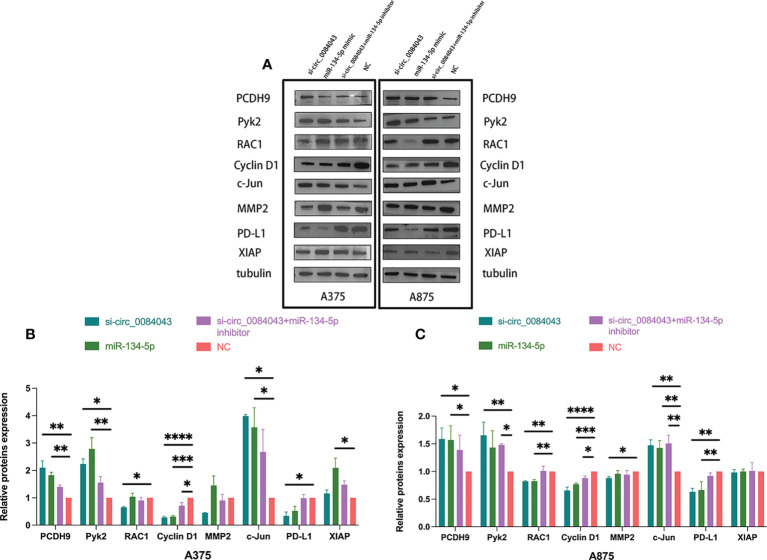
The low survival rates, poor responses, and drug resistance of patients with melanoma make it urgent to find new therapeutic targets. This study investigated whether the circ_0084043-miR-134-5p axis regulates the antitumor effect of protocadherin 9 (PCDH9) in melanoma. Ectopic expression or knock down (KD) of PCDH9 with a lentivirus vector, we explored its effects on the proliferation, invasion, and apoptosis of melanoma and verified its regulatory effect on ras-related C3 botulinum toxin substrate 1 (RAC1), proline-rich tyrosine kinase 2 (Pyk2), Cyclin D1, matrix metalloproteinase 2 (MMP2), and MMP9. We further observed the effect of KD circ_0084043 on the malignant behavior of melanoma and studied whether circ_0084043 sponged miR-134-5p and regulated PCDH9. We found that circ_0084043 was overexpressed in melanoma and associated with the malignant phenotype. PCDH9 was poorly expressed in human melanoma tissues, and overexpression of PCDH9 inhibited melanoma progression. Quantitative real-time PCR and Western blotting results showed that overexpression of PCDH9 could downregulate RAC1, MMP2, and MMP9 and upregulate Pyk2 and Cyclin D1. Circ_0084043 KD inhibited invasion and promoted apoptosis in melanoma cells. Circ_0084043 could sponge miR-134-5p and thus indirectly regulate PCDH9. Furthermore, we discovered that inhibiting circ_0084043 had an anti-PD-Ll effect. In vivo, PCDH9 overexpression inhibited melanoma tumor growth, but PCDH9 KD promoted it. In conclusion, PCDH9, which is regulated by the circ 0084043-miR-134-5p axis, can suppress malignant biological behavior in melanoma and influence the expression levels of Pyk2, RAC1, Cyclin D1, MMP2, and MMP9.
Keywords: PCDH9; Pyk2; RAC1; apoptosis; circRNAs; malignant melanoma.
Copyright © 2022 Cai, Zou, yang, Xie, Chen, Zheng, Luo, Wei, Liu and Chen.
Conflict of interest statement
The authors declare that the research was conducted in the absence of any commercial or financial relationships that could be construed as a potential conflict of interest.
Figures
Similar articles
-
PCDH9 suppresses melanoma proliferation and cell migration.Front Oncol. 2022 Nov 14;12:903554. doi: 10.3389/fonc.2022.903554. eCollection 2022. Front Oncol. 2022. PMID: 36452505 Free PMC article.
-
Circ_0016418 promotes melanoma development and glutamine catabolism by regulating the miR-605-5p/GLS axis.Int J Clin Exp Pathol. 2020 Jul 1;13(7):1791-1801. eCollection 2020. Int J Clin Exp Pathol. 2020. PMID: 32782707 Free PMC article.
-
Circ_0000620 acts as an oncogenic factor in gastric cancer through regulating MMP2 expression via sponging miR-671-5p.J Biol Res (Thessalon). 2021 Dec 31;28(1):23. doi: 10.1186/s40709-021-00154-5. J Biol Res (Thessalon). 2021. PMID: 34972532 Free PMC article.
-
Circ_0013359 facilitates the tumorigenicity of melanoma by regulating miR-136-5p/RAB9A axis.Open Life Sci. 2021 May 22;16(1):482-494. doi: 10.1515/biol-2021-0030. eCollection 2021. Open Life Sci. 2021. PMID: 34056112 Free PMC article.
-
Retracted Article: Overexpression of circ_0034642 contributes to hypoxia-induced glycolysis, cell proliferation, migration and invasion in gliomas by facilitating TAGLN2 expression via sponging miR-625-5p.RSC Adv. 2020 Jan 3;10(2):897-908. doi: 10.1039/c9ra08600e. eCollection 2020 Jan 2. RSC Adv. 2020. Retraction in: RSC Adv. 2021 Feb 1;11(10):5699. doi: 10.1039/d1ra90061g. PMID: 35494452 Free PMC article. Retracted.
Cited by
-
Curcumin targets miR-134-5p to suppress the progression of colorectal cancer through regulating the CDCA3/CDK1 pathway.Naunyn Schmiedebergs Arch Pharmacol. 2024 Jan;397(1):109-122. doi: 10.1007/s00210-023-02584-5. Epub 2023 Jun 27. Naunyn Schmiedebergs Arch Pharmacol. 2024. PMID: 37368030
-
MiR-134-5p inhibits the malignant phenotypes of osteosarcoma via ITGB1/MMP2/PI3K/Akt pathway.Cell Death Discov. 2024 Apr 25;10(1):193. doi: 10.1038/s41420-024-01946-z. Cell Death Discov. 2024. PMID: 38664375 Free PMC article.
-
circTADA2A inhibited SLC38A1 expression and suppresses melanoma progression through the prevention of CNBP trans-activation.PLoS One. 2024 Apr 18;19(4):e0301356. doi: 10.1371/journal.pone.0301356. eCollection 2024. PLoS One. 2024. PMID: 38635778 Free PMC article.
-
Exosomal circ_0084043 derived from colorectal cancer-associated fibroblasts promotes in vitro endothelial cell angiogenesis by regulating the miR-140-3p/HIF-1α/VEGF signaling axis.Heliyon. 2024 May 20;10(11):e31584. doi: 10.1016/j.heliyon.2024.e31584. eCollection 2024 Jun 15. Heliyon. 2024. PMID: 38828320 Free PMC article.
-
The lncRNA MIR22HG suppresses prostate cancer cell proliferation, migration, and epithelial-mesenchymal transition via the miR-4428/PCDH9 axis.Transl Cancer Res. 2025 May 30;14(5):3133-3148. doi: 10.21037/tcr-2024-2200. Epub 2025 May 14. Transl Cancer Res. 2025. PMID: 40530115 Free PMC article.
References
LinkOut - more resources
Full Text Sources
Research Materials
Miscellaneous