

Applications of 3D Photography in Craniofacial Surgery
- PMID: 36388007
- PMCID: PMC9648652
- DOI: 10.4103/jpn.JPN_48_22
Applications of 3D Photography in Craniofacial Surgery
Abstract
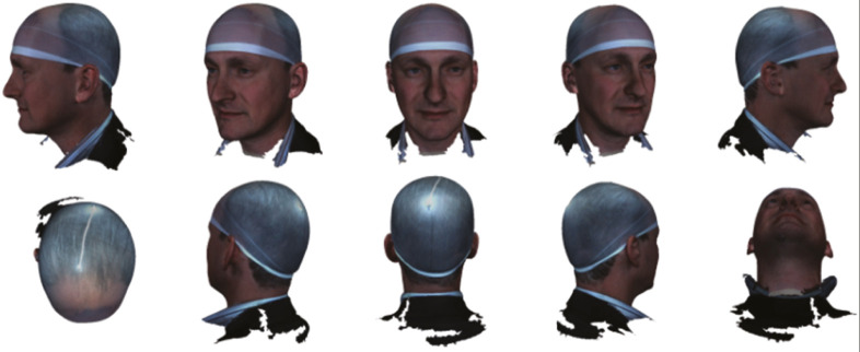
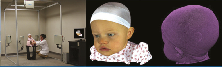
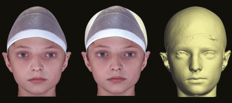
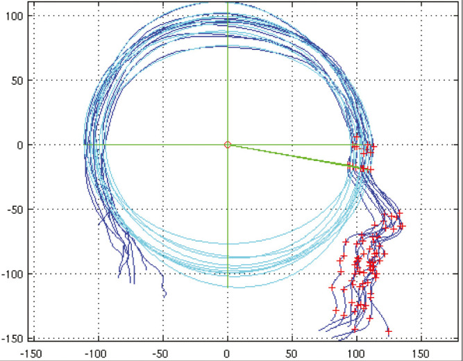
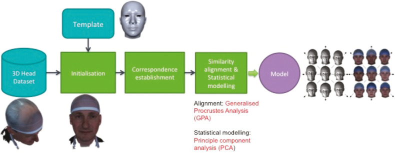
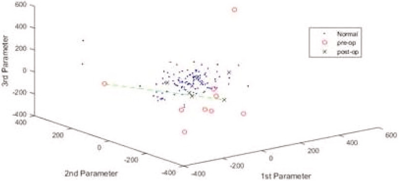
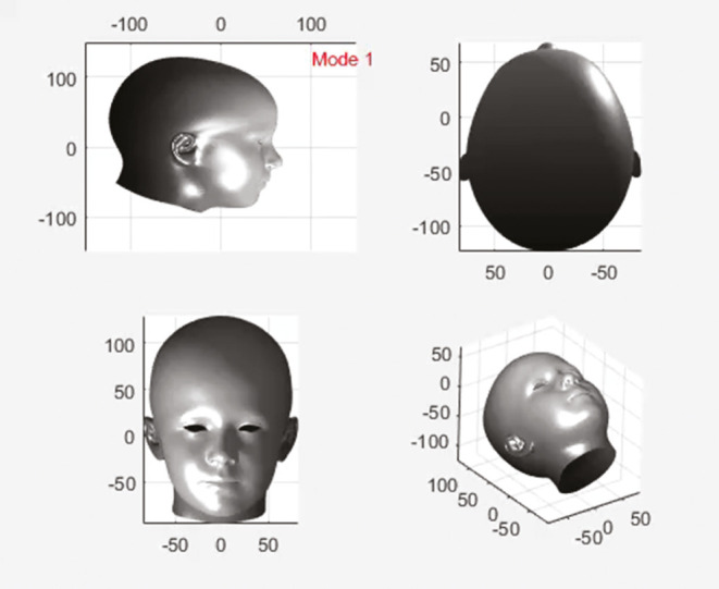
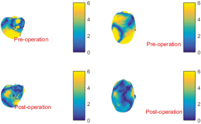
Three-dimensional (3D) photography is becoming more common in craniosynostosis practice and may be used for research, archiving, and as a planning tool. In this article, an overview of the uses of 3D photography will be given, including systems available and illustrations of how they can be used. Important innovations in 3D computer vision will also be discussed, including the potential role of statistical shape modeling and analysis as an outcomes tool with presentation of some results and a review of the literature on the topic. Potential future applications in diagnostics using machine learning will also be presented.
Keywords: 3D computer vision; 3D morphable models; 3D photogrammetry; craniosynostosis; outcomes; principal component analysis.
Copyright: © 2022 Journal of Pediatric Neurosciences.
Conflict of interest statement
There are no conflicts of interest.
Figures
References
-
- Zonnefeld FW, Lobregt S, Van der Meulen J, Vaandrager JM. Three dimensional imaging in craniofacial surgery. World J Surg. 1989;13:328–42. - PubMed
-
- Gibbons AJ, Duncan C, Nishikawa H, Hockley AD, Dover MS. Stereolithographic modelling and radiation dosage. Br J Oral Maxillofac Surg. 2003;41:416. - PubMed
-
- Nayler JR. Clinical photography: A guide for the clinician. J Postgrad Med. 2003;49:256–62. - PubMed
-
- Knoops PG, Beaumont CA, Borghi A, Rodriguez-Florez N, Breakey RW, Rodgers W, et al. Comparison of three-dimensional scanner systems for craniomaxillofacial imaging. J Plast Reconstr Aesthet Surg. 2017;70:441–9. - PubMed
Publication types
LinkOut - more resources
Full Text Sources
Other Literature Sources