Review
. 2022 Sep;17(Suppl 1):S29-S43.
doi: 10.4103/jpn.JPN_26_22. Epub 2022 Sep 19.

Multisuture and Syndromic Craniosynostoses: Simplifying the Complex

Affiliations
Review

Multisuture and Syndromic Craniosynostoses: Simplifying the Complex

Suhas Udayakumaran et al. J Pediatr Neurosci. 2022 Sep.

Abstract

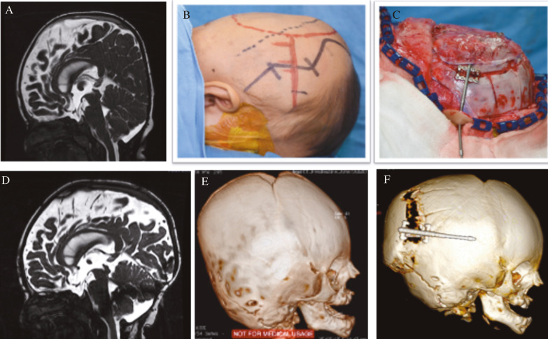
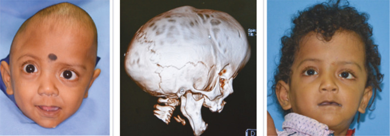
Most complex craniosynostoses are managed the same way as syndromic craniosynostoses (SCs), as these patients often experience similar problems regarding cognition and increased intracranial pressure (ICP). The evaluation and treatment plan for craniosynostoses is complex, and this, additionally, is complicated by the age at presentation. In this article, the authors review the complexity of SCs in the presentation and management. An algorithm is necessary for such multifaceted and multidimensional pathology as craniosynostoses. In most algorithms, posterior calvarial distraction is a consistent early option for complex craniosynostoses presenting early with raised ICP. Addressing the airway early is critical when significant airway issues are there. All other surgical interventions are tailored on the basis of presentation and age.

Keywords: Algorithm; complex; craniosynostoses; management; multisutural; syndromic.


Conflict of interest statement

The authors report no conflict of interest concerning the materials or methods used in this study or the findings specified in this paper.

Figures

Figure 1
Figure 1
Algorithm for less than 2-year-old (age at presentation). PVDO (PCVD) is usually the first option. This is usually followed by FOAR. Other options are as suggested in the algorithm
Figure 2
Figure 2
Algorithm for more than 2-year-old (age at presentation). PVDO (PCVD) is not usually the first option and is done only if the Chiari and hydrocephalus are a prominent presentation or if extreme turricephaly needs to be addressed. FOAR may be the initial option in this age category. Other options are as suggested in the algorithm. The algorithm is tailored depending on the patient presentation
Figure 3
Figure 3
Illustrative case 1. A and B: An 11-month-old infant presented with hydrocephalus and signs of raised ICP. He had PCVD (illustrated in Figure 4). Additionally, the child had severe airway issues with recurrent episodes of URTI. C: Subsequently, he had robotic-assisted frontofacial advancement (during the removal of posterior calvarial distractors). D: Robot-assisted insertion of transfacial pin for MD. E: MDCT showing the trajectory of the transfacial pin planned to get adequate purchase keeping avoidance to the injury globe and tooth buds in perspective
Figure 4
Figure 4
Illustrative case 2. An 11-month-old infant undergoing PCVD. A: Pre-operative MR showing tight posterior fossa and tonsillar herniation. B: Surgical planning with the patient-prone position. The incision is planned, keeping the distractor position in perspective. C: Intraoperative photograph showing the craniotomy with distractor in situ. D: Post-operative MRI improved CSF spaces in the posterior fossa and the posterior calvarium in general. E: Pre-operative MDCT showing flattened occiput, multisutural involvement, and the digital impression suggesting raised ICP. F: Post-operative MDCT showing the craniotomy with distractor in situ
Figure 5
Figure 5
Illustrative case 3. A child with Crouzons. He presented as an infant with hydrocephalus. At 3 months, he had an endoscopic third ventriculostomy. Subsequently, he had PCVD. Upper row: Post PCVD. He continued to have severe airway issues with repeated episodes of upper respiratory infections. He underwent frontofacial distraction using internal distractors. Middle row: Post frontofacial distraction. Lower row: Follow-up at 6 years. At follow-up, his aesthetic outcome seemed suboptimal, which is one of the disadvantages of early midface correction
Figure 6
Figure 6
Illustrative case 4. A: A 17-year-old, young boy with Apert syndrome presenting in a delayed manner. He was scholastically poor but his concern was aesthetics only. B: He underwent frontofacial distraction using an external distractor (RED) device. C: Post distraction, the result seemed satisfactory
Figure 7
Figure 7
Illustrative case 5. This was a 5-year-old child who presented to us with hypertelorism. He underwent frontal remodeling with box craniotomy (extreme left)
Figure 8
Figure 8
Illustrative case 6. A 5-year-old child with Apert syndrome who had an FOAR at 14 months now presented with signs of raised ICP. He would require an MD later. Presently, he has no airway issues
Figure 9
Figure 9
Illustrative case 7. A child with multisutural craniosynostoses. He presented at 2 years with signs of raised ICP. MDCT showed multiple sutural stenoses

References

    1. Tolarova MM, Harris JA, Ordway DE, Vargervik K. Birth prevalence, mutation rate, sex ratio, parents’ age, and ethnicity in Apert syndrome. Am J Med Genet. 1997;72:394–8. - PubMed
    1. Armand T, Schaefer E, Di Rocco F, Edery P, Collet C, Rossi M. Genetic bases of craniosynostoses: An update. Neurochirurgie. 2019;65:196–201. - PubMed
    1. Di Rocco F, Rothenbuhler A, Cormier Daire V, Bacchetta J, Adamsbaum C, Baujat G, et al. Craniosynostosis and metabolic bone disorder. A review. Neurochirurgie. 2019;65:258–63. - PubMed
    1. Kalmar CL, Zapatero ZD, Kosyk MS, Carlson AR, Bartlett SP, Heuer GG, et al. Elevated intracranial pressure with craniosynostosis: A multivariate model of age, syndromic status, and number of involved cranial sutures. J Neurosurg Pediatr. 2021;28:1–8. - PubMed
    1. Jabs EW, Müller U, Li X, Ma L, Luo W, Haworth IS, et al. A mutation in the homeodomain of the human Msx2 gene in a family affected with autosomal dominant craniosynostosis. Cell. 1993;75:443–50. - PubMed