. 2022 Oct 26:13:1016242.
doi: 10.3389/fphys.2022.1016242. eCollection 2022.

Stable potassium isotopes (41K/39K) track transcellular and paracellular potassium transport in biological systems

Affiliations

Stable potassium isotopes (41K/39K) track transcellular and paracellular potassium transport in biological systems

John A Higgins et al. Front Physiol. .

Abstract

As the most abundant cation in archaeal, bacterial, and eukaryotic cells, potassium (K+) is an essential element for life. While much is known about the machinery of transcellular and paracellular K transport-channels, pumps, co-transporters, and tight-junction proteins-many quantitative aspects of K homeostasis in biological systems remain poorly constrained. Here we present measurements of the stable isotope ratios of potassium (41K/39K) in three biological systems (algae, fish, and mammals). When considered in the context of our current understanding of plausible mechanisms of K isotope fractionation and K+ transport in these biological systems, our results provide evidence that the fractionation of K isotopes depends on transport pathway and transmembrane transport machinery. Specifically, we find that passive transport of K+ down its electrochemical potential through channels and pores in tight-junctions at favors 39K, a result which we attribute to a kinetic isotope effect associated with dehydration and/or size selectivity at the channel/pore entrance. In contrast, we find that transport of K+ against its electrochemical gradient via pumps and co-transporters is associated with less/no isotopic fractionation, a result that we attribute to small equilibrium isotope effects that are expressed in pumps/co-transporters due to their slower turnover rate and the relatively long residence time of K+ in the ion pocket. These results indicate that stable K isotopes may be able to provide quantitative constraints on transporter-specific K+ fluxes (e.g., the fraction of K efflux from a tissue by channels vs. co-transporters) and how these fluxes change in different physiological states. In addition, precise determination of K isotope effects associated with K+ transport via channels, pumps, and co-transporters may provide unique constraints on the mechanisms of K transport that could be tested with steered molecular dynamic simulations.

Keywords: Na-K ATPase; homeostasis; physiology; potassium; potassium channel; stable isotope.


Conflict of interest statement

The authors declare that the research was conducted in the absence of any commercial or financial relationships that could be construed as a potential conflict of interest.

Figures

FIGURE 1
FIGURE 1
The difference in δ41K values between external and intracellular K+ in growth experiments of C. reinhardtii and a diagram of K+ transport in single­celled algae grown under optimal conditions (See Methods) including major fluxes (Fin), transport machinery, and K isotope effects (εin). p = 4.5 × 10⁻⁵ by one-way ANOVA in Matlab.
FIGURE 2
FIGURE 2
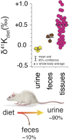
Whole-body K isotope mass balance in R. norvegicus on a controlled diet. Measured δ41K values of urine (p = 0.19) and feces (p = 0.037) relative to diet indicate a preferential uptake of 39K in the gut. The total range in δ41Kdiet values in the tissues of R. norvegicus is ∼1‰ with an estimated whole-body average δ41Kdiet value of approx. +0.5‰, consistent with preferential loss of 39K in urine.
FIGURE 3
FIGURE 3
(A) K isotopic composition of tissues/fluids R. norvegicus (casein diet) normalized to plasma (δ41Kplasma = 0‰). δ41Kplasma values that are positive indicate that net K+ transport out of the tissue/cell/fluid is enriched in 39K relative to K+ transport into the tissue/cell/fluid. δ41Kplasma values that are indistinguishable from 0 indicate that the K isotope fractionation associated with K+ transport into and out of the cell/tissue/fluid are either 0 or equal in magnitude (and thus cancel). δ41Kplasma values that are negative indicate that net K+ transport into the cell/tissue/fluid is enriched in 39K relative to K+ transport out of the cell/tissue/fluid. (B) A diagram of K isotope mass balance including transport by (1) K-channels, (2) K-pumps/co-transporters, (3) through pores in tight-junctions, and (4) glomerular filtration. Eq. 3 solves for the δ41K value of a tissue/cell relative to blood plasma assuming steady-state K isotope mass balance with bi-directional K+ transport by both channels and pumps/co-transporters. Variables include fout/in ch , the fraction of total K+ transport into/out of the tissue/cell that occurs through K+ channels, Δfch , the difference in the fraction of K transport via channels (fout ch -fin ch), εch , K isotope fractionation associated with K channels, and εp/co K isotope fractionation associated with pumps/co-transporters. K isotope fractionation associated with K+ uptake in the gut (εgut) and K+ loss in urine (εkidney) reflect the flux-weighted average of both paracellular and transcelluar K+ transport. For example, K isotope fractionation in the kidney will be associated with both re-absorption and secretion of K+ in the proximal tubule, the thick ascending limb (TAL), and cells in the collecting ducts of the kidney. (C) Correlation between intercellular [K+] and δ41Kplasma values for a subset of tissues from R. norvegicus. Also shown are the measured activities of NaKATPase (µmol P h−1 g−1 from Gick et al., (1993) and values of Δfch from Eq. 3.
FIGURE 4
FIGURE 4
K isotopic composition of cerebrospinal fluid from R. norvegicus raised on a controlled diet (vivarium chow) normalized to blood plasma (δ41Kplasma = 0‰) and brain tissues normalized to cerebrospinal fluid (δ41KCSF = 0‰) together with a diagram of K homeostasis across the choroid plexus and blood brain barrier (BBB) from Hladky and Barrand (2016) including transport by (1) K-channels, (2) K-pumps/co-transporters, and (3) transcellularly through pores in tight-junctions. δ41KCSF values that are positive indicate that net K transport out of brain tissues (spinal cord, cerebellum, and cerebrum) and into CSF is enriched in 39K relative to K transport from CSF into these tissues. The negative δ41Kplasma values for CSF indicate that net K transport into CSF from blood plasma is enriched in 39K relative to K transport from CSF to blood plasma and reflects the flux­ weighted average of the transporters that dominate K+ exchange across both the choroid plexus and BBB.
FIGURE 5
FIGURE 5
(A) The difference in δ41K values between seawater and dorsal white muscle of various stenohaline and euryhaline marine fish. Muscle [K+] and δ41Kseawater values are listed in Supplementary Table S1. p = 1.4 × 10⁻¹⁰ for the difference in δ41Kseawater value between stenohaline and euryhaline teleosts. (B) A model of K isotope mass balance in a marine fish. K+ is supplied across the gill epithelium via transport through pores in tight-junction proteins and lost from mitochondria-rich cells (MRCs) through channels Eq. 5. K+ cycling within MRCs is modeled using Eq. 3. Combining Eq. 3 and Eq. 5a yields Eq. 6, the δ41K value of plasma/muscle relative to seawater as a funciton of εch, εgill, Δfch of MRC’s and εp/co. As Δfch → 0, δ41K of fish muscle → εch - εgill (≥ 0‰, euryhaline), whereas as Δfch→ 0, δ41K of fish muscle → εp/cogill (<0‰, stenohaline). fish illustrations from Lori Moore.

References

    1. Agarwal R., Afzalpurkar R., Fordtran J. S. (1994). Pathophysiology of potassium absorption and secretion by the human intestine. Gastroenterology 107, 548–571. 10.1016/0016-5085(94)90184-8 - DOI - PubMed
    1. Barnaby C. F., Edmonds C. J. (1969). Use of a miniature GM counter and a whole body counter in the study of potassium transport by the colon of normal, sodium-depleted and adrenalectomized rats in vivo . J. Physiol. 205, 647–665. 10.1113/jphysiol.1969.sp008988 - DOI - PMC - PubMed
    1. Berneche S., Roux B. (2003). A microscopic view of ion conduction through the K+ channel. Proc. Natl. Acad. Sci. U. S. A. 100, 8644–8648. 10.1073/pnas.1431750100 - DOI - PMC - PubMed
    1. Betz A. L., Firth J. A., Goldstein G. W. (1980). Polarity of the blood-brain barrier: Distribution of enzymes between the luminal and antiluminal membranes of brain capillary endothelial cells. Brain Res. 192, 17–28. 10.1016/0006-8993(80)91004-5 - DOI - PubMed
    1. Boron W. F., Boulpaep E. L. (2005). Medical physiology. Philadelphia, Pennsylvania: Elsevier Saunders.