. 2022 Nov 7:2022:9042345.
doi: 10.1155/2022/9042345. eCollection 2022.

Exosomes Derived from Dermal Papilla Cells Mediate Hair Follicle Stem Cell Proliferation through the Wnt3a/ β-Catenin Signaling Pathway

Affiliations

Exosomes Derived from Dermal Papilla Cells Mediate Hair Follicle Stem Cell Proliferation through the Wnt3a/ β-Catenin Signaling Pathway

Jiali Li et al. Oxid Med Cell Longev. .

Abstract

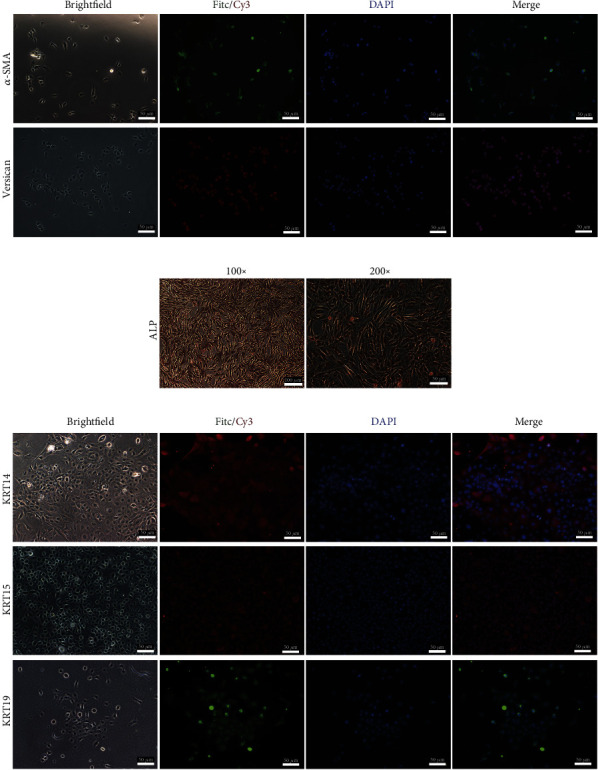
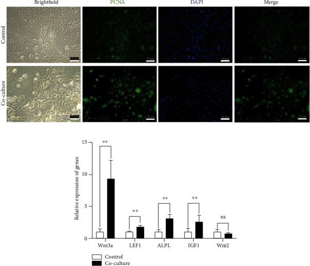
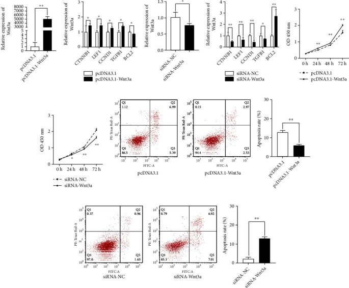
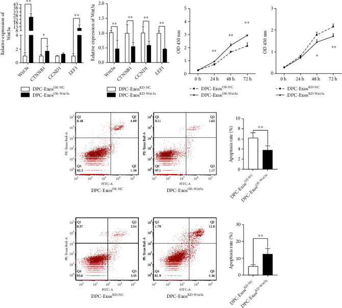
Both hair follicle stem cells (HFSC) and dermal papilla cells (DPC) are essential for hair follicle growth and proliferation. In this study, HFSCs and DPCs that made signature proteins like KRT14, KRT15, KRT19, α-SMA, and Versican were obtained. Cell coculture systems between HFSCs and DPCs were used to measure the increased PCNA protein content in HFSCs. Additionally, exosomes from dermal papilla cells (DPC-Exos), the overexpression and silencing of Wnt3a, could regulate the Wnt/β-catenin signaling pathway downstream genes. After collecting DPC-ExosOE-Wnt3a, the treatment of HFSC with DPC-ExosOE-Wnt3a showed that DPC-ExosOE-Wnt3a could upregulate the mRNA expression of downstream genes in the Wnt/β-catenin signaling pathway and that DPC-ExosOE-Wnt3a enhanced the proliferation of HFSCs while inhibiting their apoptosis. These findings suggest that DPC-Exos could regulate HFSC cell proliferation via the Wnt3a/β-catenin signaling pathway. This research offers novel concepts for the molecular breeding and efficient production of Angora rabbits, as well as for the treatment of human hair problems.


Conflict of interest statement

The authors declare that they have no competing interests.

Figures

Figure 1
Figure 1
Morphology and indirect immunofluorescence staining of HFSCs and DPCs. (a) α-SMA and Versican antibodies were incubated with DPCs (passage 2) (scale bar: 50 μm). (b) Alkaline phosphatase staining was examined with DPCs (passage 8) (scale bar: 100 μm, 50 μm). (c) KRT14, KRT15, and KRT19 antibodies were incubated with HFSCs (passage 2) (scale bar: 50 μm).
Figure 2
Figure 2
DPCs promoted the HFSC proliferation in cell cocultured system. (a) Indirect immunofluorescence staining of HFSCs. After coculturing with DPCs (passage 3) for 48 h, HFSCs (passage 3) were incubated with antibodies against PCNA (scale bar: 50 μm). The upper chamber of the control group did not contain any cells. (b) The cocultured with DPCs (passage 3) increased the expression of HFSCs (passage 3).
Figure 3
Figure 3
Wnt3a regulation of the Wnt signaling pathway. (a) The mRNA expression levels of siRNA-Wnt3a in the four groups were detected by qRT-PCR. siRNA significantly decreased the expression of the Wnt3a mRNA levels in HFSCs (passage 4). (b) The knockdown of Wnt3a decreased the expression of Wnt/β-catenin pathway. (c) pcDNA3.1-Wnt3a significantly increased the expression of the Wnt3a mRNA levels in HFSCs (passage 4). (d) The overexpression of Wnt3a increased the expression of the Wnt/β-catenin pathway. (e) siRNA-Wnt3a and NC cell proliferation estimated by CCK8 assays after 24, 48, and 72 h. (f) PcDNA3.1, pcDNA3.1-Wnt3a cell proliferation was estimated by CCK8 assays after 24, 48, and 72 h. (g) Cell apoptosis of the knockdown Wnt3a after 3 days. (h) Cell apoptosis of the overexpression Wnt3a after 3 days. P < 0.05, ∗∗P < 0.01.
Figure 4
Figure 4
The DPC-Exos identification. (a) Detection of the shape and size of the DPC-Exos by TEM (scale bars, 100 nm). (b) Detection of the particle size and concentration of the DPC-Exos by nanoparticle tracking analysis (NTA). (c) Detection of the expression of exosome-specific surface markers of the DPC-Exos by Western blotting. (d) DiI-DPC-Exos could enter HFSCs (passage 2) in the Transwell plate (scale bars, 50 μm).
Figure 5
Figure 5
DPC-Exos regulated the HF growth and HFSC proliferation through the Wnt3a/β-catenin signaling pathway. (a, b) The Wnt3a expression and mRNA expression of the genes related to HF growth and development were regulated by DPC-Exos. (c) The DPC-ExosOE-Wnt3a promoted the proliferation of HFSCs. (d) The DPC-ExosKD-Wnt3a inhibited the proliferation of HFSCs. (e) The DPC-ExosOE-Wnt3a inhibited the HFSC apoptosis. (f) The DPC-ExosKD-Wnt3a promoted the HFSC apoptosis. ∗∗P < 0.01. P < 0.05.

References

    1. Rothbauer M., Zirath H., Ertl P. Recent advances in microfluidic technologies for cell-to-cell interaction studies. Lab on a Chip . 2018;18(2):249–270. doi: 10.1039/C7LC00815E. - DOI - PubMed
    1. Hardy M. H. The secret life of the hair follicle. Trends in Genetics . 1992;8(2):55–61. doi: 10.1016/0168-9525(92)90350-D. - DOI - PubMed
    1. Oshima H., Rochat A., Kedzia C., Kobayashi K., Barrandon Y. Morphogenesis and renewal of hair follicles from adult multipotent stem cells. Cell . 2001;104(2):233–245. doi: 10.1016/S0092-8674(01)00208-2. - DOI - PubMed
    1. Gentile P., Scioli M. G., Bielli A., Orlandi A., Cervelli V. Stem cells from human hair follicles: first mechanical isolation for immediate autologous clinical use in androgenetic alopecia and hair loss. Stem cell investigation . 2017;4(7):p. 58. doi: 10.21037/sci.2017.06.04. - DOI - PMC - PubMed
    1. Chi W., Wu E., Morgan B. A. Dermal papilla cell number specifies hair size, shape and cycling and its reduction causes follicular decline. Development . 2013;140(8):1676–1683. doi: 10.1242/dev.090662. - DOI - PMC - PubMed