. 2022 Oct 26:13:998904.
doi: 10.3389/fneur.2022.998904. eCollection 2022.

Reversing painful and non-painful diabetic neuropathy with the capsaicin 8% patch: Clinical evidence for pain relief and restoration of function via nerve fiber regeneration

Affiliations

Reversing painful and non-painful diabetic neuropathy with the capsaicin 8% patch: Clinical evidence for pain relief and restoration of function via nerve fiber regeneration

Praveen Anand et al. Front Neurol. .

Abstract

Introduction: Current oral treatments for pain in diabetic peripheral neuropathy (DPN) do not affect the progression of DPN i.e., "disease modification." We assessed whether Capsaicin 8% patch treatment can provide pain relief and also restore nerve density and function via nerve regeneration, in both painful (PDPN) and non-painful (NPDPN) diabetic peripheral neuropathy.

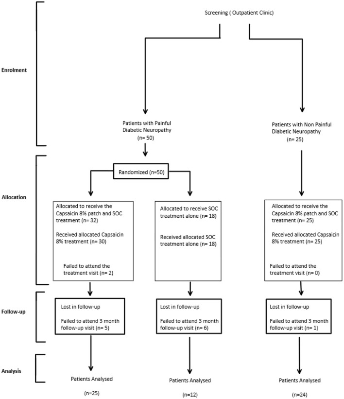
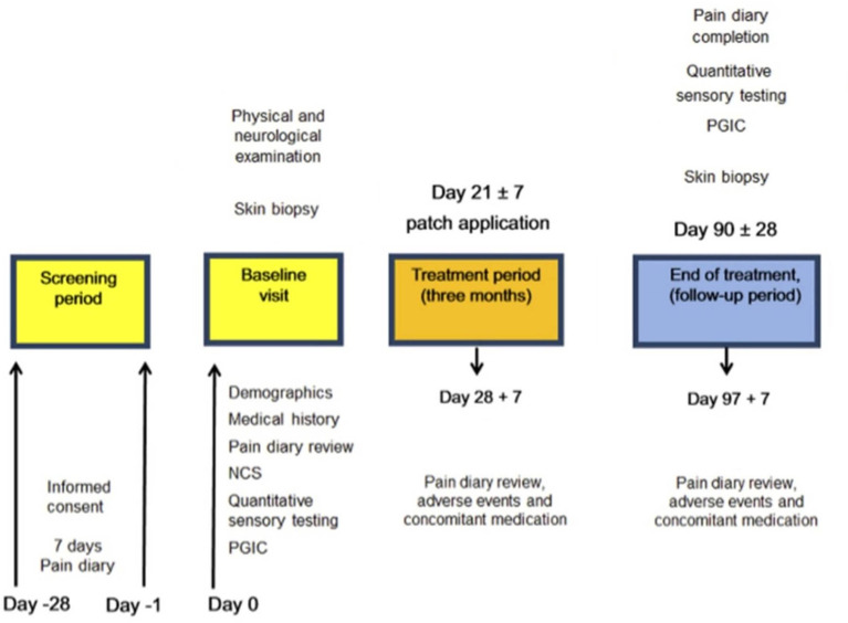
Methods: 50 participants with PDPN were randomized to receive Capsaicin 8% patch Qutenza with Standard of Care (SOC) (PDPN Q+SOC group), or SOC alone (PDPN SOC group). Pain symptoms were assessed with a diary (Numerical Pain Rating Scale, NRPS) and questionnaires. Investigations included quantitative sensory testing (QST) and distal calf skin biopsies, at baseline and 3 months after baseline visit; subsequent options were 3-monthly visits over 1 year. 25 participants with NPDPN had tests at baseline, and 3 months after all received Capsaicin 8% patch treatment.

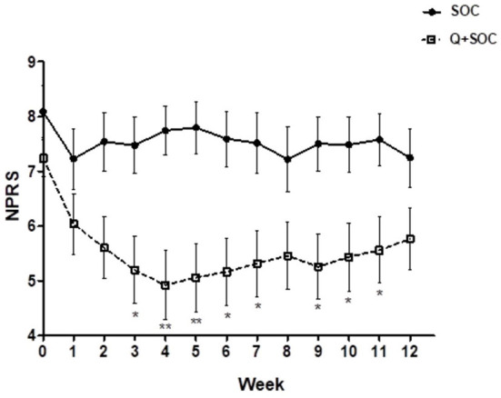
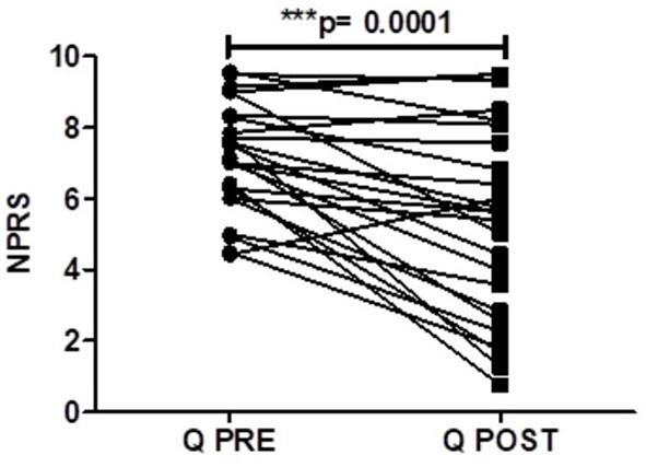
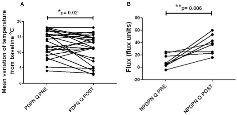
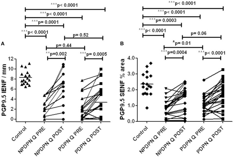
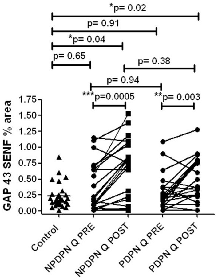
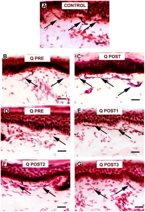
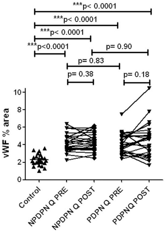
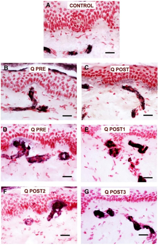
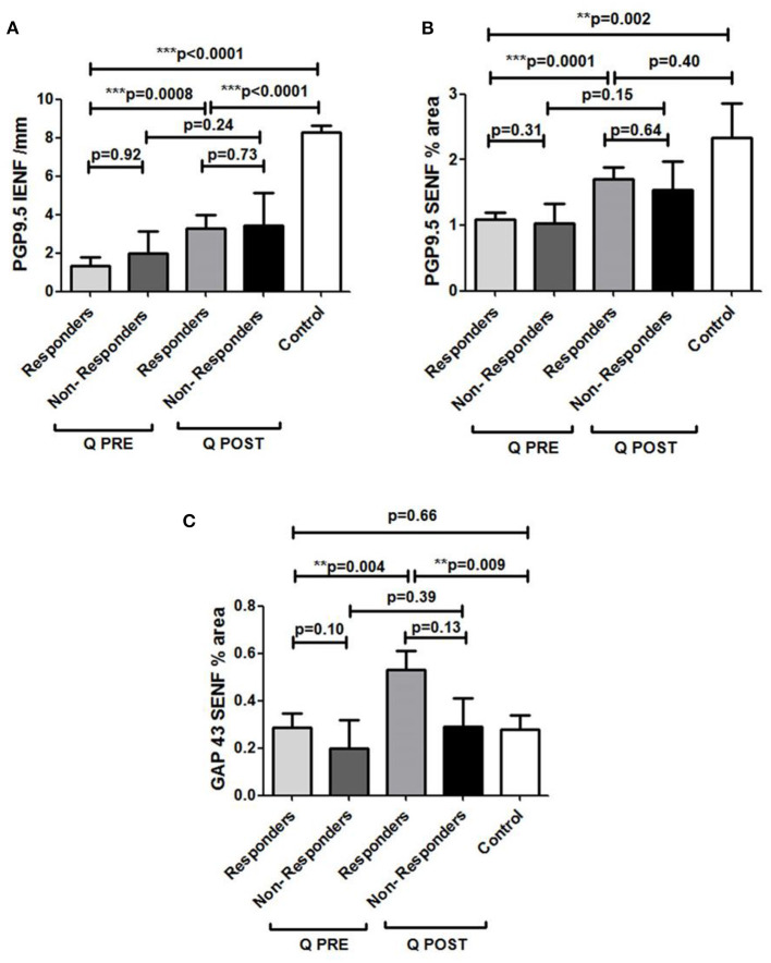
Results: At 3 months after baseline, PDPN Q+SOC group had reduction in NPRS score (p = 0.0001), but not PDPN SOC group. Short-Form McGill Pain Questionnaire (SF-MPQ) showed significant reductions in scores for overall and other pain descriptors only in the PDPN Q+SOC group. Warm perception thresholds were significantly improved only in the PDPN Q+SOC group (p = 0.02), and correlated with reduction in SF-MPQ overall pain score (p = 0.04). NPDPN Q+SOC group did not report pain during the entire study. Density of intra-epidermal nerve fibers (IENF) with PGP9.5 was increased at 3 months in PDPN Q+SOC (p = 0.0002) and NPDPN Q+SOC (p = 0.002) groups, but not in the PDPN SOC group. Increased sub-epidermal nerve fibers (SENF) were observed with GAP43 (marker of regenerating nerve fibers) only in PDPN Q+SOC (p = 0.003) and NPDPN Q+SOC (p = 0.0005) groups. Pain relief in the PDPN Q+SOC group was correlated with the increased PGP9.5 IENF (p = 0.0008) and GAP43 (p = 0.004), whereas those with lack of pain relief showed no such increase; in some subjects pain relief and increased nerve fibers persisted over months. PGP9.5 IENF increase correlated with axon-reflex vasodilatation in a NPDPN Q+SOC subset (p = 0.006).

Conclusions: Capsaicin 8% patch can provide pain relief via nerve regeneration and restoration of function in DPN (disease modification). It may thereby potentially prevent diabetic foot complications, including ulcers.

Keywords: capsaicin; clinical trial; diabetic neuropathy; pain; skin biopsy.


Conflict of interest statement

This study received supplementary funding from Grünenthal GmbH. The funder had the following involvement with the study: interpretation of data. PA has received symposia speaker fees and advisory board honoraria from Grunenthal, but no personal remuneration for conducting this study. The remaining authors declare that the research was conducted in the absence of any commercial or financial relationships that could be construed as a potential conflict of interest.

Figures

Figure 1
Figure 1
Participant flow diagram for DPN Capsaicin 8% patch studies.
Figure 2
Figure 2
Study flow diagram illustrating the sequence of assessment for participants in the study. NCS, Nerve conduction study, PGIC, Patient Global Impression of Change.
Figure 3
Figure 3
Time–course graph of spontaneous pain scores over 12 weeks in the two study groups, capsaicin 8% patch Qutenza (Q) plus Standard of Care group (PDPN Q+SOC; n = 25), and Standard of Care group (PDPN SOC alone; n = 12). Separate assessment of pain scores at each week time point showed a significant difference in NPRS scores at week 3 (*p < 0.05), week 4 (**p < 0.05), week 5 (*p < 0.05), week 6 (*p < 0.05) and week 9 (*p < 0.05; two–way ANOVA analysis).
Figure 4
Figure 4
Pain scores of patients (PDPN Q + SOC, n = 25) treated with capsaicin 8% patch Qutenza (Q), at pre–treatment visit (Q PRE) and 3 months after treatment (Q POST), on Numerical Pain Rating Scale (NPRS). Statistically significant difference at 3 months after treatment (***p = 0.0001; n = 25; paired t–test).
Figure 5
Figure 5
Warm thermal thresholds in patients with DPN. (A) Thermal thresholds for warm perception (changes from baseline at 32°C), in PDPN Q + SOC group treated with capsaicin 8% patch Qutenza (Q), at pre–treatment visit (Q PRE) and visit 3 months after treatment (Q POST). Statistically significant difference (improvement) 3 months after the treatment (*p = 0.02; paired t–test). (B) Axon reflex vasodilatation flux in patients with NPDPN group treated with capsaicin 8% patch Qutenza (Q) at pre–treatment visit (Q PRE) and visit 3 months after treatment (Q POST). Statistically significant difference (improvement) 3 months after the treatment (*p = 0.006; paired t–test).
Figure 6
Figure 6
PGP9.5 immunohistochemistry in 50 μm sections from skin biopsies in non–painful (NPDPN Q+SOC) and painful (PDPN Q+SOC) Diabetic Peripheral Neuropathy following Capsaicin 8% patch Qutenza (Q) treatment. Intra–epidermal (IENF/mm, A) and sub–epidermal fiber density (SENF, % area, B) in NPDPN and PDPN. (A) IENFs in NPDPN Q +SOC and PDPN Q+SOC at baseline (Q PRE) and statistically non–significant difference before (Q PRE, p = 0. 44; Mann–Whitney test; n = 24 NPDPN Q+SOC and n = 25 PDPN Q+SOC) or 3 months after treatment (Q POST, p = 0.52, Mann–Whitney test). Statistically significant difference between control group and baseline (Q PRE), and after treatment (Q POST) (Mann–Whitney test). (B) SENFs in NPDPN Q+SOC and PDPN Q+SOC at baseline (Q PRE) and statistically significant difference before (Q PRE, *p = 0. 01; Mann–Whitney test; n = 24 NPDPN Q+SOC and n = 25 PDPN Q+SOC) but not 3 months after treatment (Q POST, p = 0.06, Mann–Whitney test). Statistically significant decreases between control group and baseline (Q PRE) but less significance after treatment (Q POST) (Mann–Whitney test) in NPDPN Q+SOC but not PDPN Q+SOC. ***Highly significant.
Figure 7
Figure 7
Staining for PGP9.5 in control skin, NPDPN Q +SOC and PDPN Q+SOC before and after application of Capsaicin 8% patch Qutenza (Q) treatment (50 μm sections). (A) Control skin biopsy section, from a healthy human volunteer (intra–epidermal nerve fibers marked with arrowheads and sub–epidermal nerve fibers with arrows). (B) Skin biopsy section from a subject with NPDPN Q+SOC pre–treatment (Q PRE); few intra–epidermal nerve fibers and sub–epidermal nerve fibers were observed before treatment. (C) Skin biopsy section from same subject with painless DPN (NPDPN Q+SOC) as above post–treatment (Q POST): the abundance of both the IENFs and SENFs appeared restored. (Scale bar 50 μm, Original magnification x40). (D) Skin biopsy section from a subject with PDPN Q+SOC pre–treatment (Q PRE); Few intra–epidermal nerve fibers and with abnormal trajectory before capsaicin application (arrowheads). (E–G) Examples from biopsies collected 3. 6 and 9 months (Q POST 1, 2, 3) after a single Qutenza application respectively. Note the restored abundance of both IENF and SENF, and the vertical trajectory of IENF Q POST, as observed in control skin. (Scale bar 100 μm, original magnification x20).
Figure 8
Figure 8
GAP 43 immunohistochemistry from skin biopsies in non–painful (NPDPN Q+SOC) and painful (PDPN Q+SOC) Diabetic Peripheral Neuropathy following Capsaicin 8% patch Qutenza (Q) treatment. Sub–epidermal fiber density (SENF, % area) in 30 μm sections in NPDPN Q+SOC and PDPN Q+SOC at baseline (Q PRE) and statistically no significant difference in NPDPN and PDPN before (Q PRE, p = 0. 94; Mann–Whitney test; n = 24 NPDPN Q+SOC) or 3 months after treatment (Q POST, p = 0. 38, Mann–Whitney test). Statistically no significant difference between control group and baselines (Q PREs), but significant increases after treatment (Q POSTs) in both NPDPN Q+SOC and PDPN Q+SOC (*p = 0.04 and *p = 0.02 respectively, Mann–Whitney test). **Very significant, ***Highly significant.
Figure 9
Figure 9
Staining for GAP 43 in control skin, NPDPN Q+SOC and PDPN Q+SOC before and after application of Capsaicin 8% patch Qutenza (Q) (treatment Control skin biopsy section, from a healthy human volunteer (sub–epidermal nerve fiber with arrow). (A) Control skin biopsy section, from a healthy human volunteer (sub–epidermal nerve fibers marked with arrows). (B)Skin biopsy section from a participant with NPDPN Q+SOC pre–treatment (Q PRE). (C) Same participant as above 3 months post–treatment (Q POST); Note the abundance and length of SENFs which appeared increased. (D) A participant with PDPN Q+SOC pre–treatment before treatment (Q PRE). (E) Same participant as above 3 months post–treatment (Q POST1); Note the abundance and length of SENFs which appeared increased. (F,G) Same PDPN Q+SOC participant 6 months post treatment (Q POST2) and 9 months post treatment (Q POST3), note return to control level at 9 months. (Scale bar 50 μm, Magnification x40).
Figure 10
Figure 10
vWF immunohistochemistry in skin biopsies in non–painful (NPDPN Q+SOC) and painful (PDPN Q+SOC) Diabetic Peripheral Neuropathy following Capsaicin 8% patch Qutenza (Q) treatment. Vessel density (vWF, % area) in NPDPN Q+SOC and PDPN Q+SOC at baseline (Q PRE) and statistically no significant differences between NPDPN and PDPN before (Q PRE, p = 0. 83; Mann–Whitney test; n = 24 NPDPN Q+SOC) or 3 months after treatment (Q POST, p = 0. 90, Mann–Whitney test). Statistically significant increases (all ***p < 0.0001, Mann–Whitney test) between control group and baselines (Q PRE) or 3 months after treatment (Q POST) in both NPDPN Q+SOC and PDPN Q+SOC.
Figure 11
Figure 11
Staining for vWF in NPDPN Q+SOC and PDPN Q+SOC after of Capsaicin 8% patch Qutenza (Q) treatment. (A) Control skin biopsy from a human healthy volunteer (vessels stained in black). (B) NPDPN Q+SOC: Skin biopsy from subject pre–treatment (Q PRE); note increase in vessels compared to control. (C) Skin biopsy section from same NPDPN Q+SOC subject 3 months post–treatment (Q POST, blood vessels remain unchanged. (D) Skin biopsy from subject with PDPN Q+SOC pre–treatment (Q PRE); note increase in vessels compared to control and similar to NPDPN Q+SOC. (E) Skin biopsy section from same PDPN Q+SOC subject at 3 months post–treatment (Q POST1); blood vessels remain elevated and unchanged. (F,G) Further follow–ups in PDPN Q+SOC at 6 and 9 months (Q POST2 and 3, respectively) after treatment. (Scale bar 50 μm, Magnification x40).
Figure 12
Figure 12
Assessment of skin biopsy in relation to pain relief in Painful Diabetic Peripheral Neuropathy subjects treated with Capsaicin 8% patch. (A) Assessment of intra–epidermal PGP9.5 nerve fibers (IENFs/mm; 50 μm sections) in responders (n = 18) and non–responders (n = 7) at pre–treatment visit (Q PRE) and visit 3 months after treatment (Q POST). Statistically significant increase in PGP9.5 nerve fibers 3 months after capsaicin 8% patch application in responders (***p = 0.0008; paired t–test). No statistically significant difference in PGP9.5 nerve fibers 3 months after capsaicin 8% patch application in non–responders' group (p = 0.24; paired t–test). (B) Assessment of sub–epidermal PGP9.5 nerve fibers (SENFs, Area %; 50 μm sections) in responders (n = 18) and non–responders (n = 7) at pre–treatment visit (Q PRE) and visit 3 months after treatment (Q POST). Statistically significant increase in PGP9.5 nerve fibers 3 months after capsaicin 8% patch application in responders (***p = 0.0001; paired t–test). No statistically significant difference in PGP9.5 nerve fibers 3 months after capsaicin 8% patch application in non–responders group (p = 0.15; paired t–test). (C) Assessment of sub–epidermal GAP 43 nerve fibers (SENFs/mm) in responders (n = 18) and non–responders (n = 7) at pre–treatment visit (Q PRE) and visit 3 months after treatment (Q POST). Statistically significant increase in GAP 43 nerve fibers 3 months after capsaicin 8% patch application in responders (**p = 0.004; paired t–test). No statistically significant difference in GAP 43 nerve fibers in non–responder group (p = 0.39; paired t–test).

References

    1. Gylfadottir SS, Christensen DH, Nicolaisen SK, Andersen H, Callaghan BC, Itani M, et al. . Diabetic polyneuropathy and pain, prevalence, and patient characteristics: a cross-sectional questionnaire study of 5,514 patients with recently diagnosed type 2 diabetes. Pain. (2020) 161:574–83. 10.1097/j.pain.0000000000001744 - DOI - PMC - PubMed
    1. Anand P, Bley K. Topical capsaicin for pain management: therapeutic potential and mechanisms of action of the new high-concentration capsaicin 8% patch. Br J Anaesth. (2011) 107:490–502. 10.1093/bja/aer260 - DOI - PMC - PubMed
    1. Anand P, Terenghi G, Warner G, Kopelman P, Williams-Chestnut RE, Sinicropi DV. The role of endogenous nerve growth factor in human diabetic neuropathy. Nat Med. (1996) 2:703–7. 10.1038/nm0696-703 - DOI - PubMed
    1. Anand P. Nerve growth factor in diabetic neuropathy. Lancet. (1998) 352:1629–30. 10.1016/S0140-6736(05)61079-3 - DOI - PubMed
    1. Anand P. Nerve growth factor regulates nociception in human health and disease. Br J Anaesth. (1995) 75:201–8. 10.1093/bja/75.2.201 - DOI - PubMed