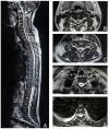
Persistent "MRI-negative" lupus myelitis-disease presentation, immunological profile and outcome
- PMID: 36388234
- PMCID: PMC9659815
- DOI: 10.3389/fneur.2022.968322
Persistent "MRI-negative" lupus myelitis-disease presentation, immunological profile and outcome
Abstract
Introduction: Myelitis is the least common neuropsychiatric manifestation in systemic lupus erythematosus (SLE). Magnetic resonance imaging (MRI)-negative myelitis is even rarer. Here, we present the largest cohort of MRI-negative lupus myelitis cases to assess their clinical and immunological profiles and outcome.
Method: A single-center, observational study conducted over a period of 5 years (2017-2021) was undertaken to evaluate patients with MRI-negative lupus myelitis for the epidemiological, clinical, immunological, and radiological features at baseline and followed up at monthly intervals for a year, and the outcomes were documented. Among the 22 patients that presented with MRI-negative myelopathy (clinical features suggestive of myelopathy without signal changes on spinal-cord MRI [3Tesla], performed serially at the time of presentation and 7 days, 6 weeks, and 3 months after the onset of symptoms), 8 patients had SLE and were included as the study population.
Results: In 8 of 22 patients presenting with MRI-negative myelopathy, the etiology was SLE. MRI-negative lupus myelitis had a female preponderance (male: female ratio, 1:7). Mean age at onset of myelopathy was 30.0 ± 8.93 years, reaching nadir at 4.9 ± 4.39 weeks (Median, 3.0; range, 1.25-9.75). Clinically, cervical cord involvement was observed in 75% of patients, and 62.5% had selective tract involvement. The mean double stranded deoxyribonucleic acid, C3, and C4 titers at onset of myelopathy were 376.0 ± 342.88 IU/ml (median, 247.0), 46.1 ± 17.98 mg/dL (median, 47.5), and 7.3 ± 3.55 mg/dL (median, 9.0), respectively, with high SLE disease activity index 2,000 score of 20.6 ± 5.9. Anti-ribosomal P protein, anti-Smith antibody, and anti-ribonuclear protein positivity was observed in 87.5, 75, and 75% of the patients, respectively. On follow-up, improvement of myelopathic features with no or minimal deficit was observed in 5 of the 8 patients (62.5%). None of the patients had recurrence or new neurological deficit over 1-year follow-up.
Conclusion: Persistently "MRI-negative" lupus myelitis presents with white matter dysfunction, often with selective tract involvement, in light of high disease activity, which follows a monophasic course with good responsiveness to immunosuppressive therapy. A meticulous clinical evaluation and a low index of suspicion can greatly aid in the diagnosis of this rare clinical condition in lupus.
Keywords: MRI-negative lupus myelitis; MRI-negative myelitis; myelitis in lupus; neuropsychiatric systemic lupus erythematosus; selective tractopathy; systemic lupus erythematosus.
Copyright © 2022 Das, Ray, Chakraborty, Banerjee, Pandit, Das and Dubey.
Conflict of interest statement
The authors declare that the research was conducted in the absence of any commercial or financial relationships that could be construed as a potential conflict of interest.
Figures
References
LinkOut - more resources
Full Text Sources
Miscellaneous


