

Optical diagnostic imaging and therapy for thyroid cancer
- PMID: 36388462
- PMCID: PMC9640994
- DOI: 10.1016/j.mtbio.2022.100441
Optical diagnostic imaging and therapy for thyroid cancer
Abstract
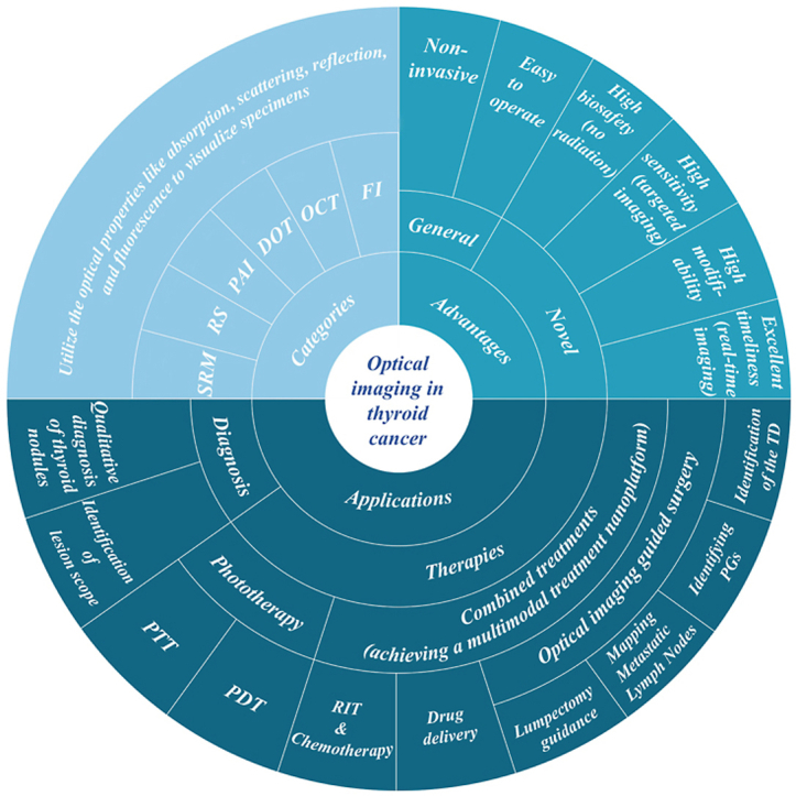
Thyroid cancer, as one of the most common endocrine cancers, has seen a surge in incidence in recent years. This is most likely due to the lack of specificity and accuracy of its traditional diagnostic modalities, leading to the overdiagnosis of thyroid nodules. Although there are several treatment options available, they are limited to surgery and 131I radiation therapy that come with significant side effects and hence cannot meet the treatment needs of anaplastic thyroid carcinoma with very high malignancy. Optical imaging that utilizes optical absorption, refraction and scattering properties, not only observes the structure and function of cells, tissues, organs, or even the whole organism to assist in diagnosis, but can also be used to perform optical therapy to achieve targeted non-invasive and precise treatment of thyroid cancer. These applications of screening, diagnosis, and treatment, lend to optical imaging's promising potential within the realm of thyroid cancer surgical navigation. Over the past decade, research on optical imaging in the diagnosis and treatment of thyroid cancer has been growing year by year, but no comprehensive review on this topic has been published. Here, we review key advances in the application of optical imaging in the diagnosis and treatment of thyroid cancer and discuss the challenges and potential for clinical translation of this technology.
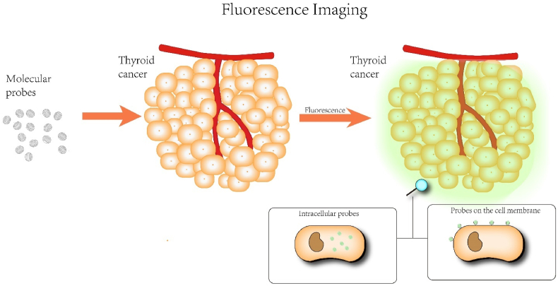
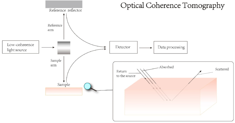
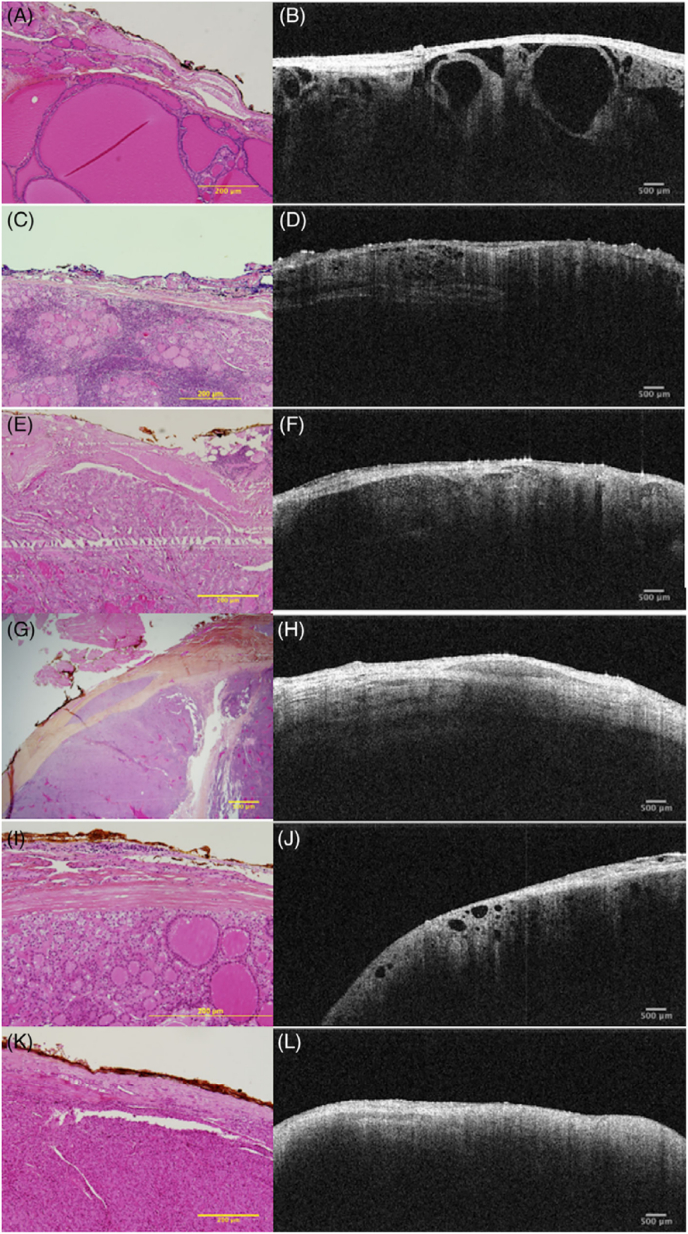
Keywords: 131I-BSA@CuS, 131I-labeled BSA-modified CuS nanoparticles; 5-ALA, 5-Aminolevulinic acid; ASIR, age-standardized rates of cancer incidence; ATC, anaplastic thyroid carcinoma; Au@MSNs, photo-triggered Gold nanodots capped mesoporous silica nanoparticles; AuNCs@BSA-I, innovative iodinated gold nanoclusters; BRAF, V-Raf murine sarcoma viral oncogene homolog B; CBDCA, Carboplatin; CDFI, color doppler flow imaging ultrasound; CLND, central compartmentalized node dissection; CPDA-131I NPs, the 131I-radiolabeled cerebroid polydopamine nano-particles; CT, Computed Tomography; DOT, Diffuse Optical Tomography; DTC, differentiated thyroid cancer; ECDT, enhanced chemodynamical therapy; EGF, epidermal growth factor; EGFR, epidermal growth factor receptor; ESMO, European Society of Medical Oncology; FDA, U.S. Food and Drug Administration; FI, fluorescence imaging; FNAB, fine-needle aspiration biopsy; FNAs, fine needle aspirations; FTC, follicular thyroid carcinoma; GC, germinal center; HAOA, Hyaluronic Acid and Oleic Acid; HYP, hypericin; ICG, indocyanine green; IJV, internal jugular vein; IR825@B-PPNs, Polymeric NPs with bevacizumab and IR825 conjugated on the surface; L-A PTA, laparoscopic photothermal ablation; MDR, multidrug resistance; MTC, medullary thyroid carcinoma; Multimodal therapy; NIR, near-infrared; NIR-FI, near-infrared fluorescence imaging; NIR-PIT, near-infrared photoimmunotherapy; NIRF, near-infrared fluorescence; NMRI, Nuclear Magnetic Resonance Imaging; OCT, Optical Coherence Tomography; OI, optical imaging; OS, overall survival; Optical imaging; Optical imaging-guided surgery; PAI, Photoacoustic Imaging; PDT, photodynamic therapy; PET, Positron Emission Tomography; PGs, parathyroid glands; PLP, porphyrin-HDL nanoparticle; PTA, photothermal reagents; PTC, papillary thyroid carcinoma; PTT, photothermal therapy; Pd-MOF, porphyrin–palladium metal–organic framework; Phototherapy; RIT, radioactive iodine therapy; ROS, reactive oxygen species; SEC, Selenocysteine; SV, subclavian vein; SiRNA, interfering RNA; TC, thyroid cancer; TD, Thoracic Duct; TF, tissue factor; Thyroid cancer; mETE, microscopic extrathyroidal extension.
© 2022 The Authors.
Conflict of interest statement
The authors declare that they have no known competing financial interests or personal relationships that could have appeared to influence the work reported in this paper.
Figures
Similar articles
-
Management of thyroid cancer: United Kingdom National Multidisciplinary Guidelines.J Laryngol Otol. 2016 May;130(S2):S150-S160. doi: 10.1017/S0022215116000578. J Laryngol Otol. 2016. PMID: 27841128 Free PMC article.
-
A sequential targeting nanoplatform for anaplastic thyroid carcinoma theranostics.Acta Biomater. 2020 Jan 15;102:367-383. doi: 10.1016/j.actbio.2019.11.043. Epub 2019 Nov 26. Acta Biomater. 2020. PMID: 31778831
-
Indocyanine Green-Loaded Silver Nanoparticle@Polyaniline Core/Shell Theranostic Nanocomposites for Photoacoustic/Near-Infrared Fluorescence Imaging-Guided and Single-Light-Triggered Photothermal and Photodynamic Therapy.ACS Appl Mater Interfaces. 2016 Dec 28;8(51):34991-35003. doi: 10.1021/acsami.6b11262. Epub 2016 Dec 13. ACS Appl Mater Interfaces. 2016. PMID: 27957854
-
NIR-activated multifunctional agents for the combined application in cancer imaging and therapy.Adv Colloid Interface Sci. 2025 Feb;336:103356. doi: 10.1016/j.cis.2024.103356. Epub 2024 Nov 22. Adv Colloid Interface Sci. 2025. PMID: 39612723 Review.
-
64Cu-Labeled bis-1,4,7,10-tetraazacyclododecane-N,N',N,N'-tetraacetic acid conjugated hypericin.2011 May 3 [updated 2011 May 26]. In: Molecular Imaging and Contrast Agent Database (MICAD) [Internet]. Bethesda (MD): National Center for Biotechnology Information (US); 2004–2013. 2011 May 3 [updated 2011 May 26]. In: Molecular Imaging and Contrast Agent Database (MICAD) [Internet]. Bethesda (MD): National Center for Biotechnology Information (US); 2004–2013. PMID: 21656982 Free Books & Documents. Review.
Cited by
-
Advances in Antioxidant Applications for Combating 131I Side Effects in Thyroid Cancer Treatment.Toxics. 2023 Jun 13;11(6):529. doi: 10.3390/toxics11060529. Toxics. 2023. PMID: 37368629 Free PMC article. Review.
-
TGF-β blocking combined with photothermal therapy promote tumor targeted migration and long-term antitumor activity of CAR-T cells.Mater Today Bio. 2023 Mar 22;20:100615. doi: 10.1016/j.mtbio.2023.100615. eCollection 2023 Jun. Mater Today Bio. 2023. PMID: 37063775 Free PMC article.
-
Non-invasive imaging with ICOS-targeting monoclonal antibody for preclinical diagnosis of rheumatoid arthritis in a humanized mouse model.J Transl Med. 2025 Feb 4;23(1):150. doi: 10.1186/s12967-024-05899-w. J Transl Med. 2025. PMID: 39905410 Free PMC article.
-
No Correlation between PD-L1 and NIS Expression in Lymph Node Metastatic Papillary Thyroid Carcinoma.Diagnostics (Basel). 2024 Aug 26;14(17):1858. doi: 10.3390/diagnostics14171858. Diagnostics (Basel). 2024. PMID: 39272644 Free PMC article.
-
Near-infrared photoimmunotherapy: mechanisms, applications, and future perspectives in cancer research.Antib Ther. 2025 Jan 20;8(1):68-85. doi: 10.1093/abt/tbaf001. eCollection 2025 Jan. Antib Ther. 2025. PMID: 39958565 Free PMC article. Review.
References
-
- Cooper D.S., Doherty G.M., Haugen B.R., Kloos R.T., Lee S.L., Mandel S.J., Mazzaferri E.L., McIver B., Pacini F., Schlumberger M., Sherman S.I., Steward D.L., Tuttle R.M. Revised American thyroid association management guidelines for patients with thyroid nodules and differentiated thyroid cancer. Thyroid. 2009;19(11):1167–1214. doi: 10.1089/thy.2009.0110. - DOI - PubMed
Publication types
LinkOut - more resources
Full Text Sources
Research Materials
Miscellaneous
